
1. Introduction
The fluorescence in situ hybridization (FISH) technique has been successfully used to investigate chromosomal structure and to obtain molecular information regarding DNA organization (Bugno-Poniewierska et al., 2009). One of the main advantages of this method is its broad applicability, as it can be used not only for the analysis of metaphase chromosomes but also for the examination of interphase nuclei, germ cells (spermatozoa and oocytes), embryos, and chromatin fibers. This wide range of applications makes the FISH technique a powerful tool for comprehensive and detailed cytogenetic studies of animal cells (Iannuzzi et al., 2023).
The sperm-FISH technique allows for the identification of fluorescent signals specific to selected chromosomes in male germ cells, enabling the detection of chromosomal number abnormalities such as aneuploidies and polyploidies. This method is valuable for the accurate assessment of the reproductive potential of carriers of chromosomal abnormalities (Anton et al., 2007). Animal studies conducted over the past two decades have demonstrated the use of sperm-FISH to examine and validate sex-sorted semen (Reinsalu et al., 2019) and for evaluating the extent of abnormal meiotic segregation leading to chromosomal aberrations. The first sperm-FISH techniques were applied in two laboratory animal species: mice (Adler et al., 1996; Hill et al., 2003) and rats (Lowe et al., 1998). In livestock species, this technique has so far been applied on spermatozoa of cattle (Hassanane et al., 1999; Kobayashi et al., 1999; Piumi et al., 2001; Rens et al., 2001; Di Berardino et al., 2004; Bonnet-Garnier et al., 2006; Bugno et al., 2008; Vozdová et al., 2008; Nicodemo et al., 2009; Pauciullo et al., 2011, 2012; Baracs et al., 2018), pig (Rubeš et al., 1999; Parrilla et al., 2003; Pinton et al., 2004), water buffalo (Di Berardino et al., 2004; Di Dio et al., 2020), sheep and goat (Di Berardino et al., 2004), and horse (Bugno-Poniewierska et al., 2009; Bugno et al., 2010). Spermatozoa of companion animals, such as the dog (Komaki et al., 2014; Oi et al., 2013) and cat (Kij-Mitka et al., 2021), have also been of interest to researchers.
A crucial step of the sperm-FISH technique is the decondensation of highly condensed sperm chromatin, the degree of which varies between species and even among individuals within the same species (Bugno-Poniewierska et al., 2009). Both excessive condensation and excessive susceptibility to decondensation of sperm nuclear chromatin impede effective hybridization of chromosome-specific probes and reduce the quality of fluorescent signals. Therefore, it is essential to apply appropriate decondensation methods that, by disrupting disulfide bridges, enable relaxation of sperm nuclear heterochromatin (Kobayashi et al., 1999). The literature also highlights methodological differences in the stages of probe denaturation and hybridization with DNA (Bugno-Poniewierska et al., 2009; Bugno et al., 2010; Pauciullo et al., 2012; Barasc et al., 2018; Reinsalu et al., 2019; Di Dio et al., 2020; Galindo et al., 2021).
Although numerous reports describe the use of the sperm-FISH technique, its broader application remains limited due to the lack of validated protocols for routine multi-bull screening. In domestic cattle, this method has primarily been applied in single cases, often involving carriers of previously identified chromosomal aberrations (Nicodemo et al., 2009; Pauciullo et al., 2011, 2012; Barasc et al., 2018; Di Dio et al., 2020). For instance, bovine-derived probes have been successfully employed to investigate chromosomal abnormalities, meiotic segregation, and sex chromosome identification in river buffalo (Révay et al., 2003; Di Berardino et al., 2004; Di Dio et al., 2020), as well as to analyze gametic segregation patterns and chromosomal rearrangements in cervids (Galindo et al., 2021). However, data on the application of this technique in large-scale studies involving breeding bulls remain scarce. Optimization of this methodology could enable its routine use in screening breeding bulls for chromosomal abnormalities, validating sex-sorted semen, and supporting cytogenetic monitoring in cattle breeding programs. We hypothesized that decondensation strategies based on distinct mechanisms of sperm chromatin relaxation (reduction-based, enzymatic, and alkaline) would differentially influence hybridization efficiency, fluorescent signal resolution, and inter-individual reproducibility, and that one approach would provide superior suitability for population-scale sperm-FISH screening in cattle. Consequently, the purpose of this study is to determine, among three protocols, which one is capable of producing the greatest number of properly decondensed cattle spermatozoa.
2. Material and methods
2.1. Semen preparation for three experiments
The study material consisted of semen samples from 10 bulls obtained from the resources of the Malopolska Biotechnic Center in Krasne. The animals were randomly selected from a population of 100 bulls and represented actively used reproductive bulls routinely included in artificial insemination programs. The selected individuals represented different breeds, including Polish Red-and-White, Polish Red, and Simmental cattle. Semen samples were collected in the form of commercial insemination doses and met the applicable quality criteria required for their use in artificial insemination. Prior to the present study, semen quality had been assessed using the computer-assisted semen analysis system SCA® CASA (Microptic, Spain), and the obtained motility and concentration parameters were within the reference ranges considered normal for this species. Two straws of frozen semen were obtained from each of the 10 bulls, then pooled and stored at −20°C. For the experiment, the samples were thawed and subsequently subjected to three types of semen preparation, decondensation, and in situ hybridization (Figure 1). Before implementation in the main experiment, all three decondensation protocols underwent a 1 year validation process. During this stage, decondensation conditions were systematically optimized by evaluating the effects of temperature, incubation time, and individual variability across several independent populations, each comprising at least 10 individuals. Based on these results, the optimal decondensation parameters were established and subsequently applied in the main study. Following completion of the validation stage, the main experiment was conducted using all three validated protocols for each of 10 individuals. Preparation of the slides and subsequent analyses were completed within approximately 1 month.
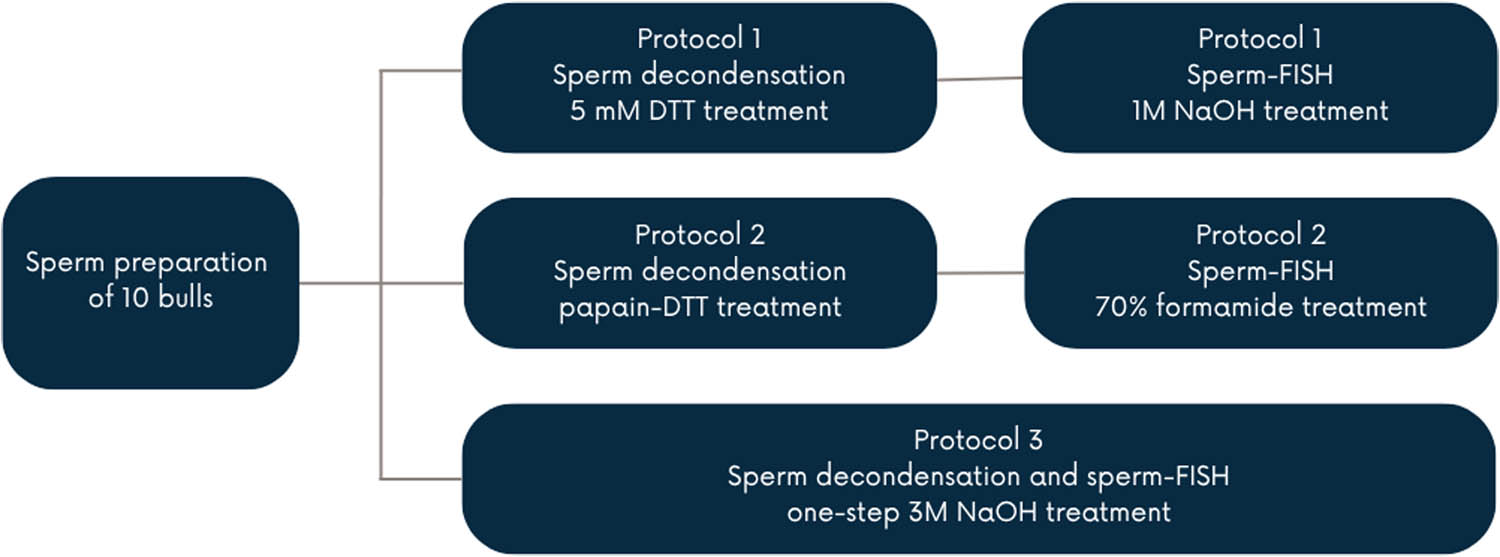
Figure 1
Experimental design.
2.2. Preparation of molecular probes
In each of the FISH protocols, the same molecular probes were used, obtained and labeled according to the following protocol. Cultivation of BAC probes obtained from INRA library specific for chromosomes 1 (INRA-989G02) and 29 (INRA-317E09). Isolation of BAC DNA was performed according to the methodology developed by BACPAC Genomics, Inc. (Emeryville, California, USA). A single bacterial colony containing DNA was inoculated into 2 mL of LB medium (Sigma-Aldrich, Germany) with chloramphenicol (Sigma-Aldrich, Germany) and cultured overnight at 37°C with shaking at 300 rpm. After incubation, the cells were centrifuged and then resuspended in lysis buffers. A neutralization buffer was added to precipitate cellular debris and genomic DNA. Samples were cooled on ice and centrifuged. The supernatant was transferred to a new tube containing chilled isopropanol to precipitate BAC DNA. After incubation on ice, DNA was centrifuged, washed with 70% ethanol, and left to air-dry. The pellet was resuspended in 20 µL TE buffer at room temperature (RT) and then stored at −20°C. Probe labeling was performed using 2 µL containing 200 ng DNA for each probe. Labeling was carried out with the BioPrime Array CGH Genomic Labeling System (Invitrogen, USA) using the Spectrum Green and Spectrum Red fluorochromes (Abbott, USA), according to the manufacturers’ instructions.
3. Protocol 1 of sperm-FISH (DTT in PBS)
3.1. Semen preparation and decondensation
The decondensation protocol was based on the protocol of Galindo et al. (2021) with minor modifications. Semen samples (50 µL each) were washed three times in 250 µL of 1× PBS to remove the extender. Each time, samples were centrifuged at 380×g, and the supernatant was removed. Finally, the pellet was resuspended in 500 µL of 5 mM DTT and incubated for 30 min at RT. Next, the semen was washed three times by adding 300 µL of 1× PBS. In the last wash, two drops of Carnoy’s fixative were added to the semen in 1× PBS, centrifuged at 380×g, and finally resuspended in Carnoy’s fixative, leaving the mixture for 30 min at −20°C. Microscope slides were prepared by dropping the pellet onto a glass slide. An area with appropriate concentration and degree of semen decondensation was selected and marked. Slides were stored at −20°C until further analysis.
3.2. FISH
The FISH protocol was performed according to the protocol of Galindo et al. (2021) with modifications. Semen slides were incubated in 2 × SSC at 70°C for 30 min, then the solution with the slides was cooled to RT, after which the slides were transferred to 0.1 × Saline-Sodium Citrate (SSC) at RT for 1 min, then to 1 M NaOH at RT for 1 min, and subsequently to 0.1 × SSC at 4°C for 1 min and 2 × SSC at 4°C for 1 min. Slides were passed through an alcohol series (70, 85, 100%) for 1 min each at RT. Slides were left to dry. Molecular probes were denatured at 92°C for 10 min and then at 37°C for 30 min. Denatured probes were applied to the slide, covered with a coverslip, sealed with glue, and incubated at 37°C for a minimum of 16 h in a humid chamber. The next day, after removing the coverslips, slides were washed in 2 × SSC at RT for 2 min, and then washed in the following solutions: 0.7 × SSC with 0.3% Octylphenoxy poly(ethyleneoxy)ethanol (IGEPAL®, Sigma-Aldrich, Germany) at 72°C for 2 min and 2 × SSC at RT for 2 min. 4′,6-Diamidino-2-phenylindole dihydrochloride (DAPI) was applied directly to the wet slide, and then a coverslip was placed.
4. Protocol 2 of sperm-FISH (Papain-DTT solution)
4.1. Semen preparation and decondensation
Decondensation was performed based on the protocol described by Bugno et al. (2010) with minor modifications. Semen samples (50 µL each) were washed three times in physiological saline (0.9% NaCl), gently mixed, and centrifuged at 1,100×g for 5 min, and the supernatant was removed. Then, 10 µL of semen was placed on a degreased glass slide, and a smear was made using a coverslip by pulling the drop behind the coverslip. Slides were dried vertically at RT, the concentration was checked, and then the slides were left in absolute ethanol at 4°C until the next day. Subsequently, the slide was dried, and decondensation was performed in a fresh papain-DTT solution (25 g papain, 0.16 g DTT, 0.5 mL dimethyl sulfoxide, 0.2 M TRIS buffer to 100 mL) at 42°C for 40 min. Next, slides were rinsed in TRIS buffer and passed through an alcohol series (95, 97, 100%) at RT for 3 min each. The degree of decondensation was checked under a phase-contrast microscope, and the best area for hybridization was marked.
4.2. FISH
The FISH method was performed according to the protocol described by Bugno et al. (2010), with minor modifications adapted for BAC probe detection. Slides with sperm smears were subjected to enzymatic digestion in a pepsin solution for 30 min at 37°C. After digestion, the slides were washed three times in 2 × SSC for 1 min at RT and then rinsed in distilled water. Samples were dehydrated in an ethanol series (70, 80, 90%; 3 min each at RT) and dried at 60°C for 30 min. Chromatin denaturation was performed in 70% formamide at 78°C for 10 min, followed by a second dehydration series (70, 80, 90%) at −20°C for 3 min each. BAC probes were denatured at 80°C for 10 min and then incubated at 37°C for 30 min. They were then applied to the previously marked, appropriately decondensed areas and sealed with glue. Hybridization was performed for a minimum of 16 h at 37°C in a humid chamber. The next day, slides were rinsed in 2 × SSC at RT until the coverslips detached, and slides were washed three times in 50% formamide at 42°C for 5 min, followed by three washes in 2 × SSC buffer at 42°C for 5 min. Slides were incubated in detergent wash solution buffer (4 × SSC, 0.01% Tween 20) for 10 min at RT. DAPI was applied to the wet slide, and then a coverslip was placed.
5. Protocol 3 of sperm-FISH (3 M NaOH)
5.1. Semen preparation
Semen samples (50 µL each) were washed 1:1 with PBS washing buffer 1× phosphate-buffered saline (PBS) supplemented with 6 mM ethylenediaminetetraacetic acid (EDTA), the supernatant was removed, and then the pellet was resuspended in PBS washing buffer three times in 200 µL to remove the extender. Each time, samples were centrifuged at 380×g, and the supernatant was removed. In the last wash, Carnoy’s fixative was added to the supernatant at a 1:1 ratio, after which samples were centrifuged twice at 380×g, the supernatant was removed and finally resuspended in 20 µL of Carnoy’s fixative. Microscope slides were then prepared by dropping the pellet onto a glass slide. An area with appropriate concentration and degree of semen decondensation was selected and marked. For fixation, slides were left at 37°C for a minimum of 4 h.
5.2. Decondensation and the FISH technique
Decondensation and the FISH technique were performed according to the protocol developed based on Pauciullo et al. (2012) and Di Dio et al. (2020) with minor modifications, using 3 M NaOH as an agent enabling both decondensation and denaturation. Semen slides were dehydrated in an alcohol series (70, 80, 90%) at RT for 3 min each, then dried, and semen decondensation was performed in 3 M NaOH at RT for 3 min, rinsed in distilled water, and then dehydrated in an alcohol series under the same parameters. BAC probes were denatured at 90°C for 10 min and then incubated at 37°C for 20 min. The probes were then applied to the slides and sealed with glue. Hybridization was performed at 37°C in a humid chamber for at least 16 h. The next day (protocol according to Bugno et al., 2010, with modifications), slides were rinsed in 2 × SSC at RT until the coverslips detached, then washed three times in 50% formamide at 42°C for 5 min, followed by three washes in 2 × SSC buffer at 42°C for 5 min. Slides were incubated in detergent wash solution buffer (4 × SSC, 0.01% Tween 20) for 10 min at RT. DAPI was applied to the wet slide, and then a coverslip was placed.
5.3. Detection of fluorescent signals
Fluorescent signals were recorded using a Zeiss Axio Imager M2 fluorescence microscope (Zeiss, Germany), equipped with an Axiocam 702 mono digital camera and ZenPro software (Zeiss, Germany). For each individual, 1,000 spermatozoa were analyzed per protocol. In total, 30,000 spermatozoa were examined (10 bulls × 1,000 spermatozoa per bull × 3 protocols), corresponding to 10,000 spermatozoa per protocol. Spermatozoa were classified into three categories based on defined morphological and hybridization criteria (properly decondensed, non-decondensed, over-decondensed) after decondensation and sperm-FISH. All scoring was performed by a single trained observer using standardized criteria based on the presence and morphology of fluorescent signals (Figure 2). Only slides with confirmed probe hybridization were included in the microscopic evaluation. Following hybridization, a 24 × 24 mm coverslip was placed centrally on each slide. All specimens were examined according to the same predefined scanning pattern, beginning from a fixed reference point and proceeding in a consistent direction across the preparation. The order of slide evaluation was randomized, and all analyses were performed blinded to the treatment groups to minimize selection and observer bias.
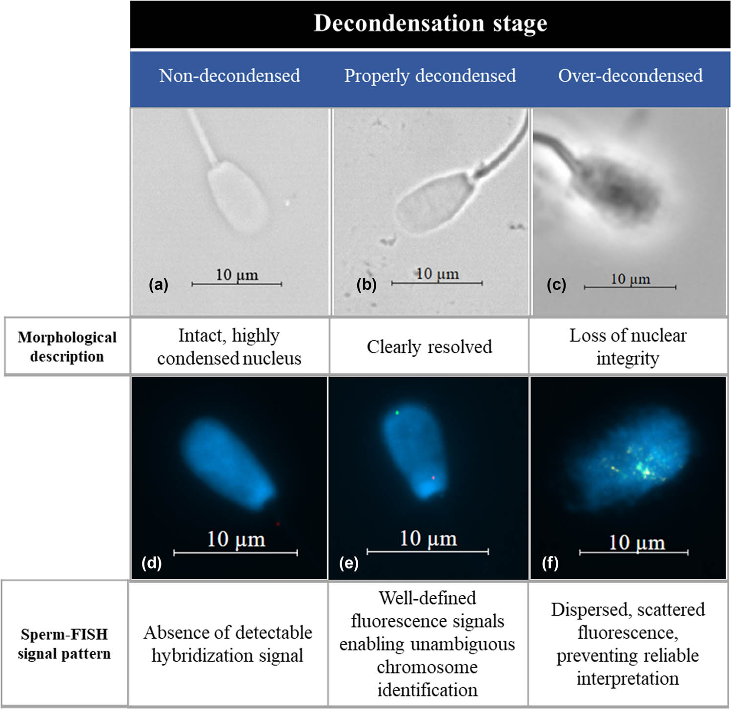
Figure 2
Representative examples of sperm nuclear chromatin decondensation and the corresponding FISH outcomes. Upper panel (a–c) shows phase-contrast microscopy images illustrating three stages of decondensation: (a) non-decondensed sperm with an intact, highly condensed nucleus; (b) properly decondensed sperm with a clearly resolved nucleus; and (c) over-decondensed sperm exhibiting loss of nuclear integrity. Lower panel (d–f) shows the corresponding FISH images: (d) absence of detectable hybridization signals in non-decondensed sperm; (e) well-defined fluorescence signals enabling unambiguous chromosome identification following proper decondensation; and (f) dispersed, scattered fluorescence signals in over-decondensed sperm, preventing reliable interpretation of the FISH results.
5.4. Statistical analysis
Statistical analysis was performed using SAS version 9.4 (SAS Institute Inc., Cary, NC, USA). Before statistical analysis, the number of non-decondensed and over-decondensed spermatozoa were summed for each animal and protocol, thereby generating two response categories: properly decondensed and improperly decondensed spermatozoa. The experimental unit was the bull, while spermatozoa were treated as binomial counts nested within each bull-protocol combination. A binomial generalized linear mixed model was fitted using PROC GLIMMIX, with a logit link function and the Laplace approximation as the estimation method. The number of properly decondensed spermatozoa was used as the numerator and the total number of evaluated spermatozoa as the denominator. Protocol was included as a fixed effect, and bull was included as a random intercept to account for the repeated-measures structure of the study and for inter-individual variability among bulls. Least squares means were backtransformed from the logit scale to the probability scale and are reported with their standard errors. Pairwise comparisons among protocols were performed using the DIFF option, with Tukey adjustment for multiple comparisons. Statistical significance was declared at P ≤ 0.05.
6. Results
Each of the applied protocols enabled the production of good proportion of proper sperm chromatin decondensation, confirming that an appropriate level of nuclear chromatin relaxation had been achieved and thereby ensuring hybridization specificity for accurate assessment of fluorescent signal copy number.
In each decondensation protocol, three categories of spermatozoa were observed: non-decondensed, properly decondensed, and over-decondensed (Figure 2). Assessment based solely on the degree of chromatin decondensation was insufficient to reliably distinguish all three categories, as only over-decondensed spermatozoa could be readily identified morphologically. Clear discrimination between the three categories was only possible after sperm-FISH hybridization. Non-decondensed spermatozoa were characterized by the absence of fluorescent signals, adequately decondensed spermatozoa exhibited distinct and well-defined fluorescent signals, whereas over-decondensed spermatozoa displayed dispersed fluorescent signals that precluded reliable interpretation of the hybridization results.
Statistical analysis showed significant differences among the protocols (P < 0.001). Protocol 3 resulted in the highest proportion of properly decondensed spermatozoa (89.7%), followed by Protocol 2 (77.4%), and Protocol 1 (74.9%) (Table 1).
Table 1
Effect of protocol on the percentage of properly decondensed spermatozoa, expressed as least-squares means ± standard error, of the total spermatozoa analyzed
| Protocol 1 | Protocol 2 | Protocol 3 | |
|---|---|---|---|
| Properly decondensed spermatozoa | 74.9 ± 0.554 A | 77.4 ± 0.518 B | 89.7 ± 0.344 C |
A–C – Mean values in the same row with different letters significantly differ (P < 0.05).
7. Discussion
Among the most important factors for the success of the sperm-FISH technique is the effective decondensation of sperm nuclear chromatin, which allows fluorescent probes to hybridize with the target DNA sequences. In the case of cattle spermatozoa, this process is particularly demanding due to the high degree of sperm chromatin condensation resulting from the formation of protamine-DNA complexes linked by disulfide bridges (Kobayashi et al., 1999). Compared with human and mouse spermatozoa, bovine sperm chromatin is more condensed and structurally stable, resulting in slower decondensation, which reflects stronger disulfide bond-mediated protamine interactions (Perreault et al., 1988). Similarly, pig and cattle spermatozoa exhibit higher chromatin condensation than human or horse spermatozoa (Ribas-Maynou et al., 2021).
These differences are rooted in the distinct molecular organization of sperm chromatin among species. In humans and horses, sperm DNA is condensed with the participation of protamine 1 and protamine 2, whereas in pigs and cattle, only protamine 1 is present in the chromatin (Corzett et al., 2002). Although protamine 1 has a lower ability to condense DNA compared with protamine 2, bulls show stronger interactions between protamine proteins, resulting from a higher number of cysteine residues capable of forming disulfide bonds compared with other species (Ribas-Maynou et al., 2021). This mechanism ensures high stability of sperm chromatin, which is reflected both in difficult decondensation and in sperm nuclear morphology: in pigs and cattle, nuclei are larger and disc-shaped, while in humans and horses they are smaller and more oval (Gu et al., 2019). Disulfide bonds stabilize sperm chromatin, which requires chemical or physical reducing agents, such as thiols, for decondensation (Jager et al., 1990).
In sperm-FISH, hybridization efficiency depends on chromatin decondensation; therefore, three established and standardized protocols were evaluated. Despite prior validation, differences in efficiency and signal clarity were observed among individuals. Decondensation was performed using three chemical agents: in Protocol 1: DTT, a strong reducing agent that reduces disulfide bridges and prevents the formation of intra- and extracellular disulfide linkages, destroying the quaternary structure of proteins; in Protocol 2: papain, which, due to its enzymatic properties, digests the structure of protein bonds between lysine and arginine; and in Protocol 3: NaOH, which, through alkaline DNA denaturation, causes the breakdown of hydrogen bonds of guanine and thymine.
Regarding Protocol 1, the technique proved unsuitable due to performing decondensation in tubes and the requirement for a substantially larger amount of material compared to the other two protocols. Achieving an appropriate degree of decondensation necessitated multiple attempts, at least 3–4 per sample, as initial preparations frequently contained a high proportion of aggregated and improperly decondensed spermatozoa, resulting in an insufficient number of nuclei suitable for scoring. It should be noted that this protocol employed only DTT, whose effectiveness in decondensing bull sperm chromatin may be limited. This is supported by the findings of Motoishi et al. (1996), which indicate that DTT alone does not consistently achieve complete relaxation of sperm chromatin. Similar observations were reported by Suga et al. (2000), who demonstrated that the use of DTT without additional supporting agents results only in partial decondensation, highlighting the limited efficacy of DTT when used in isolation. One of the challenges was the high sensitivity of semen to washing in the tube following decondensation and fixation with Carnoy’s solution, as fixation after decondensation caused spermatozoa to aggregate, resulting in improper spreading on slides. The study describing the applied protocol on deer semen (Galindo et al., 2021) involved up to 10 animals; however, the hybridization results were comparable to those reported by the authors. Although Bovidae and Cervidae are phylogenetically related, interspecies differences in sperm chromatin organization may influence the efficiency and reproducibility of decondensation and subsequent molecular cytogenetic techniques. The requirement for larger semen volumes and tube-based processing further limits the suitability of Protocol 1 for extensive cytogenetic analyses in cases of limited semen availability, particularly in individuals of high reproductive value, where minimal material consumption is critical.
Protocol 2 reduced reproducibility in subsequent experiments due to its enzymatic properties, which required freshly prepared solutions and introduced inherent variability in the procedure. Semen was already fixed on the slides; therefore, any repetition of the protocol required starting from the beginning each time, taking 3 days per iteration, and up to 4 days when combined with the sperm-FISH procedure to obtain results. With a limited amount of semen material, this could be problematic due to the risk of poor repeatability. Such limitations reduce its usefulness in population-level screening, where repeatability and the ability to reanalyze archived material are essential. In the FISH technique, formamide was used to induce denaturation of sperm DNA, constituting an additional step applied prior to hybridization and important for method validation. It should be noted that, in the FISH procedure, formamide is employed primarily to denature DNA strands and facilitate probe hybridization rather than to induce sperm chromatin decondensation. As reported by Shim et al. (2024), formamide lowers the melting temperature of DNA and stabilizes the transition from the helical to the denatured state, without disrupting protamine-DNA complexes or reducing disulfide bonds responsible for the highly compact sperm chromatin structure. In the context of the authors’ studies on which the protocol was based, the obtained results were consistent with studies conducted by Bugno et al. (2008), Bugno-Poniewierska et al. (2009) and Bugno et al. (2010), however, on a scale of a few individuals, with a different type of painting probes, and also conducted in another species.
Protocol 3 proved to be the most effective and adaptable technique, allowing multiple experimental attempts within a short timeframe, totaling 2 days, including the FISH procedure. A major advantage of this protocol was that the prepared semen in the first step was sufficient for 4–6 subsequent experiments. It demonstrated the highest repeatability in achieving proper decondensation and producing readable fluorescent signals for each individual. This enabled reliable analysis of hybridization signals and facilitated screening studies in the cattle population for chromosomal aberrations in spermatozoa. Other studies have also shown that using NaOH as a denaturing agent, compared with DTT, provides easier-to-control denaturation, better preservation of sperm morphology, and more consistent hybridization results on microscope slides, with the intensity of hybridization in sperm chromatin comparable to that observed in metaphase chromosomes (Habermann et al., 2005; Di Dio et al., 2020). NaOH-based denaturation will outperform DTT-based reduction due to bovine protamine structure. Crucially, Protocol 3 complies with the methodological requirements for large-scale sperm-FISH applications, as it enables repeated analyses from a single semen preparation and ensures high inter-individual repeatability. A comparison of three different methods of sperm chromatin decondensation and the obtained hybridization signals showed a statistically significant difference in the quality of results between protocols, indicating Protocol 3 as the most suitable, particularly when a large number of slides must be analyzed.
The practical relevance of sperm-FISH optimization should be considered in the context of cytogenetic diagnostics applied in cattle breeding, particularly in Poland. The introduction of Instruction No. 1/89 (May 29, 1989) established mandatory cytogenetic testing of breeding bulls, aiming to prevent the dissemination of chromosomal abnormalities through artificial insemination programs. Early studies within Polish breeding programs (Słota et al., 1999) estimated chromosomal abnormalities at 1%, later decreasing to ∼0.5% (Sysa et al., 2002); however, large-scale population data remain limited. Following the implementation of genomic evaluation based on SNP microarrays in 2014 (Gurgul et al., 2015; Strabel, 2025), the routine use of conventional cytogenetics has declined, resulting in reduced availability of recent data on balanced chromosomal aberrations. Although genomic tools are increasingly applied in this field (Iannuzzi et al., 2024), they are not yet fully validated for large-scale detection of structural chromosomal abnormalities in spermatozoa.
In this context, optimized sperm-FISH protocols offer a valuable complementary approach, enabling high-throughput assessment of meiotic segregation (Pinton et al., 2004; Bonnet-Garnier et al., 2008) and supporting the detection of abnormalities affecting fertility. Their application at a population scale could improve estimates of chromosomal aberration prevalence and support cytogenetic monitoring in breeding programs and artificial insemination centers (Ducos et al., 2008). Furthermore, considering species-specific sperm chromatin and its response to reducing agents enables the development of repeatable and reliable protocols, particularly for large-scale studies. Therefore, selecting protocols adapted to species and study scale is essential for consistent cytogenetic data and accurate assessment of reproductive potential.
Acknowledgements
We sincerely thank Malopolska Biotechnic Center in Krasne, for their generous support in providing materials essential for our research.
Funding information
This research was supported by the Chromobull project, grant number 00101.DDD.6509.00197.2022.06.
Author contributions
Conceptualization: M.B.-P. and W.K.-M.; Methodology: W.K.-M., P.P., M.B., A.I., R.P., and A.S.; Formal analysis: W.K.-M. and B.K.-M.; Investigation: W.K.-M., P.P., and M.B.; Writing – original draft: M.B.-P. and W.K.-M.; Writing – review and editing: M.B.-P., P.P., M.B., and W.K.-M.; Funding acquisition: M.B.-P.; and Supervision: M.B.-P.
Conflict of interest statement
The authors declare no conflicts of interest.