
1. Introduction
Over recent decades, rabbit production has undergone notable transformations, largely driven by evolving consumer preferences and changes in production practices. Although rabbit meat consumption remains high or is increasing in countries such as China and Mexico, several European nations with traditionally strong rabbit production sectors, including Italy, Poland, France, and Spain, have experienced a significant decline in both production levels and consumption (Krunt et al., 2022). These developments have been accompanied by the implementation of alternative housing systems designed to improve animal welfare, such as group housing and modified enclosure structures. While these systems may offer welfare benefits, they are often associated with higher production costs (Szendrő et al., 2020) and may also give rise to new behavioural and social issues among animals. In addition, the planned phase-out of conventional cage systems in some countries (Valkova et al., 2021) highlights the growing need to adapt rabbit production to more welfare-oriented housing approaches.
The impact of housing conditions on rabbit performance and meat quality is well documented (Combes et al., 2010; Loponte et al., 2018; Krunt et al., 2022). Housing systems have been shown to affect growth rate, carcass traits, and the major physicochemical properties of meat (Mattioli et al., 2016; Dalle Zotte et al., 2015). Overall, rabbits reared in pens tend to exhibit poorer growth performance and lower dressing percentages, though they often develop more substantial hind limb musculature, reflecting increased physical activity (Combes et al., 2010). Housing conditions may also influence the chemical composition of meat, particularly fat deposition and fatty acid profiles, which are closely associated with muscle development and the intensity of locomotor activity (Szabó et al., 2002).
Alongside production efficiency and meat quality, growing emphasis has been placed on animal welfare, which remains a central challenge in modern rabbit farming. Welfare status is strongly shaped by housing conditions, as the quality of the environment determines animals’ comfort and their opportunities to express natural behaviours. Suboptimal housing, in turn, can increase exposure to social and environmental stressors (Trocino & Xiccato, 2006; Trocino et al., 2022). Recent reviews have highlighted that housing conditions and environmental enrichment can influence stress responsiveness, behaviour, health status, and immune-related processes in rabbits. Environmental stress may affect physiological regulation and immune function, potentially contributing to changes in the expression of genes involved in inflammatory and adaptive responses (El-Sabrout et al., 2024). Ensuring high welfare standards is not only an ethical requirement but also a key factor influencing animal health, resilience, and productivity (Trocino et al., 2022). As housing systems are increasingly modified to meet welfare regulations, it becomes essential to assess how such changes affect rabbits – not only in terms of performance and carcass traits but also at physiological and molecular levels (Ozella et al., 2024).
In this context, recent research has increasingly focused on genetic and molecular indicators that reflect the animals’ responses to environmental and management conditions (Hoffmann & Willi, 2008). Genes involved in immune regulation, inflammation, and stress responses, such as cytokines and chemokine receptors, play a crucial role in animals’ adaptation to their housing environment (Hansen et al., 2012). Alterations in the expression of genes associated with pro- and anti-inflammatory pathways may provide valuable insights into the biological mechanisms underlying rabbits’ adaptation to diverse environments (Rossnerova et al., 2020). Consequently, investigating the expression of such genes may improve our understanding of the biological mechanisms underlying animal adaptation, resilience, and welfare under different housing conditions.
The genes selected for analysis represent key components of inflammatory and regulatory immune pathways. Pro-inflammatory markers (IL6, IL1B, TNF, IL8, and PTGS2) are involved in the initiation and amplification of innate immune responses, whereas IL10 and TGFB1 play important roles in immune regulation and the resolution of inflammation. In addition, CXCR1 is associated with chemokine-mediated immune cell recruitment and migration. Together, these genes provide insight into the balance between immune activation and regulation, allowing assessment of physiological responses to different housing environments (Kim & Moudgil, 2008; Hansen et al., 2012; Hu et al., 2023, 2024).
Popielno White (PW) rabbits were selected for this study because, despite being a native breed maintained within a conservation programme, they exhibit productive performance comparable to that of commercial meat-type rabbits, including satisfactory growth rate and carcass yield. In addition, their good adaptability and disease resistance, characteristic of many native rabbit populations, make them a suitable model for evaluating physiological responses to different housing conditions (Kowalska & Bielański, 2010).
It was hypothesised that different housing systems, providing varying environmental conditions and levels of physical activity, would influence the physiological and immunological status of rabbits, resulting in alterations in the expression of selected molecular markers associated with inflammation, immune regulation, and adaptation. Blood was selected as an indicator of systemic immune responses, whereas the spleen was included as a major secondary lymphoid organ reflecting local immune regulation and lymphocyte activity. The expected effects would be reflected through changes in the expression of genes related to inflammation, immune regulation, and adaptation (CXCR1, IL10, IL8, IL1B, TNF, PTGS2, TGFB1, IL6) in blood and spleen samples. The aim of the present study was to evaluate the impact of housing systems on the expression of selected genes associated with stress response, inflammation, and adaptation processes in PW rabbits.
2. Material and methods
2.1. Animals and experimental design
The experiment was conducted at the National Research Institute of Animal Production in Aleksandrowice and at the Experimental Station of the National Research Institute of Animal Production in Chorzelów, Poland. The experimental material consisted exclusively of PW female rabbits. At weaning (35 days of age), animals were allocated to five housing systems differing in structural design and environmental exposure. Each housing system comprised 16 females. Rabbits were housed in groups of four animals per cage or pen. Consequently, each housing system comprised four cages or pens, which were regarded as independent housing units. The housing systems were as follows: A – hutches with an outdoor run; B – rabbit tractor cages with an outdoor run; C – single-floor cages without bedding inside a livestock building; D – pens on deep litter inside a livestock building; E – modified cages inside a livestock building (two cages connected with a plastic pipe). The images illustrating the housing systems have been previously published and are available in the study by Siudak et al. (2026). The mean body weight at weaning was 951 ± 98 g.
Standard cages measured 60 × 80 cm, whereas pens measured 120 × 80 cm. Outdoor enclosures measured 200 × 100 cm. Indoor rabbits were maintained under natural gravitational ventilation, with ambient temperatures ranging from 14 to 18°C, relative humidity of 50–65%, and a photoperiod of 14 h light and 10 h dark (14L:10D). The mean outdoor temperature ranged from 14 to 22°C. All housing conditions complied with Polish regulations concerning the keeping of farm and fur animals (Journal of Laws, item 1966). Animals were included in a veterinary prophylaxis programme including vaccination against myxomatosis and rabbit haemorrhagic disease. No infectious disease outbreaks were recorded during the experimental period, and mortality did not exceed 3%. For gene expression analysis, eight rabbits per housing system were selected. Rabbits were chosen randomly, with two individuals sampled from each cage or pen to ensure representation across all housing units.
2.2. Feeding and slaughter procedure
During the rearing period, rabbits were fed ad libitum with a complete pelleted diet. The chemical composition of the diet was analysed at the Central Laboratory of the National Research Institute of Animal Production. Digestible energy (DE) of the experimental diets was estimated based on their proximate composition using standard energy coefficients, according to the following equation: DE (kcal/kg dry matter [DM]) = 4.0 × CP + 9.4 × EE + 4.15 × NFE + 1.9 × CF, where CP, EE, CF, and NFE represent crude protein, ether extract, crude fibre, and nitrogen-free extract, respectively (de Blas and Wiseman, 2020). The obtained values were converted to MJ/kg DM (1 MJ = 239 kcal). The ingredients and chemical composition of the diet is presented in Tables 1 and 2.
Table 1
Component composition of feed mixture fed to rabbits
| Component | % |
|---|---|
| Post-extraction soybean meal | 13.00 |
| Alfalfa meal | 20.10 |
| Wheat bran | 18.10 |
| Ground barley | 28.80 |
| Ground maize | 11.00 |
| Rapeseed oil | 2.00 |
| Phosphate | 1.00 |
| NaCl | 0.50 |
| Mineral and vitamin premix | 1.00 |
| Arbocel | 4.50 |
Table 2
Chemical composition of feed mixture
| Chemical composition | Content |
|---|---|
| DM (%) | 90.64 |
| Crude ash (%) | 6.92 |
| Crude fat (%) | 4.19 |
| CP (%) | 15.20 |
| CF (%) | 14.2 |
| NDF (%) | 25.6 |
| ADF (%) | 14.4 |
| ADL (%) | 2.95 |
| NFE (%) | 59.49 |
| DE (MJ/kg DM) | 15.65 |
Note: NDF – neutral detergent fibre; ADF – acid detergent fibre; ADL – acid detergent lignin; NFE – nitrogen-free extract; DE – digestible energy, DP – digestible protein.
At a body weight of 2,600–2,900 g, selected animals were used for gene expression analysis as described above; this resulted in an age range of 90–120 days due to differences in growth rates between housing systems. Animals were fasted for 16 h with free access to water prior to slaughter. Slaughter procedures were carried out according to Blasco et al. (1993). Rabbits were humanely stunned before exsanguination. Peripheral whole blood samples were collected during bleeding from the severed carotid artery into Tempus™ Blood RNA Tubes (Thermo Fisher Scientific, Waltham, MA, USA). Spleen samples were collected immediately post mortem, snap-frozen in liquid nitrogen, and stored at −80°C until further analysis. Blood samples were stored at −20°C prior to RNA isolation.
2.3. Quantitative real-time PCR
Total RNA was isolated from blood samples using the PureLink™ RNA Mini Kit with the PureLink™ DNase Set (Invitrogen, Carlsbad, CA, USA). RNA samples were stored at –80°C until analysis. RNA concentration and purity were assessed with a NanoDrop 2000 spectrophotometer (Thermo Scientific, Waltham, MA, USA), and integrity was verified by 2% agarose gel electrophoresis. cDNA was synthesised from 500 ng of total RNA using the High-Capacity RNA-to-cDNA Reverse Transcription Kit (Applied Biosystems, Foster City, CA, USA) according to the manufacturer’s protocol. Expression of IL6, CXCR1, IL10, TGFB1, IL8, PTGS2, IL1B, and TNF was quantified using TaqMan assays (Applied Biosystems) on a QuantStudio 7 Flex Real-Time PCR System (Thermo Fisher Scientific, Waltham, MA, USA). Reactions were carried out in triplicate using the RT HS-PCR Mix Probe kit (A&A Biotechnology, Gdańsk, Poland) with β-actin (ACTB) and beta-2-microglobulin (B2M) as reference genes (Ni et al., 2022). The TaqMan Gene Expression Assays (Thermo Fisher Scientific) used were: ACTB (Assay ID: Oc03824857_g1-ENDO), B2M (Assay ID: Oc06779339_m1-ENDO), CXCR1 (Assay ID: Oc04250543_s1), IL10 (Assay ID: Oc03396940_m1), IL8 (Assay ID: Oc03397860_m1), IL1B (Assay ID: Oc03823250_s1), TNF (Assay ID: Oc03397715_m1), PTGS2 (Assay ID: Oc03398295_m1), TGFB1 (Assay ID: Oc04176122_u1), and IL6 (Assay ID: Oc04097053_m1). Thermal cycling conditions were 95°C for 5 min, followed by 95°C for 15 s and 60°C for 45 s. Gene expression was analysed using the ΔΔCt method (Livak & Schmittgen, 2001). Relative quantification (RQ) values were calculated as 2−ΔΔCt. Amplification efficiency for each assay was evaluated using standard curves according to the formula E = 10(−1/slope) − 1 and was within the acceptable range of 90–110%. Only assays with comparable amplification efficiencies were included in the analysis. No-template controls were included and showed no amplification. Relative transcript abundance was normalised to reference genes and expressed relative to the calibrator sample with the lowest expression (Pfaffl, 2001).
2.4. Statistical analysis
Statistical analyses were carried out using Statistica 13.1 software (TIBCO Software Inc., Palo Alto, CA, USA). The data were analysed using a mixed-effects model, in which housing system was included as a fixed factor, while cage or hutch nested within housing system was treated as a random effect. The model can be expressed as follows:
where Y ijk denotes the observed gene expression value, μ represents the overall mean, H i corresponds to the effect of housing system, C j (i) is the random effect of cage/hutch nested within housing system, and e ijk is the residual error. The inclusion of cage or hutch as a random effect was intended to account for the hierarchical structure of the data and to minimise the risk of pseudoreplication arising from the group housing of animals. The statistical evaluation was based on relative gene expression values (RQ; 2−ΔΔCt). Although RQ data may not always follow a normal distribution, both normality (assessed using the Shapiro–Wilk test) and homogeneity of variances (tested with Levene’s test) were examined prior to analysis and showed no significant deviations. Consequently, the use of parametric methods was considered appropriate, and no data transformation was required. In addition, exploratory analyses performed on ΔCt values yielded comparable results, supporting the reliability of the analytical approach. Individual rabbits were treated as experimental units, with observations nested within cages or hutches. Differences between housing systems were further examined using pairwise post hoc comparisons, with the level of statistical significance set at P ≤ 0.05. To account for multiple testing across the eight analysed genes within each tissue, P-values for the main effect of housing system were adjusted using the Benjamini–Hochberg false discovery rate (FDR) procedure. FDR correction was performed separately for blood and spleen datasets, and statistical significance after correction was accepted at q ≤ 0.05. Pairwise post hoc comparisons were used to identify differences between housing systems and were interpreted in relation to the FDR-adjusted main effects. Data are presented as mean values ± standard deviation (SD).3. Results
3.1. Blood gene profile
The relative expression levels of selected inflammatory and regulatory genes were analysed in the blood of rabbits kept under different housing systems (A–E) and are presented in Figures 1–8. Results of the FDR-adjusted analysis are presented in Table 3. After FDR correction, significant effects of housing system were confirmed for IL6, CXCR1, IL10, and PTGS2, whereas TGFB1, IL1B, IL8, and TNF did not retain significance. The expression of IL6 differed significantly among the housing systems. Higher expression was observed in groups A and B compared with group C (P ≤ 0.05), whereas expression in group E was significantly lower than in groups A–D (P ≤ 0.05). The expression of CXCR1 varied significantly between housing systems, with significantly higher expression in group E compared with groups A–D (P ≤ 0.01). The IL10 gene exhibited significant variability among groups. Expression was highest in group D and significantly higher than in groups A, B, C, and E (P ≤ 0.05). Expression in groups A and B was higher than in groups C and E (P ≤ 0.05). For TGFB1, pairwise comparisons indicated lower expression in group A than in groups B, C, D, and E (P ≤ 0.05). The PTGS2 gene showed pronounced variation between housing systems. Expression was significantly higher in group D compared with groups A, C, and E (P ≤ 0.05), and in group B compared with group C (P ≤ 0.05). For IL8, pairwise comparisons revealed lower expression in group E compared with groups A, B, C, and D (P ≤ 0.05). For TNF, pairwise differences were observed, with lower expression in group E than in groups A–D (P ≤ 0.05).
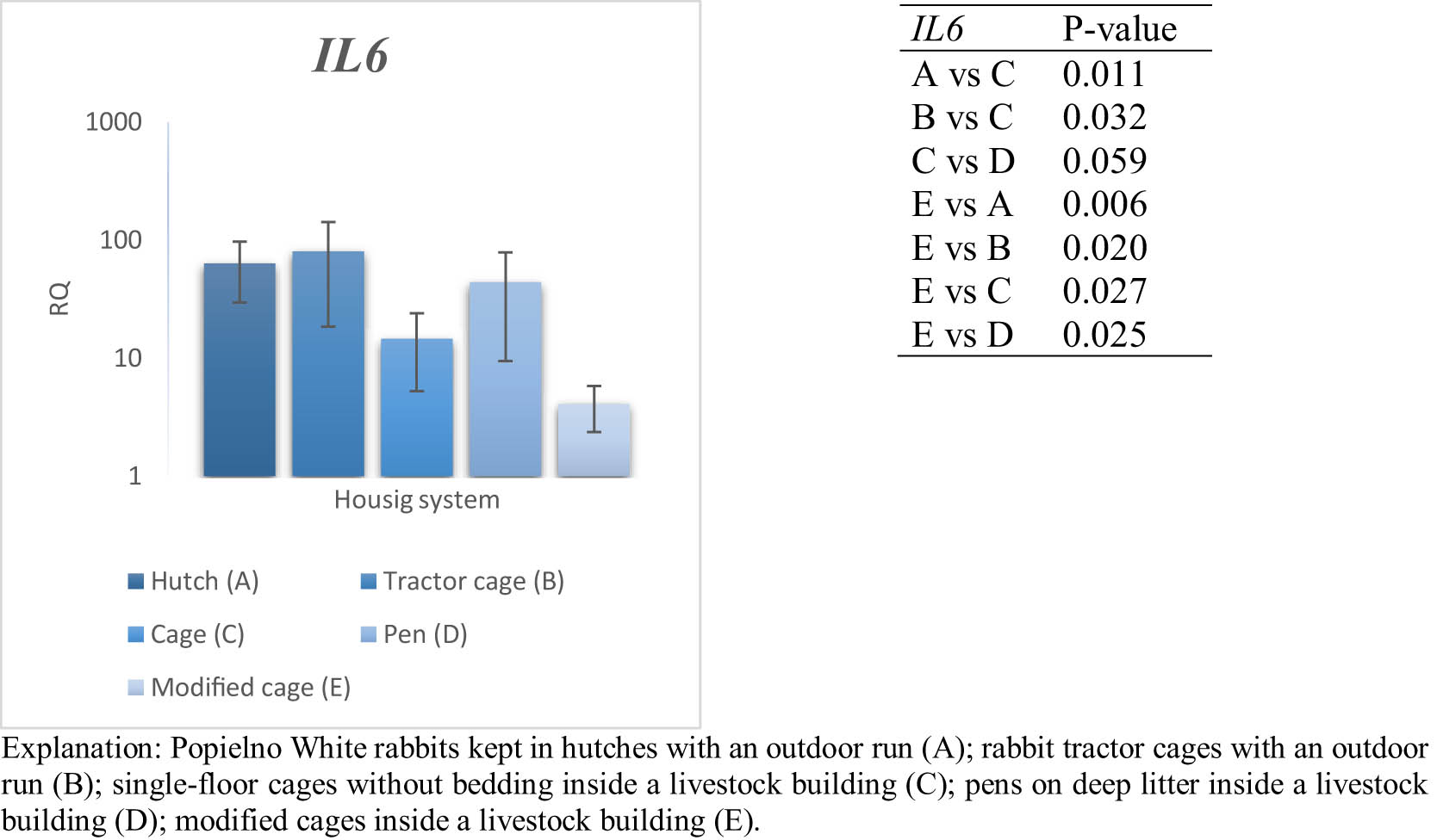
Figure 1
Effect of housing system on the relative expression of the IL6 gene in the blood of PW rabbits (n = 8 rabbits per housing system; mean values ± SD) (RQ – relative quantity normalised to ACTB and B2M).
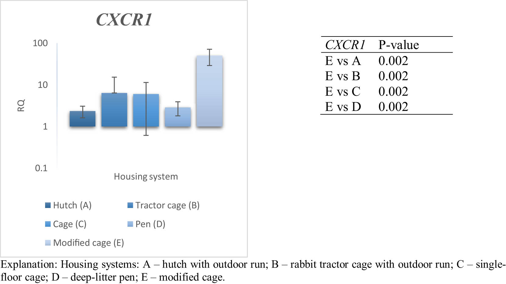
Figure 2
Effect of housing system on the relative expression of the CXCR1 gene in the blood of PW rabbits (n = 8 rabbits per housing system; mean values ± SD) (RQ – relative quantity normalised to ACTB and B2M).
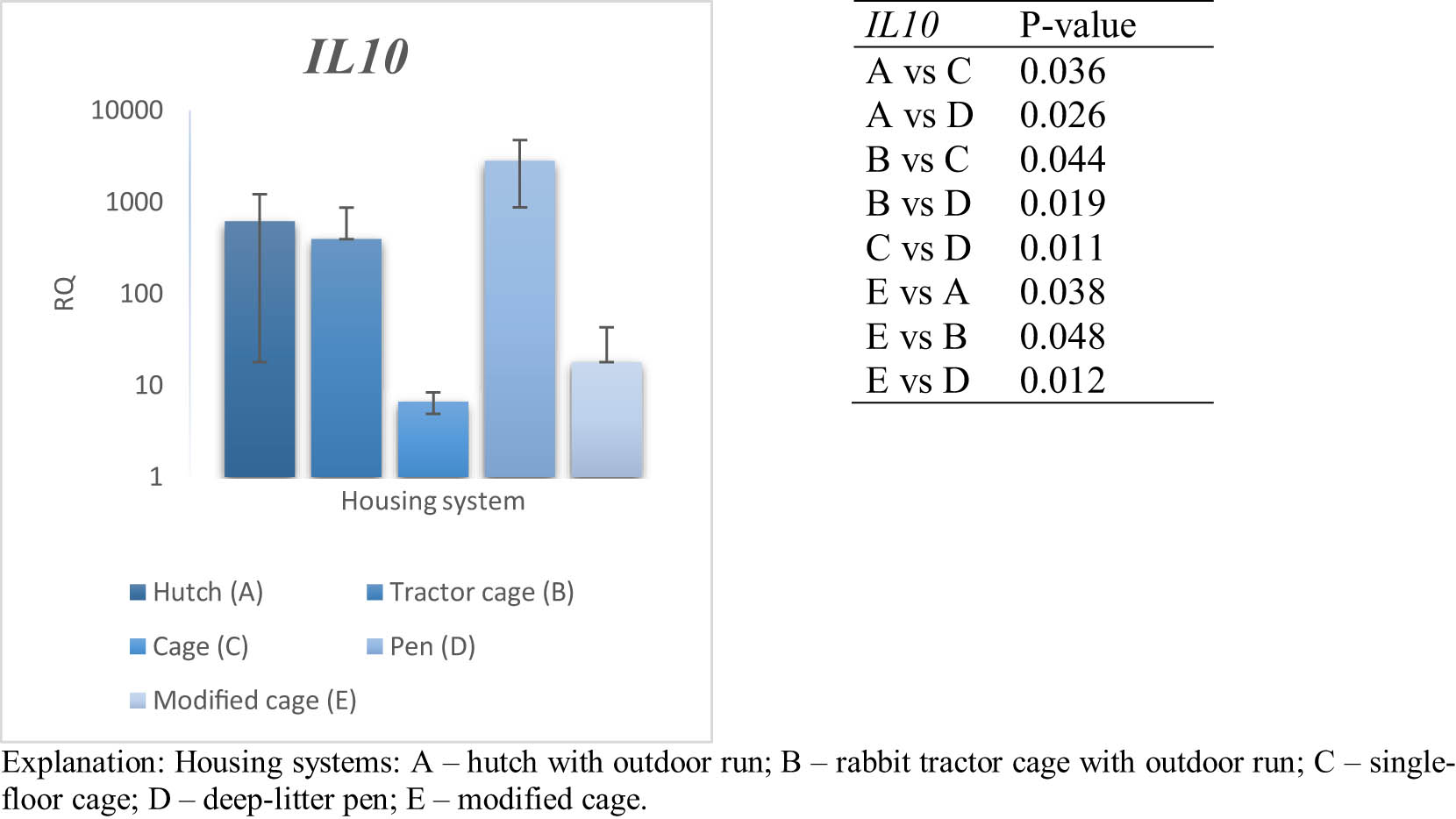
Figure 3
Effect of housing system on the relative expression of the IL10 gene in the blood of PW rabbits (n = 8 rabbits per housing system; mean values ± SD) (RQ – relative quantity normalised to ACTB and B2M).
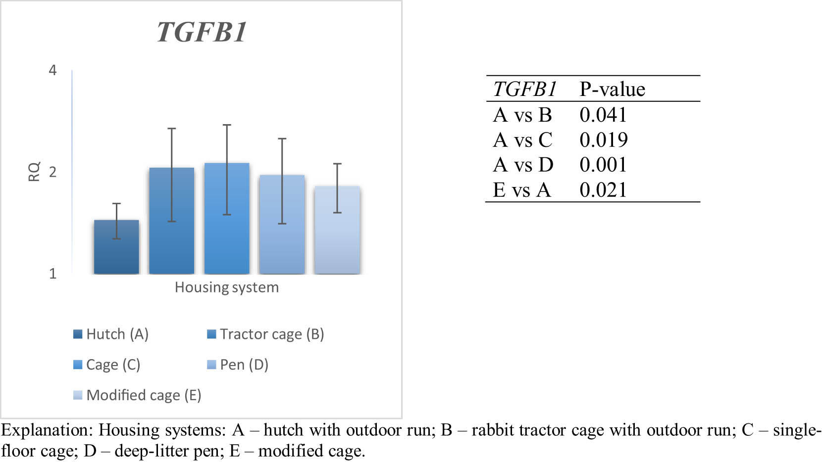
Figure 4
Effect of housing system on the relative expression of the TGFB1 gene in the blood of PW (PW) rabbits (n = 8 rabbits per housing system; means ± SD) (RQ – relative quantity normalised to ACTB and B2M).
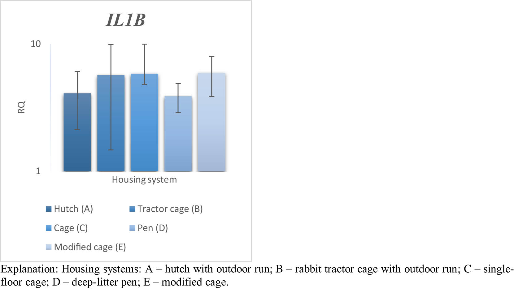
Figure 5
Effect of housing system on the relative expression of the IL1B gene in the blood of PW rabbits (n = 8 rabbits per housing system; mean values ± SD) (RQ – relative quantity normalised to ACTB and B2M). Explanation: Housing systems: A – hutch with outdoor run; B – rabbit tractor cage with outdoor run; C – single-floor cage; D – deep-litter pen; and E – modified cage.
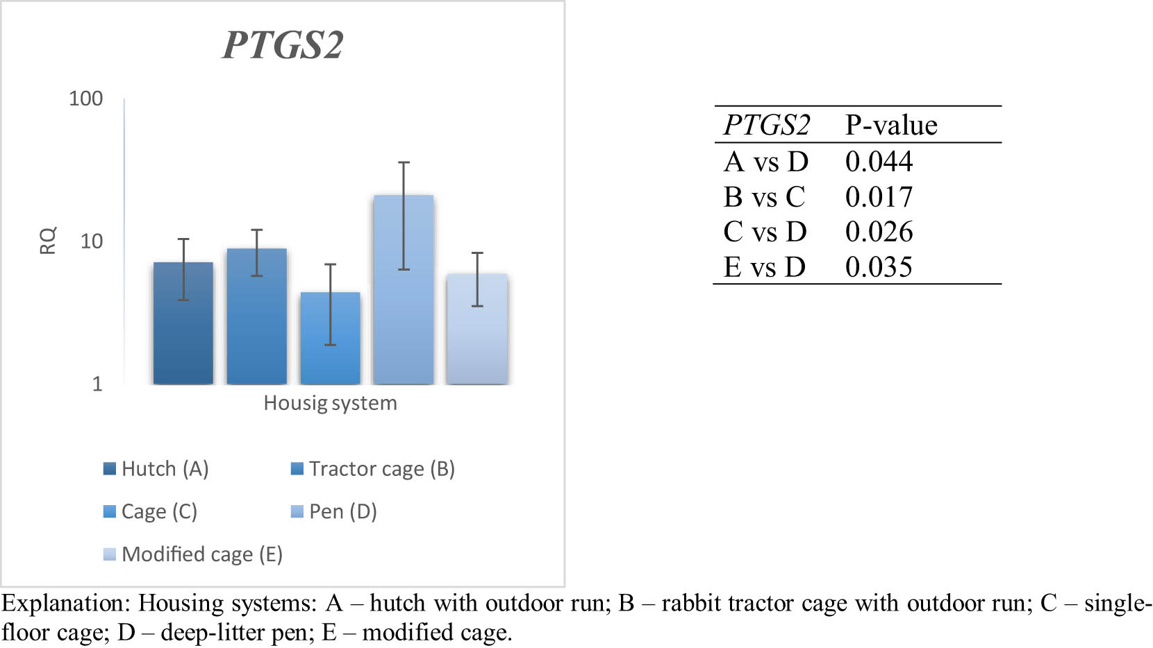
Figure 6
Effect of housing system on the relative expression of the PTGS2 gene in the blood of PW rabbits (n = 8 rabbits per housing system; mean values ± SD) (RQ – relative quantity normalised to ACTB and B2M).
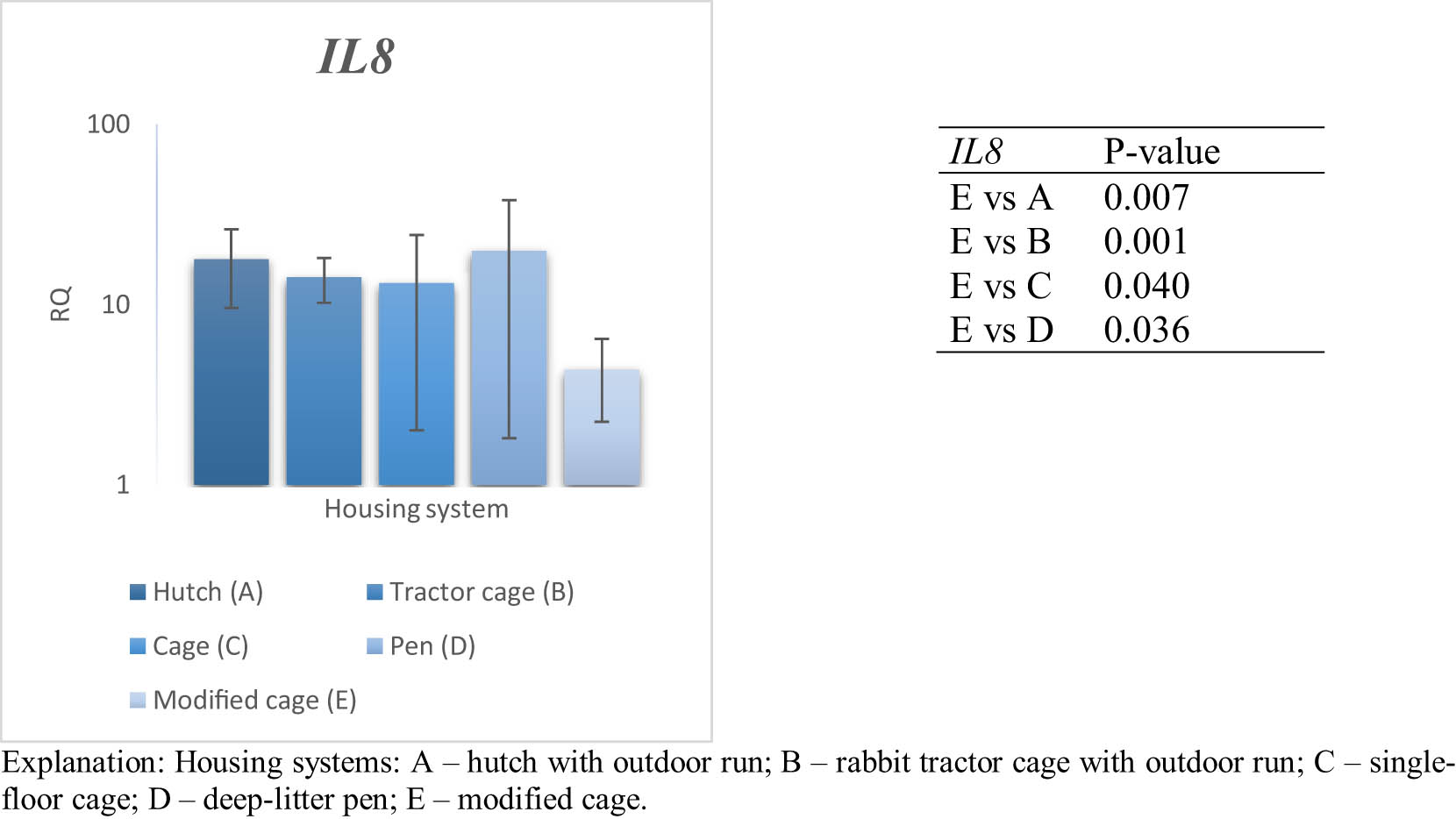
Figure 7
Effect of housing system on the relative expression of the IL8 gene in the blood of PW rabbits (n = 8 rabbits per housing system; mean values ± SD) (RQ – relative quantity normalised to ACTB and B2M).
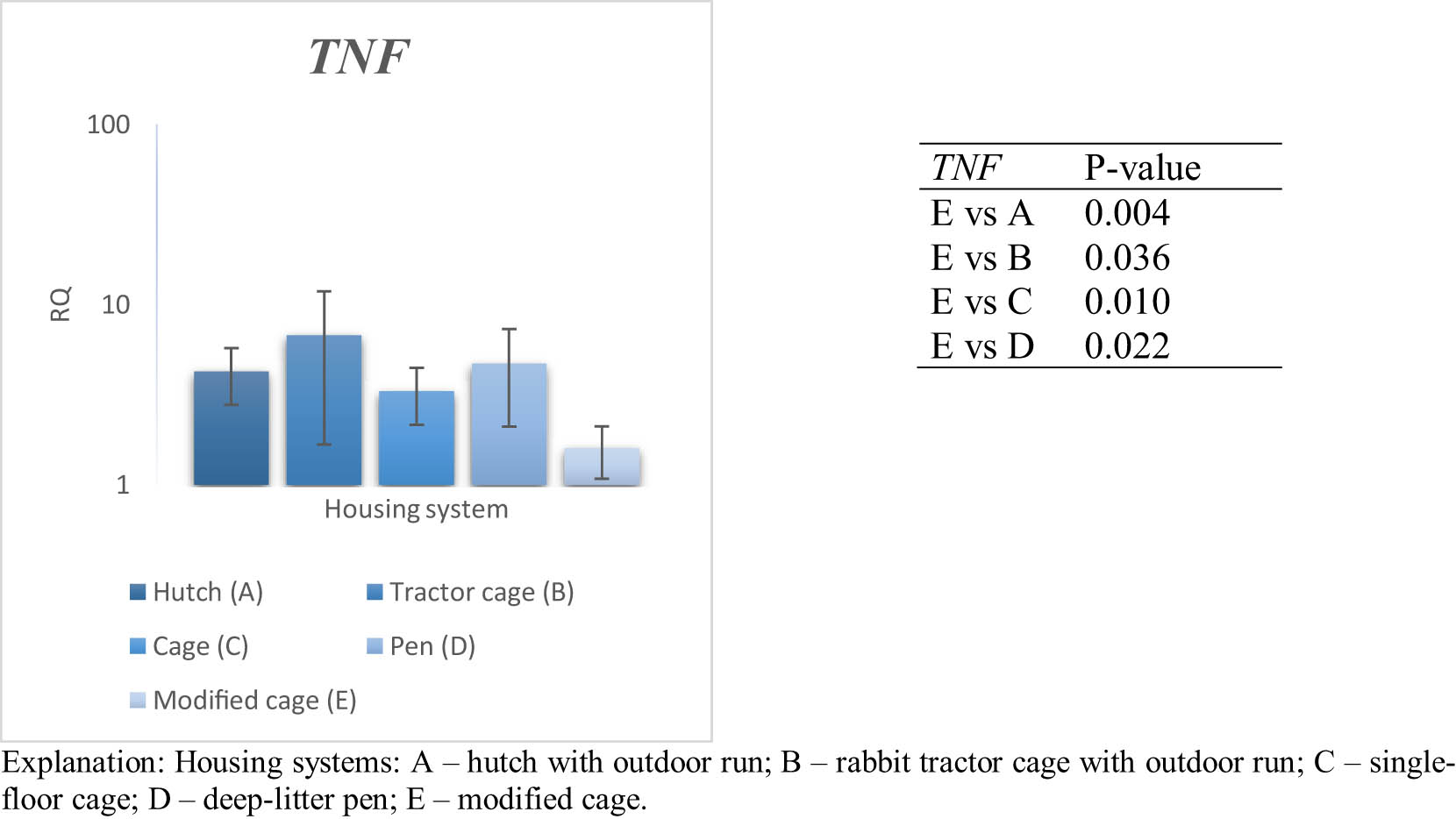
Figure 8
Effect of housing system on the relative expression of the TNF gene in the blood of PW rabbits (n = 8 rabbits per housing system; mean values ± SD) (RQ – relative quantity normalised to ACTB and B2M).
Table 3
Effect of housing system on gene expression after Benjamini–Hochberg FDR correction applied separately within each tissue (n = 8 rabbits per housing system)
| Tissue | Gene | Raw P-value | FDR-adjusted p-value | Significant after FDR (q ≤ 0.05) |
|---|---|---|---|---|
| Blood | IL6 | 0.011 | 0.021 | Yes |
| Blood | CXCR1 | <0.001 | <0.001 | Yes |
| Blood | IL10 | 0.001 | 0.001 | Yes |
| Blood | TGFB1 | 0.108 | 0.144 | No |
| Blood | IL1B | 0.746 | 0.746 | No |
| Blood | PTGS2 | 0.007 | 0.019 | Yes |
| Blood | IL8 | 0.191 | 0.218 | No |
| Blood | TNF | 0.071 | 0.114 | No |
| Spleen | IL6 | 0.096 | 0.192 | No |
| Spleen | TGFB1 | 0.372 | 0.372 | No |
| Spleen | IL1B | 0.261 | 0.372 | No |
| Spleen | PTGS2 | 0.012 | 0.050 | Yes |
| Spleen | IL8 | 0.260 | 0.372 | No |
| Spleen | TNF | 0.012 | 0.050 | Yes |
| Spleen | CXCR1 | 0.296 | 0.372 | No |
| Spleen | IL10 | 0.023 | 0.062 | No |
3.2. Spleen gene profile
The relative expression levels of inflammatory and regulatory genes were analysed in the spleen of rabbits maintained under different housing systems (A–E) and are presented in Figures 9–16. Results of the FDR-adjusted analysis are presented in Table 3. After FDR correction, significant effects of housing system were confirmed for PTGS2 and TNF, whereas the remaining genes did not retain significance. Pairwise comparisons indicated differences in IL6 expression between selected housing systems, with higher transcript levels in group B than in group E (P ≤ 0.05). For TGFB1, higher expression was recorded in group A compared with groups B and C (P ≤ 0.05), whereas group E showed higher expression than group A (P ≤ 0.05). Pairwise comparisons for IL1B showed higher expression in group E than in groups A and B (P ≤ 0.01). The PTGS2 gene showed considerable variability in expression levels. Expression in group A was higher than in group C (P ≤ 0.05), and expression in group B was higher than in group C (P ≤ 0.05). Group E showed markedly higher expression than groups A, B, and D (P ≤ 0.01). For IL8, pairwise comparisons indicated higher expression in group E than in group C (P ≤ 0.05). The TNF gene displayed significant variation among housing systems. Expression was higher in group A compared with group D (P ≤ 0.05), and in group E compared with group D (P ≤ 0.001). Expression of CXCR1 was higher in group E compared with groups A and B (P ≤ 0.01). The IL10 gene showed pairwise differences across housing systems. Higher transcript levels were observed in group D compared with group A (P ≤ 0.05), and group E showed elevated expression compared with groups A–C (P ≤ 0.05).
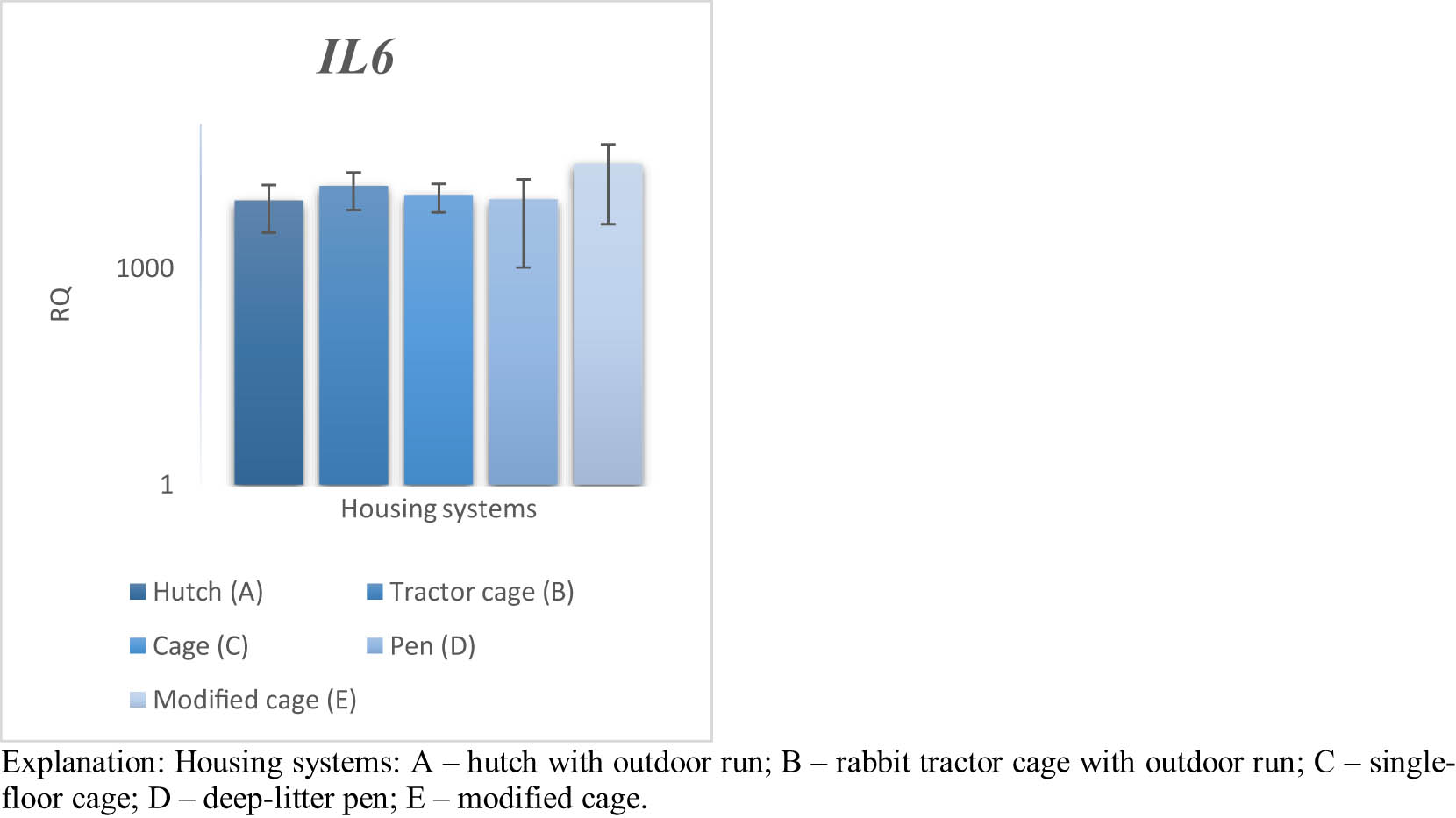
Figure 9
Effect of housing system on the relative expression of the IL6 gene in the spleen of PW rabbits (n = 8 rabbits per housing system; mean values ± SD) (RQ – relative quantity normalised to ACTB and B2M).
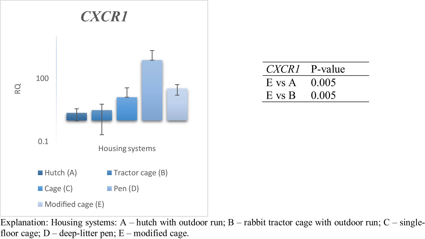
Figure 10
Effect of housing system on the relative expression of the CXCR1 gene in the spleen of PW rabbits (n = 8 rabbits per housing system; mean values ± SD) (RQ – relative quantity normalised to ACTB and B2M).
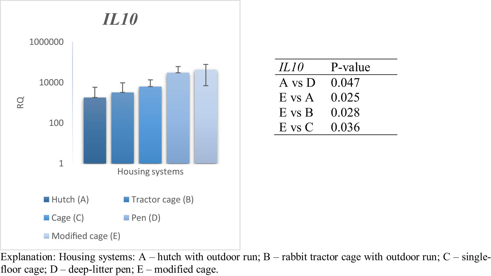
Figure 11
Effect of housing system on the relative expression of the IL10 gene in the spleen of PW rabbits (n = 8 rabbits per housing system; mean values ± SD) (RQ – relative quantity normalised to ACTB and B2M).
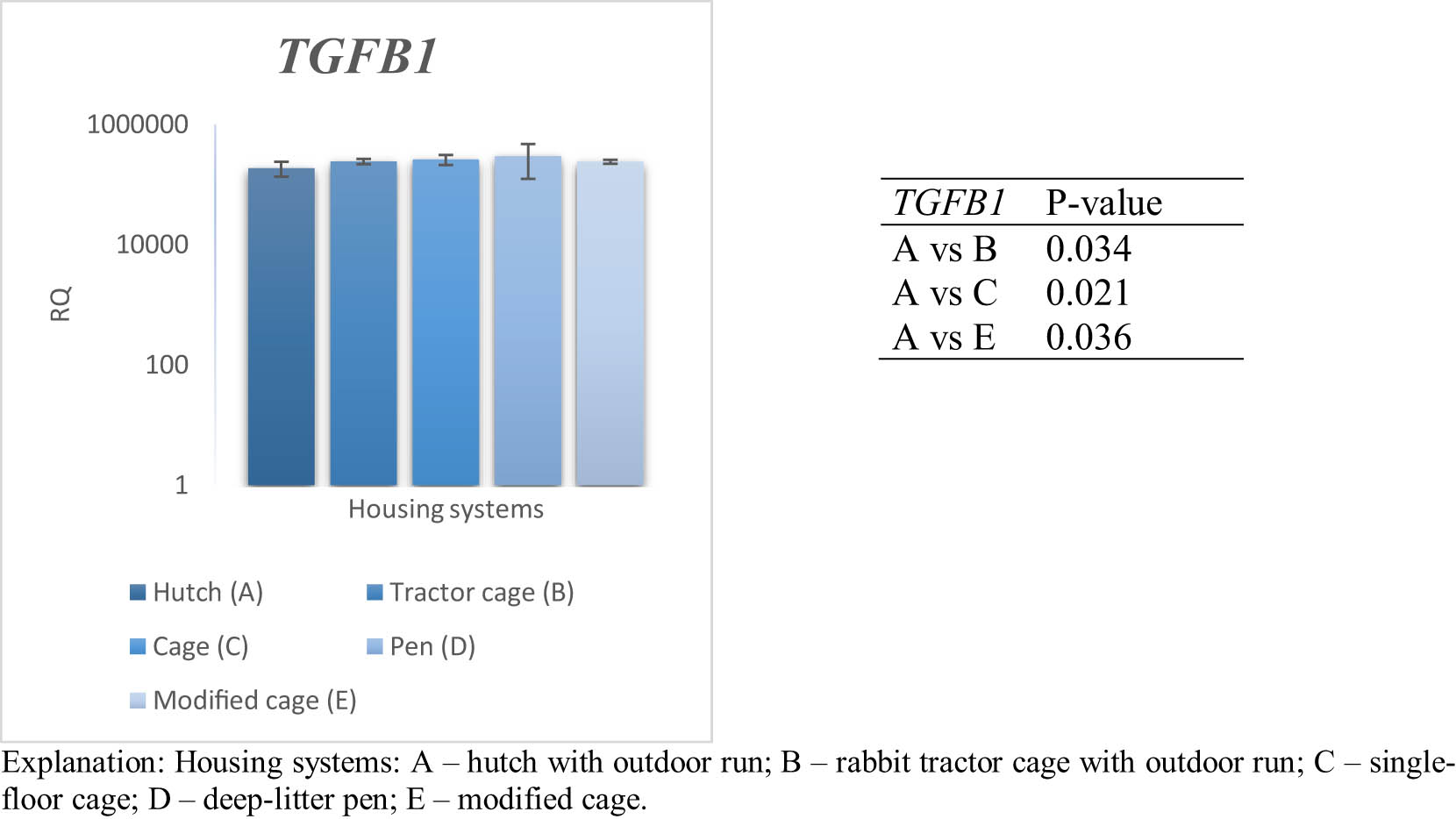
Figure 12
Effect of housing system on the relative expression of the TGFB1 gene in the spleen of PW rabbits (n = 8 rabbits per housing system; mean values ± SD) (RQ – relative quantity normalised to ACTB and B2M).
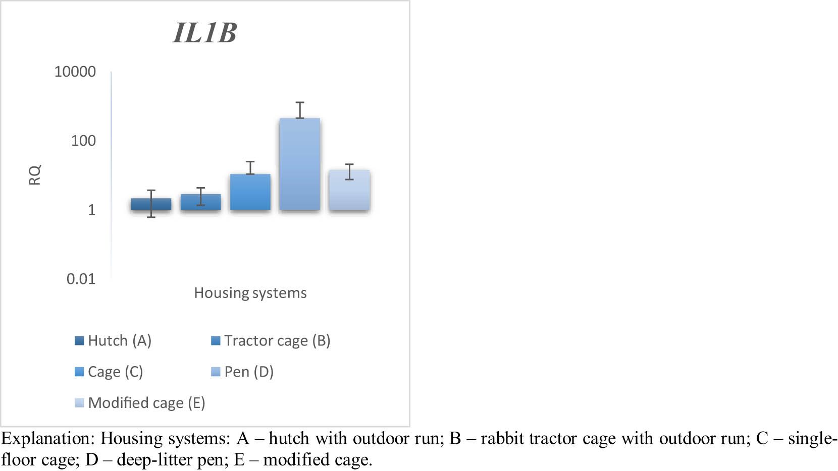
Figure 13
Effect of housing system on the relative expression of the IL1B gene in the spleen of PW rabbits (n = 8 rabbits per housing system; mean values ± SD) (RQ – relative quantity normalised to ACTB and B2M).
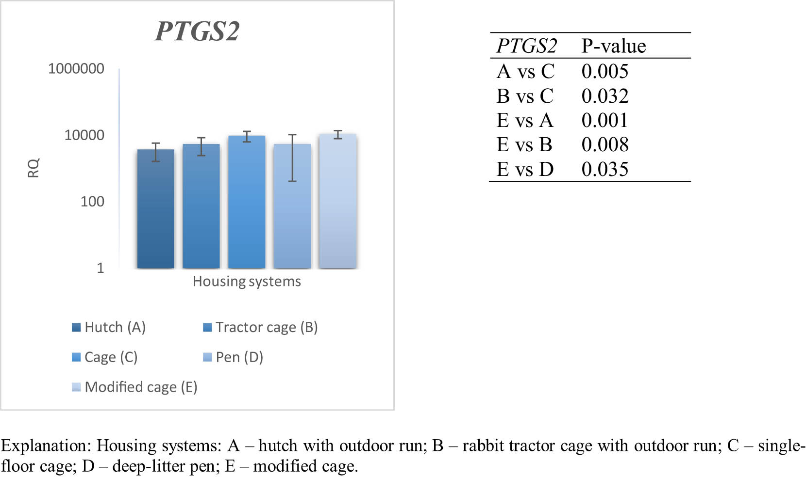
Figure 14
Effect of housing system on the relative expression of the PTGS2 gene in the spleen of PW rabbits (n = 8 rabbits per housing system; mean values ± SD) (RQ – relative quantity normalised to ACTB and B2M).
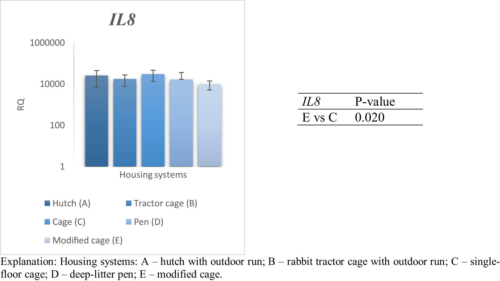
Figure 15
Effect of housing system on the relative expression of the IL8 gene in the spleen of PW rabbits (n = 8 rabbits per housing system; mean values ± SD) (RQ – relative quantity normalised to ACTB and B2M).
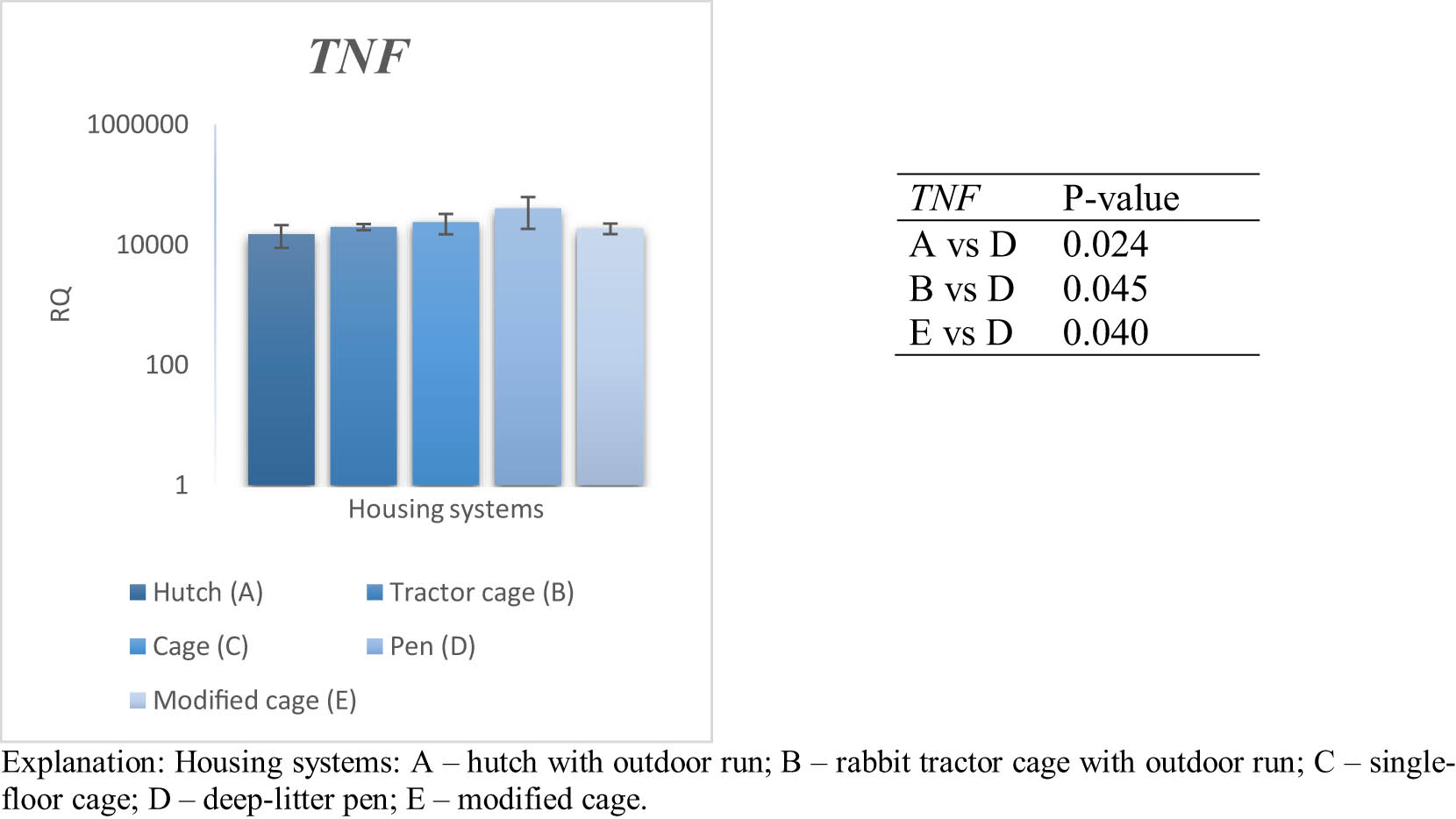
Figure 16
Effect of housing system on the relative expression of the TNF gene in the spleen of PW rabbits (n = 8 rabbits per housing system; mean values ± SD) (RQ – relative quantity normalised to ACTB and B2M).
4. Discussion
Nutritional strategies, including dietary fibre sources, probiotics, prebiotics, phytobiotics, and other functional feed additives, have been extensively studied as modulators of rabbit health and immune function (Abd El-Rahim, 2017; Belhassen, 2023; Saghir et al., 2023). In contrast, considerably less attention has been paid to the potential role of housing environment as a factor influencing immune-related physiological responses. The present study demonstrates that housing conditions can significantly influence the expression of genes involved in inflammatory and immunoregulatory processes in rabbits, both in peripheral blood and in the spleen. The observed variation between housing systems indicates that environmental factors may induce distinct physiological responses, potentially reflecting differences in stress exposure, behavioural activity, and adaptive capacity (Lopez et al., 2023). The diversity of expression patterns further supports the concept that the housing microenvironment can modulate immune function at multiple regulatory levels (Devenish et al., 2021). A clear distinction was observed between peripheral blood and spleen responses, suggesting tissue-specific sensitivity to environmental conditions (Jiang and Chen, 2022). Peripheral blood exhibited a more pronounced and consistent response across all analysed genes, indicating that circulating immune cells are highly responsive to environmental variation. In contrast, the spleen showed a more selective and regulated transcriptional response, which is consistent with its role as a central lymphoid organ involved in controlled immune activation and lymphocyte differentiation.
In blood, the variability in IL6 expression suggests that certain housing conditions were associated with differences in inflammatory signalling pathways, while others may attenuate systemic inflammatory responses. Given the key role of IL6 in mediating inflammation, these differences likely reflect variation in systemic responses to environmental stimuli (Hu et al., 2024). Pairwise differences in the expression of IL8 and TNF were also observed, suggesting potential variation in pro-inflammatory signalling intensity; however, these effects should be interpreted cautiously. Interestingly, the elevated expression of CXCR1 under specific housing conditions suggests that immune modulation is not only limited to cytokine production but also involves alterations in chemokine receptor signalling and immune cell migration dynamics (Kim & Moudgil, 2008; Xiao et al., 2021). This may indicate a shift in how immune cells are recruited and distributed in response to environmental factors, rather than a simple increase or decrease in inflammatory activity. The observed variation in PTGS2 expression further supports the involvement of housing conditions in modulating inflammatory and stress-related pathways. As PTGS2 plays a central role in prostaglandin synthesis, its increased expression may reflect enhanced inflammatory signalling and stress responses under certain environmental conditions (Hu et al., 2023). At the same time, differences in the expression of regulatory genes such as IL10 and TGFB1 indicate that immune responses are balanced by anti-inflammatory mechanisms. The elevated expression of these genes in selected housing systems suggests activation of compensatory processes aimed at maintaining immune homeostasis under varying environmental pressures (García-González et al., 2000; Hoffmann & Willi, 2008). However, it should be emphasised that gene expression patterns alone do not provide direct evidence of animal welfare status and should be interpreted as molecular indicators of physiological responses to housing conditions. Although pairwise differences were observed for IL8, TNF, and TGFB1, the overall effects of housing system on the expression of these genes were not significant; therefore, these results should be interpreted with caution and regarded as exploratory observations requiring further confirmation.
In the spleen, gene expression patterns indicate a more tissue-specific immune response to housing conditions than that observed in blood. Following FDR correction, significant effects of housing system were confirmed for PTGS2 and TNF, suggesting that local inflammatory pathways may be particularly responsive to environmental conditions. The increased expression of PTGS2 and TNF in selected housing systems may reflect activation of innate immune mechanisms at the level of lymphoid tissue (Xiao et al., 2021). As PTGS2 is involved in prostaglandin synthesis and TNF plays a central role in inflammatory signalling, variation in the expression of these genes may indicate differences in local immune activity and physiological adaptation to housing conditions (Zelová & Hošek, 2013; Xiao et al., 2021). Pairwise differences were also observed for IL1B, IL8, CXCR1, IL10, and TGFB1. These genes are associated with immune cell recruitment, cytokine signalling, and anti-inflammatory regulation, suggesting that housing conditions may influence multiple aspects of splenic immune function (Guo et al., 2020). However, the overall effects of housing system on these genes were not significant after FDR correction, and therefore these observations should be interpreted with caution. Overall, the results suggest that housing conditions may affect both inflammatory and regulatory pathways within the spleen. Nevertheless, the coexistence of pro-inflammatory and anti-inflammatory signals should be interpreted as evidence of differential immune responsiveness rather than as a direct indicator of animal welfare status (Makris et al., 2022; Mota & Madden, 2022).
The comparison between blood and spleen responses indicates that environmental conditions can differentially modulate systemic and local immune processes. Peripheral blood appears to reflect more rapid and dynamic changes in immune status, suggesting high sensitivity of circulating immune cells to environmental variation (Devenish et al., 2021). In contrast, the spleen exhibits a more selective and regulated response, consistent with its role as a central lymphoid organ involved in maintaining immune homeostasis and coordinating adaptive responses (Makris et al., 2022). The gene expression patterns observed across both tissues further demonstrate that housing conditions influence the balance between pro-inflammatory and regulatory mechanisms in rabbits. Some environmental conditions appear to promote stronger activation of inflammatory pathways, whereas others favour regulatory responses associated with anti-inflammatory control. This tissue-dependent and context-specific modulation of immune activity suggests that the biological response to housing conditions is complex and may have important implications for physiological adaptation and immune regulation, although direct conclusions regarding animal welfare cannot be drawn from gene expression data alone.
Beloumi et al. (2020) reported relationships between the ability of rabbits to cope with environmental stress and the activation of the immune system, analysing lines selected for divergent variability in litter size. The authors demonstrated that animals characterised by greater environmental variability exhibited increased concentrations of TNF-α and CRP, accompanied by reduced leukocyte counts, indicating a reduced capacity to maintain immune homeostasis under stressful conditions. These findings suggest that inflammatory responses in rabbits are shaped by both environmental and genetic factors, as well as differences in stress sensitivity. In agreement with Beloumi et al. (2020), housing systems associated with greater environmental variability and activity (e.g. outdoor systems A and B) appeared to promote stronger systemic immune activation. In contrast, more confined indoor systems, particularly the modified cages (E), were characterised by reduced pro-inflammatory signalling in blood, which may indicate lower environmental stimulation or altered immune responsiveness. At the same time, the increased expression of CXCR1 under these conditions suggests that immune modulation may involve changes in cell migration rather than a simple suppression of inflammation. Systems providing more complex environments, such as deep-litter pens (D), were associated with higher IL10 expression, suggesting activation of regulatory pathways potentially involved in immune homeostasis. Compared with the results of the study by Beloumi et al. (2020), the present results suggest that PW rabbits exhibit concurrent changes in both pro-inflammatory and regulatory markers, which may reflect breed-specific differences in adaptation to environmental conditions.
Trocino and Xiccato (2006) emphasised that housing conditions play a critical role in shaping physiological stress responses, behaviour, and disease susceptibility in rabbits. Suboptimal environments, including restricted space, limited behavioural opportunities, and inadequate environmental enrichment, may lead to chronic stress and disruption of homeostasis, ultimately impairing immune function. In line with these observations, the present study demonstrated clear differences in the expression of key inflammatory markers (IL6, IL8, TNF, and PTGS2) between housing systems. Systems associated with lower cytokine expression, such as group E (modified cages), may reflect differences in immune responsiveness under the studied conditions. Conversely, higher expression levels observed in other groups may indicate increased activation of immune pathways in response to environmental constraints. These findings support the view that housing conditions can influence immune-related molecular responses in rabbits.
Another factor that may contribute to the observed differences is the interaction between the gut microbiota and the immune system. The microbiome–immune axis has recently been recognised as a key regulator of inflammatory processes in rabbits, and its composition is highly sensitive to environmental conditions, stress, and behaviour (Combes et al., 2017). Alterations in microbial communities have been linked to changes in the expression of cytokines such as IL6, TNF, and IL1B, as well as regulatory mediators including IL10 and TGFB1 (Plaza-Diaz et al., 2014). In the present study, the coexistence of elevated pro-inflammatory and regulatory gene expression in certain housing systems may reflect microbiome-driven modulation of immune responses. Wu et al. (2024) demonstrated that rearing conditions can significantly influence the gut microbiome and its functional interactions with host immunity, with more complex environments promoting distinct immune-related transcriptional profiles. This supports the hypothesis that differences in immune gene expression observed in PW rabbits may be partially mediated by environment-induced changes in microbial composition.
Despite the numerous significant differences observed between housing systems, the patterns of gene expression were not identical between blood and spleen. Blood showed a broader and more pronounced response to housing conditions, whereas spleen responses were more selective, with PTGS2 showing the clearest housing-related differences after FDR correction. These findings further support the tissue-specific nature of immune responses to environmental conditions.
Genetic factors may further contribute to the observed variability in immune responses. Neves et al. (2015) demonstrated that cytokine genes in lagomorphs are generally highly conserved but still exhibit structural and sequence variability that may influence immune function. Even subtle genetic differences, such as variations in glycosylation sites or cysteine residues, can affect cytokine activity and the balance between pro- and anti-inflammatory pathways. In the case of PW rabbits, such genetic characteristics may interact with environmental conditions to shape the observed gene expression profiles, leading to differences in immune responsiveness compared with other rabbit populations. The results obtained in this study demonstrate that housing conditions were associated with differences in the expression of selected immune-related genes, although the magnitude and consistency of these effects varied among markers and tissues. Following FDR correction, significant housing-related effects were confirmed primarily for IL6, CXCR1, IL10, and PTGS2 in blood, and for PTGS2 and TNF in the spleen. These findings suggest that housing conditions may influence both inflammatory and regulatory pathways, with blood generally showing a broader response than the spleen. The observed tissue-specific expression patterns indicate that environmental conditions can differentially modulate systemic and local immune processes. However, gene expression profiles should be interpreted as indicators of physiological and immunological responses to housing conditions rather than direct measures of animal welfare status.
Several limitations of the present study should be considered. Gene expression data do not necessarily correspond directly to protein levels, and therefore future studies should include the determination of cytokine concentrations in plasma and tissues. In addition, although ACTB and B2M were used as reference genes, their stability was not formally validated under the specific experimental conditions, which may influence the accuracy of normalisation. Although FDR correction was applied to the main effects of housing system, the interpretation of pairwise comparisons should still be approached with caution. Additional data on animal behaviour, microclimatic conditions and oxidative stress parameters would help to better explain the mechanisms underlying the observed differences. Factors not evaluated in the present study, including potential differences in growth performance among housing systems, may also have contributed to the observed variation in gene expression. Therefore, the relationship between productive performance and immune-related transcriptional responses requires further investigation. Furthermore, the age at slaughter ranged from 90 to 120 days due to differences in growth performance among housing systems. Therefore, age-related variation in gene expression cannot be completely excluded and should be considered when interpreting the results. Future research should also consider changes in immune cell populations and the temporal dynamics of immune responses during the rearing period.
The evaluated housing systems exhibited distinct immunological profiles overall. Outdoor systems (A and B) were associated with increased pro-inflammatory activity, while modified cages (E) were characterised by reduced inflammatory signalling and altered immune dynamics. Deep-litter pens (system D) were associated with distinct expression patterns of selected immune-related genes, particularly IL10 and PTGS2. However, the biological significance of these differences and their relationship to animal welfare require further investigation.
5. Conclusion
This study demonstrates that housing conditions are associated with differences in the expression of selected inflammatory and regulatory genes in the blood and spleen of PW rabbits. The effects were more pronounced in peripheral blood, indicating greater responsiveness of circulating immune cells to environmental variation, whereas the spleen exhibited a more selective and tissue-specific pattern of gene expression. Differences between housing systems were reflected in the expression of genes involved in both pro-inflammatory and regulatory pathways, suggesting that housing conditions may influence immune-related physiological processes. Outdoor systems (A and B), modified cages (E), and deep-litter pens (D) were associated with distinct expression profiles, indicating differences in immune responsiveness under the evaluated environmental conditions. However, gene expression data alone do not allow direct assessment of animal welfare status and should be interpreted as indicators of physiological adaptation rather than definitive measures of welfare. Overall, gene expression profiling may serve as a sensitive tool for detecting biological responses to housing conditions and may complement conventional indicators used in rabbit welfare and production studies. Future research integrating molecular, behavioural, physiological, microbiome, and protein-level data is needed to better understand the biological significance of these responses and their relationship to animal welfare.
Funding information
This research was funded by the statutory activity of the National Research Institute of Animal Production (project no. 504-140-211).
Author contributions
Conceptualization, P.B. and D.K.; Methodology, P.B., D.K., K.R.-M. and K.P.; Investigation, Z.S., P.B., D.K., K.R.-M. and K.P.; Data curation, Formal analysis, Writing – original draft, Z.S.; Writing – review and editing, P.B., D.K., K.R.-M. and K.P.; Supervision, P.B. and D.K.
Conflict of interest statement
Authors state no conflict of interest.
Data availability statement
The data supporting this study are available from the corresponding author upon reasonable request.