
Canine mammary tumours (CMTs) are among the most commonly diagnosed neoplasms in dogs, occurring predominantly in intact females due to prolonged exposure to endogenous ovarian steroid hormones which stimulate epithelial proliferation in mammary gland (Vazquez et al., 2023). CMTs pose a serious challenge in veterinary oncology, as it is indicated that approximately half of all CMTs are malignant (Moe, 2001; Salas et al., 2015; Zheng et al., 2022), although it should be noted that there are also studies from Spain in which the rate of malignant tumour diagnosis was approximately 90%. However, the authors themselves point out that this fact may be due, for example, to the approach of the veterinary surgeons, who may only report tumours with a more alarming phenotype (Pastor et al., 2018; Rodríguez et al., 2022).
Epidemiological studies indicate that the risk and malignancy of CMTs are influenced by breed, body size and age. Small-breed dogs, such as Dachshunds, Yorkshire Terriers or Shih Tzus, show a higher overall incidence of CMTs than large-breed dogs, whereas malignant tumours are proportionally more frequent in some large breeds, e.g., Alaskan Malamute, Samoyed, Schnauzer, Doberman, Husky or German Shepherd (Salas et al., 2015; Vascellari et al., 2016; Edmunds et al., 2023). Mammary tumours are most commonly diagnosed in middle-aged and older dogs, typically between 5 and 13 years of age, and malignant CMTs are usually larger at the time of diagnosis than benign lesions (Ežerskytė et al., 2011; Salas et al., 2015; Vascellari et al., 2016; Sorenmo et al., 2009; Burrai et al., 2020; Zheng et al., 2022; Edmunds et al., 2023). Consequently, both the age of the dog and the tumour size are considered potentially sufficient parameters for a preliminary assessment of CMT malignancy (Burrai et al., 2020).
Hormonal status is another key determinant of CMT risk. Numerous studies have demonstrated that ovariohysterectomy reduces the overall incidence of CMTs, and unspayed bitches account for the majority of diagnosed cases (Salas et al., 2015; Vascellari et al., 2016; Rodríguez et al., 2022; Zheng et al., 2022; Edmunds et al., 2023). At the same time, neutering appears to modify the pattern of disease. Edmunds et al. (2023) reported that, although spayed dogs are less likely to develop CMTs or multiple tumours, malignant lesions may be proportionally more common when tumours do occur.
Surgical excision of the tumour, often accompanied by the removal of local lymph nodes, remains the standard and most effective treatment for CMTs (Papazoglou et al., 2014). Dogs diagnosed with benign CMTs have a very good prognosis and a high five-year survival rate. However, malignant tumours are associated with a significantly poorer prognosis, as life expectancy decreases with increasing tumour malignancy and invasiveness, often not exceeding three months following diagnosis in severe cases (Rasotto et al., 2017).
The development of CMTs reflects a complex interaction of genetic, epigenetic, hormonal and environmental factors. At the cellular level, DNA mutations that disrupt cell cycle control and genome stability play an important role in CMT initiation and progression (Fymat, 2017; Hu and Baeg, 2017; Compton, 2020). Conceptually, it follows the canonical sequence of initiation-promotion-progression – the emergence and consolidation of mutations (e.g., in p53, BRCA1/2) with avoidance of apoptosis and clonal expansion, the acquisition of autonomous growth capabilities, angiogenesis and metastasis (Klopfleisch et al., 2011; Gama et al., 2004; Kaszak et al., 2018, 2022; Hernández-Suárez et al., 2022; Grizzi et al., 2006). Detailed discussions of long-known (‘classical’) biomarkers associated with these stages (e.g., Ki-67, PCNA, apoptosis-related markers, ER/PR, COX-2, VEGF, CEA, CA 15-3, HER-2, E-cadherin) are available in other publications and are not the subject of this review (Kumaraguruparan et al., 2006; Yang et al., 2006; Klopfleisch et al., 2011; Kaszak et al., 2018, 2022; Cao et al., 2019; Hernández-Suárez et al., 2022; Vazquez et al., 2023).
Based on these observations, early diagnosis of CMT is an important factor influencing the expected prognosis. To improve not only diagnosis but also prognosis and gain a deeper understanding of the molecular mechanisms underlying CMT development and progression, research efforts are focused on identifying effective biomarkers of tumourigenesis. Tumour biomarkers are defined as traits that indicate a patient's risk of cancer, disease progression or expected therapeutic outcome. These characteristics can be subcellular, cellular or systemic, and include nucleic acids (DNA, RNA), proteins and metabolites detected in tissues or body fluids (Sarhadi and Armengol, 2022). Current research on CMT biomarkers primarily examines the potential applicability of validated biological markers already utilised in human medicine. This approach is driven by the significant similarities between mammary tumour development in dogs and breast cancer in women. These parallels not only highlight the potential for cross-species application of biomarkers but also position the dog as an excellent research model for studying the biology of breast cancer in humans (Vascellari et al., 2016; Ferreira et al., 2023).
Several review articles have analysed the molecular mechanism of tumourigenesis and the well-established proteins involved in this process in dogs (Klopfleisch et al., 2011; Kaszak et al., 2018, 2022; Cao et al., 2019; Hernández-Suárez et al., 2022; Vazquez et al., 2023). However, as canine tumourigenesis is not yet fully understood, and considering the extensive body of literature on this topic, this review concentrates on selected, less extensively characterised or emerging protein biomarkers detectable in CMT tissue and/or blood, with particular emphasis on their potential application in the diagnosis, prognosis and monitoring of CMTs.
Proteins are among the most promising molecules for biomarker discovery. As essential structural components of cells, they play a key role in regulating various processes, acting as enzymes, receptors and transcription factors. Their expression is often specific to the type and state of the cell, including cancer cells. Modern proteomic tools enable the characterisation of complex protein expression profiles in tumour tissue and in body fluids such as plasma or saliva, revealing alterations associated with tumour presence and behaviour (Li and Chan, 2014; Marques et al., 2017; Franco-Martínez et al., 2020; Pöschel et al., 2021). These findings expand our understanding of the molecular mechanism underlying tumourigenesis and the identification of proteins whose expression changes may serve as indicators of disease processes. Discovering and analysing novel protein biomarkers holds promise for future applications, such as identifying the type and subtype of CMT, monitoring disease course, determining the efficacy of treatments and predicting clinical outcomes (Li and Chan, 2014). Most of the proteins discussed below have already been applied to Figure 1 in order to illustrate the usefulness of the proteins discussed in this text. We cannot propose markers for initiation stage because this phase reflects rare, early driver mutations occurring in single cells long before a detectable mass exists. The literature on CMTs discussed here analyses samples of developed tumours rather than precancerous lesions and measures changes in expression rather than initial mutational events. In contrast, the analysed proteins reflect the consequences of tumour development and therefore correspond to later stages: promotion, progression, invasion and metastasis.
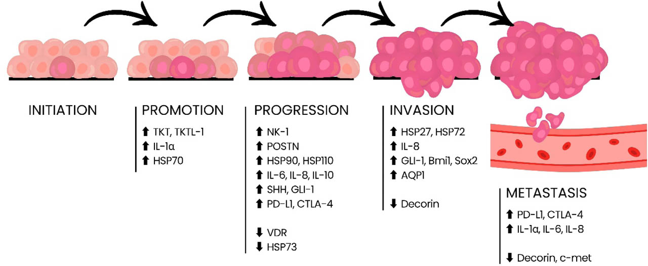
Figure 1.
Schematic representation of the development of malignant CMTs, together with the proteins associated with them, which are likely to be key to the given stage of carcinogenesis. The relationships between proteins and stages are suggested by the authors based on the evidence gathered in this review.
Search strategy
This article is a review that synthesises and analyses current literature on proteins with potential utility in the diagnosis, prognosis and monitoring of CMTs. Literature searches were performed in the PubMed (http://www.ncbinlm.nih.gov/pubmed) and Google Scholar (https://scholar.google.com/) databases. The search terms included: “canine mammary gland tumour”, “biomarkers in canine mammary tumour”, “biomarkers in CMT”, “proteins in CMT” and similar phrases.
Additionally, studies were included if (1) they involved dogs with mammary tumours, (2) evaluated protein biomarkers measured in tumour tissue and/or body fluids (e.g. serum/plasma/saliva), (3) contained data allowing for comparison between groups (e.g. benign and malignant), and (4) were written in English to ensure that the cited evidence remains accessible and verifiable for an international readership. In addition, the names of specific proteins considered relevant based on the collected literature were also used as search terms. Protein-related keywords included: interleukins (ILs), heat shock proteins (HSPs), vitamin D receptor (VDR), c-met, neurokinin-1 (NK-1), sonic hedgehog (SHH), glioma-associated oncogene 1 (GLI-1), polycomb complex protein BMI-1 (Bmi1), transcription factor Sox-2 (Sox2), monocyte chemotactic protein 1 (CCL2), stromal cell-derived factor 1 (CXCL12), perostin (POSTN), decorin (DCN), programmed cell death ligand 1 (PD-L1), cytotoxin T-lymphocyte protein 4 (CTLA-4), transketolase (TKT) protein, transketolase-like 1 protein (TKTL-1) and aquaporins (AQPs). Study selection, data extraction and synthesis were performed manually by the authors according to the above criteria.
In addition, artificial intelligence (AI) tools available through ChatGPT's paid subscription (https://chatgpt.com/) were used on a trial basis to search for additional literature. In this context, ‘search’ refers to the identification and potential inclusion of scientific publications cited by AI in response to specific prompts. It should be emphasised that AI was not directly involved in the writing of this publication or the analysis of scientific studies. It functioned solely as an auxiliary tool, aiding in the search for additional scientific articles beyond those retrieved from PubMed and Google Scholar.
Potential new protein biomarkers of CMT
The development of a tumour, though initially asymptomatic, significantly impacts many processes in the body. To reach its full potential, the tumour recruits neighbouring stromal and immune cells and remodels the extracellular matrix and local microenvironment (Khan et al., 2018; Tao et al., 2018). These interactions, together with the tumour's capacity to infiltrate tissues and disseminate via lymphatic and blood vessels, allow proteins associated with CMT development to enter the circulation, creating opportunities for circulating protein biomarkers useful for diagnosis, prognosis and disease monitoring. To date, several indicator proteins used clinically or investigationally have been reported, including Ki-67, CEA and CA 15-3, whose comprehensive characterisation and applications in human and veterinary oncology are reviewed elsewhere (Kaszak et al., 2018, 2022). Accordingly, the following section focuses on emerging protein candidates in blood and/or tumour tissue that may complement routine assessment (Li and Chan, 2014).
A summary of the findings describing potential new protein markers in blood in CMT is presented in Table 1, while protein markers in tissues are listed in Table 2.
Table 1.
Summary of the collected study results of changes in protein concentrations and suggestion of their potential clinical use as CMTs blood biomarkers. More detailed data can be found in supplementary Table S1
| Group | Function | Full name | Short name | Change in concentration | Suggested clinical use of potential blood biomarker for assessment of | References |
|---|---|---|---|---|---|---|
| 1 | 2 | 3 | 4 | 5 | 6 | 7 |
| Proteins of the immune system | Interleukins are signalling proteins that coordinate innate and acquired immunity. They regulate the development, recruitment and activation of leukocytes. They also influence tissue repair and the resolution of inflammation. | Interleukin-1 | IL-1 | Higher in M than in N. | i.d. | Machado et al., 2015 |
| Interleukin-1α | IL-1α |
|
| Szczubiał et al., 2022; de Andrés et al., 2013 | ||
| Interleukin-6 | IL-6 |
|
| Ren et al., 2023; de Andrés et al., 2013; Szczubiał et al., 2018; Machado et al., 2015 | ||
| Interleukin-8 | IL-8 |
|
| Ren et al., 2023; de Andrés et al., 2013; Szczubiał et al., 2022 | ||
| Interleukin-10 | IL-10 |
|
| Ren et al., 2023; de Andrés et al., 2013; Szczubiał et al., 2018 | ||
| Chemokines are chemoattractant cytokines that orchestrate leukocyte trafficking and, in cancer, shape the tumour microenvironment by promoting cell migration, invasion, and angiogenesis through receptor-guided signalling. | Monocyte chemotactic protein 1 | CCL2 | Higher in MM than in N. | i.d. | Szczubiał et al., 2023 | |
| Stromal cell-derived factor 1 | CXCL12 | |||||
| Hedgehog signalling pathway protein | Hedgehog pathway proteins regulate tissue development and homeostasis by controlling proliferation, differentiation, and maintenance of stem cells. | Sonic hedgehog | SHH |
|
| |
| Glioma-associated oncogene 1 | GLI-1 |
|
| Koo et al., 2023 | ||
| Heat shock proteins | HSPs are stress-induced chaperone proteins that maintain proteostasis by assisting protein folding, stabilising proteins, and modulating apoptosis, cell cycle control, and signalling. | Heat shock protein 27 | HSP 27 |
| histological grade | Birdi et al., 2019 |
Table 2.
Summary of the collected study results of changes in protein expression and suggestion of their potential clinical use as CMTs tissue biomarkers. More detailed data can be found in supplementary Table S2.
| Group | Function | Full name | Short name | Change of expression | Suggested clinical use of potential tissue biomarker for assessment of | References |
|---|---|---|---|---|---|---|
| 1 | 2 | 3 | 4 | 5 | 6 | 7 |
| Proteins of the immune system | Interleukins are signalling proteins that coordinate innate and acquired immunity. They regulate the development, recruitment and activation of leukocytes. They also influence tissue repair and the resolution of inflammation. | Interleukin-6 | IL-6 |
|
| Ren et al., 2023; de Andrés et al., 2013 |
| Interleukin-8 | IL-8 |
|
| Ren et al., 2023; de Andrés et al., 2013 | ||
| Interleukin-10 | IL-10 |
|
| Ren et al., 2023; de Andrés et al., 2013 | ||
| Immune checkpoints are inhibitory pathways that restrain T-cell activity to maintain self-tolerance and limit immunopathology. |
| PD-L1 |
|
| Ariyarathna et al., 2020 | |
| Cytotoxic T-lymphocyte protein 4 | CTLA-4 | |||||
| Hedgehog signalling pathway protein | Hedgehog pathway proteins regulate tissue development and homeostasis by controlling proliferation, differentiation, and maintenance of stem cells. | Sonic hedgehog | SHH |
| tumour progression | Koo et al., 2023 |
| Glioma-associated oncogene 1 | GLI-1 |
|
| Koo et al., 2023 Liu et al., 2022 | ||
| Polycomb complex protein BMI-1 | Bmi1 | Higher in iM than in M and N. | tumour invasiveness | Liu et al., 2022 | ||
| Transcription factor Sox-2 | Sox2 | |||||
| Proteins of the extracellular matrix | The ECM proteins regulate adhesion, migration, proliferation and survival through integrin and growth factor signalling. | Periostin | POSTN |
|
| Borecka et al., 2020 a, b; Alani and Al-Biaty, 2024 |
| Decorin | DCN | Higher in nMM and B than in N and MM. |
| Szczubiał et al., 2020 | ||
| Heat shock proteins | HSPs are stress-induced chaperone proteins that maintain proteostasis by assisting protein folding, stabilising proteins, and modulating apoptosis, cell cycle control, and signalling. | Heat shock protein 27 | HSP27 |
| tumour invasiveness | Romanucci et al., 2006 |
| Heat shock protein 70 | HSP70 |
|
| Szczubiał et al., 2015 | ||
| Heat shock protein 72 | HSP72 |
| tumour invasiveness | Romanucci et al., 2006 | ||
| Heat shock protein 73 | HSP73 | Lower in M than in N. | i.d. | Romanucci et al., 2006 | ||
| Heat shock protein 90 | HSP90 | Higher in M than in N. | i.d. | Romanucci et al., 2006 | ||
| Heat shock protein 110 | HSP110 | Expression is dependent on tumour subtype and due to histological grade. |
| Okada et al., 2021 | ||
| Protein receptors | Protein receptors are cellular sensors that detect extracellular signals – soluble ligands, matrix components, or cell-to-cell signals and translate them into intracellular responses through diverse structures. By regulating pathways of proliferation, survival, differentiation, migration, and immunity, they coordinate tissue homeostasis. | Substance-P receptor/neurokinin-1 receptor | NK-1 |
| tumour progression | Song et al., 2020 |
| Vitamin D receptor | VDR |
|
| Sánchez-Céspedes et al., 2018, 2021 | ||
| Hepatocyte growth factor receptor | c-met |
|
| Chen et al., 2018 | ||
| Pentose phosphate pathway enzymes | The pentose phosphate pathway diverts glucose metabolism through oxidative and non-oxidative branches to generate NADPH (for antioxidant defence and reductive biosynthesis) and ribose-5-phosphate (for nucleotide synthesis). Cancer cells often up-regulate this pathway to buffer oxidative stress and sustain anabolic growth. | Transketolase | TKT |
|
| Burrai et al., 2017 |
| Transketolase-like 1 | TKTL-1 |
|
| Burrai et al., 2017 | ||
| Aquaporins | AQPs are cell membrane channel proteins that facilitate the rapid, selective transport of water (and in some cases also small solutes such as glycerol) across biological membranes, to maintain cellular water homeostasis by regulation of osmotic balance, cell volume and fluid flow in tissues. | Aquaporin 1 | AQP1 |
|
| Freeman et al., 2010; Fujimoto et al., 2024 |
| Aquaporin 3 | AQP3 | Lower due to higher histological grade. | histological grade | Fujimoto et al., 2024 | ||
| Aquaporin 5 | AQP5 | Absent in grade III samples. | histological grade | Fujimoto et al., 2024 |
[i] M – malignant tumour; iM – invasive malignant tumour; nM – non-invasive malignant tumour; MM – metastatic malignant tumour; nMM – non-metastatic malignant tumour; IMC – inflammatory mammary cancer; nIMC – non-inflammatory mammary cancer; B – benign tumour; N – normal (control); i.d. – insufficient data.
Proteins of the immune system
Proteins associated with immune system activity are the first group with potential bio-marker value. As mentioned earlier, tumours are able to recruit immune cells, which secrete various proteins, including cytokines, into the microenvironment. This recruitment, particularly in inflammatory tumours, often leads to increased secretion of immune-related proteins in the tumour environment and, consequently, changes in their blood concentrations (Cohen et al., 2015; Tao et al., 2018). Taken together, these characteristic features of cancer cells provide a consistent basis for evaluating the same proteins in the venous blood and tumour tissue within a single set of biomarkers.
Interleukins (IL-1α/β, IL-6, IL-8, IL-10, IL-17)
Cytokines, including pro- and anti-inflammatory ILs, are secreted by immune cells and can also be produced by tumour cells and cancer-associated stroma (CAS), where they intensify inflammation, promote neoangiogenesis and support tumour proliferation and metastasis (Fisher et al., 2014; Salvatore et al., 2017; Samadi et al., 2023).
Across studies on serum and plasma, IL-6 and IL-8 concentrations are repeatedly higher in dogs with malignant CMTs than in dogs with benign tumours and healthy controls. Both ILs are also associated with higher histological grade and show higher concentrations in metastatic compared with non-metastatic malignant cases (de Andrés et al., 2013; Machado et al., 2015; Szczubiał et al., 2018, 2022; Ren et al., 2023). IL-10 is generally elevated in malignant groups relative to controls but does not allow reliably distinguishing between animals bearing benign and malignant tumours, as shown by several studies (de Andrés et al., 2013; Szczubiał et al., 2018; Ren et al., 2023). Across studies focusing on both serum and plasma, IL-1α tends to be higher in malignant CMTs compared to benign tumours and the healthy dogs, especially in metastatic tumours and those with a higher histological grade (de Andrés et al., 2013; Szczubiał et al., 2022). By contrast, IL-1β shows no significant differences in the single available serum study (de Andrés et al., 2013), and IL-17 shows no plasma difference between malignant CMTs and healthy dogs in a single report (Bujak et al., 2020).
Immunohistochemistry (IHC) and expression analyses (including Western blot and sandwich chemiluminescent immunoassay) indicate overexpression of IL-6, IL-8 and IL-10 in tumour tissue relative to healthy mammary gland tissue. IL-6 and IL-8 increase with higher histological grade, and IL-6 is higher in metastatic than in non-metastatic malignant tumours (de Andrés et al., 2013; Ren et al., 2023). IL-1α and IL-1β show limited discriminatory value in tissues in the reported study (de Andrés et al., 2013). IHC evaluation of IL-17 expression demonstrated a significant increase in protein levels in tumours (regardless of malignancy or metastasis) compared to healthy tissues (Bujak et al., 2020). These findings indicate that the evaluation of gene expression does not provide a complete picture of protein levels in the sample.
In summary, IL-6 and IL-8 are the leading ILs for an integrated blood and tissue biomarker approach in CMTs. IL-10, IL-1α and IL-17 provide contextual information on inflammation and tumour biology but are not consistent primary discriminators. However, as interleukin concentrations can vary in many different inflammatory conditions unrelated to cancer, these results should be considered more as an additional tool for clinical assessment and staging of the disease rather than as stand-alone diagnostic tests.
Chemokines (CCL2, CXCL12 and related axes)
Following cytokines, chemokines represent another critical group of proteins to consider within the discussed topic of CMTs. Chemokines belong to a subset of cytokines that act chemotactically, attracting immune cells to areas of infection, inflammation, or injury (Mackay, 2001; Murphy, 2001). Studies indicate that chemokines are produced by tumour cells and cells in the tumour microenvironment, affecting proliferation, neoangiogenesis, metastasis, and tumour cell survival (Lazennec and Richmond, 2010). Promising results regarding chemokines and their receptors have already been documented in human oncology (Lubowicka et al., 2018; Feng et al., 2020).
In the single plasma-based study, CCL2 and CXCL12 concentrations were significantly increased in bitches with metastatic malignant CMTs compared with healthy controls. In non-metastatic malignant and benign groups the differences did not reach significance, although both chemokines showed an upward trend, and values tended to be higher in G2 and G3 vs G1 tumours without statistical significance (Szczubiał et al., 2023).
CCR2 expression in tissues did not differ between inflammatory and non-inflammatory CMTs (Raposo et al., 2016). However, gene-level analyses indicate higher expression of several chemokines and receptors in metastatic tumours – most commonly CXCR4, CXCR7, CCR9, CCL5 and CXCL12 – while CXCL10 and CCR4 did not show such an increase (Ariyarathna et al., 2020 b; Bujak et al., 2020). Furthermore, the expression of CXCR4, CCR9, CCL5, and CXCL12 correlated with shorter survival times, and CCL5 acted as an independent prognostic factor for survival, regardless of tumour size, histological malignancy grade, and other chemokine-related variables (Ariyarathna et al., 2020 b).
Briefly, the CXCL12–CXCR4 axis and CCL5-centred signalling emerge as the most informative chemokine pathways for tissue-level prognosis, while plasma CCL2/CXCL12 provide a plausible metastasis signal that requires independent confirmation. Like interleukins, circulating chemokine test results are not specific to cancer and are best considered as adjunctive tools for staging/prognostication rather than as standalone diagnostic tools.
Immune checkpoints (PD-1/PD-L1, CTLA-4)
As is already known, T-lymphocyte activation is inhibited in many types of cancer. This phenomenon is mediated by the expression of several proteins, including PD-L1, PD-1 and CTLA-4. The interaction between PD-1 and PD-L1 impairs effector functions of T-cells, such as cytokine secretion and cell proliferation, both of which are crucial for a robust immune response (Chen and Flies, 2013; Topalian et al., 2015). In human medicine, it has been observed that the expression of both PD-1L and CTLA-4 increases with tumour aggressiveness. Moreover, selective blockade of these immune checkpoints has been shown to enhance the immune system to target and eliminate tumour cells (Topalian et al., 2015).
The literature on CMT reviewed here does not include studies describing measurements of PD-1, PD-L1 or CTLA-4 in the serum or plasma, so there is currently a lack of evidence based on blood biomarkers. However, data from tissues and cell lines analyses indicate increased expression of checkpoint proteins in CMT. PD-L1 expression was observed in almost all dog tumour cell lines tested, including those derived from CMT (CIPp, CIPm, CTBp, CTBm, CHMp, CHMm and CNMm), although the authors did not specify which lines showed expression and whether there was a change in expression. (Shosu et al., 2016). In tumour tissue, PD-L1 showed very high positivity rates in mammary adenocarcinomas (80% in one cohort and 100% in another) (Maekawa et al., 2016, 2021). In the only clinical-pathological comparative study, both PD-L1 and CTLA-4 showed higher expression in malignant tumours than in benign CMTs and higher in metastatic tumours than in non-metastatic tumours (Ariyarathna et al., 2020 a). CTLA-4 did not show a pattern similar to PD-L1 in cell lines (no comparable overexpression was reported in vitro) (Shosu et al., 2016).
In short, PD-L1 is consistently upregulated in CMT cell lines and tumour tissue and, together with CTLA-4, shows higher expression in malignant and metastatic tumours. However, their current value as bio-markers remains limited by a lack of data, so they should currently be regarded as additional markers of metastasis.
Heat shock proteins (HSP27, HSP70, HSP72, HSP73, HSP90, HSP 110)
HSPs are a large and diverse group of proteins involved in cell signalling, cell cycle regulation and apoptosis. Their expression is regulated in response to cellular stress, and their primary function is to regulate stress responses through chaperone activity. HSPs are categorised into several families named according to their approximate molecular weight (Whitley et al., 1999; Chatterjee and Burns, 2017). These proteins are being intensively studied in human medicine as potential targets for anticancer therapy (Shrestha et al., 2016).
HSP27 concentration was elevated in the serum of dogs with malignant CMT compared to healthy dogs. Moreover, the high serum HSP27 level were correlated with higher histological malignancy grade (G2 vs G1), however no apparent correlation with tumour subtype has been observed (Birdi et al., 2019). In contrast, HSP110 showed no significant differences in serum concentration between healthy, benign and malignant tumour groups in the described cohort (Okada et al., 2021).
In tissues, seven HSPs were examined in tumour tissue in IHC studies: HSP27, HSP70, HSP72, HSP73, HSP90, HSP90α and HSP110 (Romanucci et al., 2006; Szczubiał et al., 2015; Okada et al., 2021). In one study, HSP72 and HSP90 were overexpressed in malignant tumours compared to healthy tissue, HSP72 was higher in invasive than non-invasive lesions, and HSP27 was detected only in invasive tumours. HSP73 did not show this pattern of upregulation, on the contrary, its expression was lower in malignant tumours than in healthy tissues. (Romanucci et al., 2006). In another study, HSP70 was higher in both benign and malignant tumours compared to normal tissue, did not differ between benign and malignant groups, and decreased with increasing malignancy (G3 < G1) (Szczubiał et al., 2015). In the case of HSP110, no significant differences were generally observed between malignant, benign and healthy tumour groups, but expression was higher than in healthy tissue in each histological grade (G1–G3) and higher in G2 than in benign tumours. No differences were detected between the grades themselves, and the highest expression was found in simple carcinomas, complex carcinomas and simple adenomas (Okada et al., 2021).
Among HSPs, serum HSP27 is up-regulated in malignant CMTs and increases with grade, suggesting that blood HSP27 levels may reflect the tumour's dependence on stress adaptation and heat shock protein activity as the tumour progresses. In tissue, HSP72 and HSP90 are overexpressed (with HSP72 higher in invasive lesions), which is consistent with the intensification of proteostasis requirements in aggressive tumours, as these phenotypes are expected to function under conditions of increased proteotoxic and metabolic stress. HSP70 levels, on the other hand, are elevated in tumours but decrease with higher malignancy, suggesting that HSP70 provides more information about early tumour-related stress responses than about late-stage progression. HSP73 shows the opposite direction of expression in tumours, while HSP110 does not provide a clear distinction between benign and malignant tumours, but can capture differences related to histotype, which is consistent with its role in phenotypic heterogeneity rather than malignancy per se. Overall, HSP profiling appears to be most useful for capturing the stress adaptation profile of a tumour and the risk of invasiveness, with HSP27 (serum) and HSP72/HSP90 (tissue) emerging as the most promising candidates for panel-based assessment.
Hedgehog signalling pathway proteins (SHH, GLI-1, Bmi1, Sox2)
Hedgehog signalling pathway is particularly active during embryonic development, its activity being associated with stem cell survival, proliferation, and tissue homeostasis (Briscoe and Thérond, 2013). Due to its involvement in cellular proliferation, the Hh pathway has become a focus of oncological research, including studies on breast cancer in women (Riobo-Del Galdo et al., 2019; Lu et al., 2016). The key Hh pathway proteins of interest in cancer research are SHH and GLI-1. SHH is a ligand that initiates the signalling cascade, leading to the activation of GLI transcription factors (including GLI-1) to stimulate the expression of target genes, e.g. Snail (Li et al., 2006; Briscoe and Thérond, 2013; Riobo-Del Galdo et al., 2019).
Circulating SHH and GLI-1 show a higher concentration in malignant than in benign tumours and in benign in comparison to healthy controls. In malignant tumours, both proteins were associated with the presence of metastases, and GLI-1 additionally correlated with a higher histological tumour grade (G2 vs G1). Interestingly, the concentrations of these proteins showed no correlation with the age or breed of the dogs or the histological type of the tumour (Koo et al., 2023). Tissue study results mirror serum data: SHH and GLI-1 expression is lowest in healthy controls, moderate in benign tumours, and highest in malignant tumours, indicating consistent directionality of blood and tissue within the same pathway (Koo et al., 2023). Other studies further confirm the association with tumour aggressiveness, showing higher expression of GLI-1 along with Bmi1 and Sox2 in invasive CMTs than in their non-invasive counterparts and normal tissue (Liu et al., 2022). Despite this observation, a similar correlation has not been confirmed in metastatic tumours (Koo et al., 2023).
The Hedgehog pathway appears to be the most promising as a stratification marker, supporting the differentiation between non-cancerous, benign and malignant tumours and helping to confirm cancer progression, particularly in cases of metastasis and higher grades of malignancy. The consistent directionality between circulating concentrations and tissue expression of these proteins supports a combined sampling approach, in which blood tests can provide a minimally invasive signal of tumour, and tissue staining for GLI-1 can complement histopathological classification and metastasis risk assessment.
Extracellular matrix proteins (POSTN, DCN)
The extracellular matrix (ECM) is a network of proteins and other macromolecules located in the extracellular space of tissues. Its primary function is to maintain tissue structure and regulate various cellular biological processes, including cell proliferation, adhesion, migration, differentiation and apoptosis (Yue et al., 2014). Given its involvement in these critical processes, it is unsurprising that ECM proteins have emerged as potential cancer biomarkers. Based on the available literature, POSTN (Borecka et al., 2020 a, b; Alani and Al-Biaty, 2024) and DCN (Szczubiał et al., 2020) could serve as potential ECM biomarkers useful for supporting tumour stratification and prognosis.
POSTN is a glycoprotein secreted by fibroblasts into the extracellular matrix. By activating multiple integrin receptors, this protein can modify intracellular signalling pathways involving protein kinases such as PI3-K/Akt (Ratajczak-Wielgomas and Dziegiel, 2015; Nuzzo et al., 2016). In addition, it regulates cell migration and adhesion processes, potentially contributing to the formation of metastases in CMT (Horiuchi et al., 1999; Ratajczak-Wielgomas and Dziegiel, 2015; Nuzzo et al., 2016). In CMTs, POSTN has been examined in both tumour cells and the stromal compartment. Two studies reported overexpression of POSTN in malignant tumours compared to benign ones, with higher expression occurring in cases of higher histological grades (Borecka et al., 2020 a). Analysis of the stroma in cancer-associated fibroblasts similarly showed higher POSTN levels in high-grade tumours (Borecka et al., 2020 b). In addition, POSTN expression was not detected in healthy mammary gland tissues (Borecka et al., 2020 a). However, in another study, no differences between benign and malignant tumours were confirmed, weak POSTN positivity was observed in normal tissue, and higher POSTN levels were found in malignant CMTs than in healthy tissue, but no association with subtype or grade was found (Alani and Al-Biaty, 2024). In summary, the direction of change compared to normal tissue is generally upward for malignant tumours, but the distinction between benign and malignant tumours and the associations with malignancy grade are not fully consistent across studies.
DCN, a pluripotent molecule synthesised by fibroblasts in stroma cells (Mohan et al., 2011), plays a role in regulating cell adhesion, migration, growth, proliferation and survival. Through its influence on various signalling pathways, it exhibits pro-inflammatory effects and is involved in anti-fibrillogenesis (Vu et al., 2018; Zhang et al., 2018). In CMTs, DCN expression is higher in benign and non-metastatic malignant tumours than in normal mammary tissue and metastatic malignant tumours, while histological type and grade do not significantly alter its level (Szczubiał et al., 2020). This reduction in DCN levels in metastatic disease, despite maintained or increased levels in benign and non-meta-static tumours, suggests a potential prognostic and disease monitoring role distinct from the other proteins discussed in this article.
Overall, ECM profiling in CMTs indicates POSTN up-regulation with malignancy and DCN down-regulation with metastases. These complementary behaviours support a combined ECM readout – high POSTN with low DCN – as a plausible signature of aggressive biology of the tumour and poor prognosis.
Protein receptors (VDR, c-met, NK-1)
Protein receptors form the backbone of signalling cascades that are essential to all cellular processes. Mutations in genes encoding these receptors can significantly alter signal transduction, making them interesting targets for identifying novel protein markers of CMTs or therapeutic targets. Among the well-established receptor biomarkers are vascular endothelial growth factor (VEGF) and epidermal growth factor receptor (EGFR/HER1). Additionally, researchers have also explored the potential of other receptors in this regard, including the VDR (Sánchez-Céspedes et al., 2018, 2021), c-met (Chen et al., 2018) and NK-1 (Song et al., 2020).
Vitamin D levels in dogs are mainly dependent on the diet (Pineda et al., 2024). While vitamin D is well-known for regulating calcium metabolism, it also modulates cancer cell growth, differentiation, apoptosis and angiogenesis via its receptor, VDR (Duffy et al., 2017). Numerous studies in human medicine have explored the therapeutic potential of vitamin D and its possible use in various disease entities, showing that its supplementation can improve disease-free survival in several cancers, including breast cancer (Griffin and Dowling, 2018). However, studies on vitamin D's therapeutic potential in canine oncology remains limited. Preliminary research suggests that vitamin D metabolites exert cytotoxic and inhibitory effects on canine cancer cell lines (Kunakornsawat et al., 2002; Kaewsakhorn et al., 2005). Despite these promising reports, only two studies have investigated VDR expression in CMTs to date (Sánchez-Céspedes et al., 2018, 2021). In CMTs, VDR expression in tissues decreases with disease progression, and its abundance varies depending on the molecular subtype: it is maintained in triple-negative tumours, reduced or partially present in luminal A/B tumours, and absent in tumours overexpressing HER2 (Sánchez-Céspedes et al., 2018, 2021). Lower VDR expression is associated with advanced age and tumour malignancy, which is consistent with reduced tissue sensitivity to vitamin D signalling in more advanced disease (Sánchez-Céspedes et al., 2018). In contrast, epidemiological data indicate that vitamin D deficiency is not a general pre-disposing factor for CMT, although older, obese female dogs with tumours may have lower blood vitamin D levels (Pineda et al., 2024; Nascimento-Hama et al., 2022). The above suggests strong implication of response to vitamin D in cancerous tissue linking it with cancer development and progression.
The oncogenic receptor tyrosine kinase c-met transduces signals from the extracellular matrix to the cytoplasm by binding to its ligand, hepatocyte growth factor (HGF). It plays a key role in key cellular processes, regulating cell migration, proliferation and survival, which are essential for tissue development and wound healing (Maina et al., 1996; Trusolino et al., 2010). A single study has shown reduced c-met expression in malignant tumours compared to benign and normal mammary tissue, with lower levels correlating with higher malignancy and the presence of metastases (Chen et al., 2018). This inverse relationship suggests that loss of c-met signalling accompanies progression and may have prognostic value, with reduced expression indicating greater aggressiveness and poorer outcomes in affected animals (Chen et al., 2018).
NK-1, a G protein-coupled receptor, is expressed in nerve and immune cells. The receptor is also referred to as the substance P receptor, as this neurotransmitter (substance P, SP) has a high affinity for NK-1. SP and NK-1 are involved in multiple signalling cascades regulating inflammation and wound healing processes (Muñoz et al., 2011; Suvas, 2017). Emerging evidence suggests that NK-1 and SP may play a key role in cancer development and progression by stimulating cancer cell proliferation and migration, inhibiting apoptosis and stimulating neo-angiogenesis. Additionally, NK-1 receptor antagonists have shown the ability to inhibit angiogenesis while reducing tumour cell proliferation and migration (Muñoz et al., 2011; Coveñas and Muñoz, 2014). Only one study has investigated NK-1 expression in CMTs (Song et al., 2020). Unlike the two previously described protein receptors, NK-1 was over-expressed in malignant CMTs compared to healthy tissues and benign tumours. However, no correlation was found between NK-1 expression and the histological grade of the tumours. Despite the relatively small sample size, these findings align with previous reports in human medicine conducted on tumour tissues and breast cancer cell lines (Huang et al., 2010; Davoodian et al., 2019). Notably, NK-1 expression was observed in the cytoplasm, cell membrane and nucleus in both CMT and human breast cancers, highlighting similarities in its sub-cellular localization in different species.
In brief, the receptor landscape in CMTs shows VDR down-regulation with disease progression (varying by molecular subtype), c-met reduction in malignant and meta-static tumours, and NK-1 overexpression in malignancy without grade association. The loss of VDR, particularly in specific molecular subtypes, may support tissue-based progression stratification and provide a biological rationale for investigating vitamin D-related therapeutic responsiveness, rather than serving as a general risk marker at the population level. Reduced c-met expression may serve as a prognostic indicator of aggressive and metastatic behaviour, while NK-1 overexpression appears more suitable for distinguishing cancerous from non-cancerous tissue and for capturing tumour-associated neuroimmunological signalling.
Proteins of the pentose phosphate pathway (TKT, TKTL-1)
Cancer cells preferentially utilise glycolysis over oxidative phosphorylation for energy production, a phenomenon known as the Warburg effect. An important metabolic pathway that links to glycolysis is the pentose phosphate pathway (PPP), an alternative route for glucose metabolism. Reactions in the PPP, mediated by proteins such as TKT, produce ribose-5-phosphate and NADPH. Ribose-5-phosphate is a key substrate for nucleotide synthesis, while NADPH is a pleiotropic molecule with anti-oxidant properties in cancer cells (Murphy, 2012; Xu et al., 2016). The PPP is considered crucial for providing energy for rapidly proliferating cancer cells. Moreover, PPP proteins, such as TKT can promote cancer cell growth by mitigating oxidative stress. Consequently, selective inhibitors of PPP pathway proteins are being investigated for their potential in anticancer therapy (Xu et al., 2016). Particular interest has focused on TKTL-1, which is overexpressed in many types of human cancers, including cervical cancer and breast cancer (Chen et al., 2009; Schmidt et al., 2010; Xu et al., 2016). However, the precise function of TKTL-1 has not yet been elucidated. Some evidence suggests it may alter the enzymatic activity of TKT (Coy et al., 1996). In CMT tissue, both TKT and TKTL-1 are upregulated relative to normal mammary gland, indicating metabolic reprogramming consistent with proliferative demand (Burrai et al., 2017). Expression varies depending on histotype: TKTL-1 tends to be most highly expressed in simple tumours (benign and malignant), while TKT shows lower expression in simple carcinomas with higher histological grade, suggesting that aggressive phenotypes may downregulate TKT while maintaining or redirecting flux through alternative pathways (Burrai et al., 2017). Complementing tissue data, TKT has also been detected in benign mixed tumours and complex carcinomas in cell culture, supporting its presence across diverse CMT lineages (Fhaikrue et al., 2020).
Both TKT and TKTL-1 are generally up-regulated in CMT tissue relative to normal mammary gland. Their overall upregulation indicates tumour-associated metabolic remodelling, while divergent behaviour within simple tumours suggests potential utility in histotype and grade-specific profiling rather than in the general distinction between benign and malignant tumours.
Aquaporins (AQP1, AQP3, AQP5)
AQPs are a family of transmembrane water channel proteins that facilitate transport of water (and some small solutes such as glycerol, ammonia, urea and H2O2) across cell membranes, thereby regulating cellular water homeostasis (Michałek et al., 2020; Pimpão et al., 2025). In addition to their classic role in regulating osmosis, several AQPs also act as “transceptors” functioning in cell signalling pathways that stimulate the proliferation, migration and invasion of cancer cells. Among the thirteen known mammalian AQPs, isoforms AQP1, AQP3, and AQP5 have garnered particular interest in oncology. These three AQPs are often overexpressed in various human malignancies, and their upregulation is strongly associated with tumour progression, metastasis and poor patient prognosis (Pimpão et al., 2025). Such findings highlight the potential of AQPs as bio-markers and therapeutic targets in cancer biology.
To date, no studies have been published that measure AQPs concentration in the serum or plasma of dogs with mammary tumours, reflecting a gap in current research. In contrast to the lack of blood data, there is growing evidence from tissue studies regarding AQP expression in CMT. IHC analyses evaluated AQP1, AQP3 and AQP5 in normal and malignant CMTs. Freeman et al. (2010) found that AQP1 immunoreactivity was absent in most CMT samples. However, the authors of this publication did not provide data about histological malignancy grade. In a more recent study, Fujimoto et al. (2024) also showed that AQP1 was largely absent in G1 and G2 CMTs, but importantly, it appeared in a subset of G3 tumours. AQP1 in these tumours was detected in polygonal tumour cells and lymphatic vessel-infiltrating cells, suggesting that this protein is also involved in metastasis. On the other hand, AQP3 and AQP5 show the opposite expression pattern. AQP3 was observed in more than 90% of CMT G1 and G2 tumours (combined), while in G3 tumours, the expression of this protein was almost completely lost. Similarly, AQP5 was present in every G1 and G2 sample but was absent in G3 tumours. It is worth noting that both of these proteins are abundantly expressed in normal mammary epithelium, while AQP1 is expressed in erythrocytes and vascular endothelial cells. Interestingly, areas of lobular hyperplasia adjacent to tumours retained strong expression of AQP3 and AQP5, emphasising that the expression of these channels is associated with the differentiated phenotype of mammary epithelium (Fujimoto et al., 2024).
Despite limited scientific evidence, aquaporins appear to be a useful panel of markers in the differentiation of malignant tumours. High levels of AQP3 and AQP5 and the absence of AQP1 in cells may indicate a well-differentiated, potentially less aggressive cancer, while the absence of AQP3 and AQP5 and the presence of AQP1 in CMT may indicate a highly malignant and aggressive cancer. This suggests potential utility in grade-specific profiling.
Large-scale protein studies in search of CMT markers
Large-scale proteomic studies represent advanced analytical approaches that enable simultaneous investigation of thousands of proteins, providing a comprehensive overview of the proteome in specific biological or pathological conditions. These studies facilitate the detailed observation of alterations in signalling and metabolic pathways, as well as protein interactions and their potential roles in disease processes.
Among the few large-scale proteomic studies focusing on CMT protein expression profiles, two were conducted by the same research group (Klopfleisch et al., 2010; Klose et al., 2011). Both studies hypothesised that there is a linear progression in protein expression from healthy tissue to malignant metastatic tumours. By conducting comparative analyses of the protein profiles of healthy mammary gland tissue, benign CMTs and malignant CMTs without meta-static potential, and metastatic tumours, the authors identified 2,014 proteins, of which only 48 significantly differentiated the tissues analysed. Among these, the expression of 16 proteins differed between healthy tissue and benign tumours, 14 proteins had significantly different expression between benign and non-metastatic tumours (Klose et al., 2011), and the expression of 24 proteins differentiated non-metastatic from metastatic tumours (Klopfleisch et al., 2010). Proteins significantly overexpressed in benign CMTs compared to healthy tissue included KRT8, KRT17, KRT19, ACTB, and PGAM, while proteins with significantly reduced expression were siderophilin, HPX, FGB, FGG, CALU, and EEF1D. In non-metastatic tumours, the following proteins were overexpressed compared to benign tumours: VCL, HPX, EIF4A3, CKB, TPM1, and 14-3-3-zeta, while GSN, PEPD, and FTL showed reduced expression. Proteins overexpressed in metastatic carcinomas compared to nonmetastatic carcinomas included PCNA, FTL, TXNDC5, ADA, OAT, CORO1A, BOMAPIN, RANBP1, TPM3, PGDH and EEF1D. Conversely, proteins with significantly lower expression in metastatic versus non-metastatic carcinomas were CALB2, MYL2, PRDX6, MASPIN, FGB, VCL, IDH1, TPM1, ANXA5, and ARHGAP1. Overall, it can be indicated that the changes in expression are mainly related to the cytoskeleton, cell-cell adhesion, translation and oxidative stress. These data suggest a gradual change in the proteome throughout the entire process of carcinogenesis rather than a complete proteome overhaul and prioritise a short list of proteins/pathways for further investigation, perhaps via IHC. In general, the importance of cytoskeleton/adhesion and translation is consistent with the motility, invasiveness and metastatic capacity associated with malignant tumours, while redox changes are consistent with oxidative stress, which acts both as a factor promoting carcinogenesis and as a consequence of tumour metabolism and inflammation.
A separate study compared protein profiles between well-differentiated and poorly differentiated subpopulations of CMTs cells. The proteomic profile comparison of these subpopulations revealed that the malignant phenotype might arise from altered expression of key proteins such as FNDC1, A1BG, CANX, HSPA5, and PDIA3 (Cordeiro et al., 2021). That observation underscored the data from the previous studies discussed, suggesting that the malignant phenotype of CMTs is primarily modulated by proteins associated with stress adaptation and immunomodulatory mechanisms. It is worth noting that oxidative stress is a common denominator in these studies: redox-related changes accompany the transition from healthy tissue to benign and malignant tumours, while sub-population analysis points to stress-responsive proteins that may support a poorly differentiated cancer phenotype.
Another large-scale analysis of protein expression changes in CMT was performed by Pöschel et al. (2021). The study compared CAS protein profiles with those of healthy mammary gland stroma. The researchers identified 2,371 proteins in both CAS and healthy stroma, of which 282 showed significant differential expression. Authors found broad ECM proteins up-regulation, primarily matrix proteins (e.g., FN1, THBS1/2), fibrillins/selected collagens, lysyl oxidase-like enzymes and concomitant down-regulation of proteins linked to contractile stroma and lipid metabolism (Pöschel et al., 2021). This pattern corresponds to an activated matrix-forming niche, consistent with invasion and with signalling triggered by increased matrix rigidity. Those findings also overlap with previously described study of Klopfleisch et al. (2010) and Klose et al. (2011), who have noticed modification of cytoskeletal proteins, adhesion molecules and trans-location proteins which are remodelled in invasive cancer phenotypes.
Franco-Martínez et al. (2020) performed a proteomic analysis of serum obtained from dogs with malignant CMTs, identifying 379 proteins, of which 35 were significantly altered. The malignant profile featured increases in acute-phase and complement components (e.g., HP, fibrinogen, α-chain, C5/C6) alongside decreases in transport/structural proteins (e.g. ALB, GSN) (Franco-Martínez et al., 2020). However, while HP has been previously reported at higher concentrations in dogs with CMTs, serum HP testing lacks diagnostic specificity, serving primarily as an indicator of tumour-associated inflammation (Planellas et al., 2009; Tecles et al., 2009). Overall, serum discovery data are dominated by inflammation and coagulation signatures typical for acute phase response. Therefore, clinically useful tests will likely require complex oncology-specific panels rather than relying on single acute phase readings.
Exosomes are 30–150 nm extracellular vesicles released by many cells including cancer cells that carry proteins reflective of their cell of origin and are detectable in biofluids, making them attractive liquid-biopsy sources (Zhang et al., 2019). In models derived from CMT, the proteomes of exosomes from primary tumour lines compared to metastatic lines showed divergence: exosomes similar to primary tumours were enriched with glycolytic enzymes, while exosomes similar to metastatic tumours were enriched with extracellular matrix-organising proteins and adhesion proteins (interacting with integrin/laminin/cadherin) (Kim and Cho, 2024). This again highlights the shift in metabolism to the matrix as the disease progresses and points to exosome proteins as a potential site for the search for circulating biomarkers.
Recurrent changes in cytoskeletal organisation and intercellular adhesion in tissue and exosome data are consistent with a phenotypic shift towards increased cell motility, which is critical for invasion and metastatic spread. Additionally, the characteristics of acute phase and complement components observed in serum proteomics are consistent with a systemic pro-inflammatory state that may support extracellular matrix remodelling and tissue invasion, although they remain too non-specific to function as standalone oncological markers.
Autoantibodies
Proteins that are overexpressed or mislocalised in tumour cells can act as autologous immunogenic antigens, referred to as tumour-associated antigens (TAAs). The humoral immune response to TAAs results in the production of tumour-associated autoantibodies (AAbs), which show greater resistance to proteolysis and higher stability in serum compared to soluble TAAs. This characteristic enables AAbs to remain stable in the serum even when their target TAAs are no longer detectable (Sexauer et al., 2022). In CMTs, three studies illustrate how AAbs can be taken from concept to candidate markers while avoiding redundancy with primary studies (Hussain et al., 2018; Chang et al., 2021; Yuan et al., 2022).
The first study evaluated a panel of AAbs targeting proteins involved in glycolytic metabolism: TPI, MNSOD, PGAM1, CMYC and MUC1 (Lincet and Icard, 2015; Hussain et al., 2018). That panel showed that single AAbs have limited sensitivity, whereas combining multiple AAbs markedly improves case detection relative to healthy dogs (Hussain et al., 2018). In practice, this means that several antibodies are superior to a single antibody, which is consistent with the heterogeneity of CMT biology. Based on tissue proteomics findings that identified overexpression of TYMS, HAPLN1, and IGFBP5 in CMT tissue (Wu et al., 2019), a subsequent study found that serum TYMS-AAb, HAPLN1-AAb and IGFBP5-AAb levels in serum are higher in malignant CMT than in benign and control groups, and selected pairs improve diagnostic discrimination: TYMS-IGFBP5 for malignant tumours compared to benign tumours, and IGFBP5-HAPLN1 for early-stage malignant tumours compared to benign tumours. It is worth noting that autoantibody levels were not significantly dependent on age, castration status or body weight, suggesting that serum TYMS-AAb, HAPLN1-AAb and IGFBP5-AAb levels were closely associated with the progression of CMT from mild to malignant forms (Chang et al., 2021). Further studies confirmed that TYMS-AAb and IGFBP5-AAb are the most reproducible components of the diagnostic panel. The results confirmed previous outcomes for these AAbs in the context of differentiation of canine groups. Surprisingly, while HAPLN1-AAb levels were elevated only in malignant vs. control dogs, AGR2-AAb failed to differentiate between any groups. Importantly, higher levels of TYMS-AAb and IGFBP5-AAb were associated with shorter overall survival, and both AAbs decreased after tumour resection, indicating potential utility in prognosis and postoperative monitoring (Yuan et al., 2022). Taken together, these studies move AAbs beyond simple case-control contrasts towards clinically meaningful endpoints. The current evidence supports a panel-based AAb strategy in CMTs, with TYMS-AAb and IGFBP5-AAb as lead candidates for diagnostic triage (malignant vs benign), early-stage detection in combination with HAPLN1-AAb, and risk stratification/monitoring. The future of AAb utilisation currently depends on test standardisation, predefined cut-off values, and multicentre validation including disease control, and most importantly, on demonstrating that serum AAbs signals track the presence and biology of their target TAAs in tumour tissue.
Summary and conclusions
CMTs remain a significant challenge in veterinary medicine. The literature synthesised in this review shows that the central obstacle is not a lack of candidate biomarkers, but the fragmentation of evidence across matrices, cohorts and endpoints. Most studies analyse single proteins in small and heterogeneous groups, often without harmonised controls or consistent representation of invasive and metastatic disease. As a result, the field has generated many signals, but comparatively few markers have progressed towards clinically actionable assays (Kaszak et al., 2018, 2022).
A key message emerging from the included studies is that biologically meaningful biomarker signals in CMTs are frequently composite. Tumour development is accompanied by coordinated changes in tumour cells and their microenvironment, and the most repetitive patterns tend to reflect these system level shifts rather than isolated molecular events. For this reason, panel-based approaches are likely to outperform single analytes (Sexauer et al., 2022). In practical terms, the review supports a framework in which biomarkers are selected to represent complementary modules of tumour biology. Importantly, several candidates show concordant directionality in tissue and blood when assessed within the same study, supporting the mechanistic interpretation that the circulating signal/signals reflects tumour pathogenesis and progression and therefore providing a rationale for integrated blood and tissue panel design.
The evidence indicates that the most realistic near-term applications are triage of CMTs based on malignancy risk and assessment of metastatic and invasion risk. In this context, circulating measurements are attractive because they enable repeat sampling, whereas tissue markers are most useful when they improve histopathology based decision making or capture biological features that correlate with clinical behaviour.
The expression patterns reported by Klose et al. (2011) provide an effective framework for standardising the analysis of CMTs protein profiles. By examining how protein expression profiles shift from benign to malignant and metastatic disease in CMTs, these authors identified three main patterns of protein expression changes during tumour development and progression, including benign (adenomatous), malignant (cancerous) and metastatic. In these patterns, proteins exhibit either decreased expression or overexpression specific to the tissue type corresponding to each pattern, while maintaining stable expression levels both before and after these alterations. Several of the proteins analysed conform to the patterns described. Analysis of Figures 2 (A) and 3 (A) revealed an additional fourth pattern i.e. the linear pattern, characterised by a gradual increase or decrease in expression/concentration. The proteins showing expression corresponding to these patterns are listed below:
Benign pattern:
Tissue biomarkers: HSP70, TKT, TKTL-1, c-met
Malignant pattern:
Blood biomarkers: IL-1α, IL-10
Tissue biomarkers: NK-1
Metastatic pattern:
Blood biomarkers: CCL2, CXCL12
Tissue biomarkers: GLI-1, Bmi1, Sox2
Linear pattern:
Blood biomarkers: IL-6, IL-8, SHH, GLI-1
Tissue biomarkers: IL-6, IL-8, SHH, GLI-1, VDR
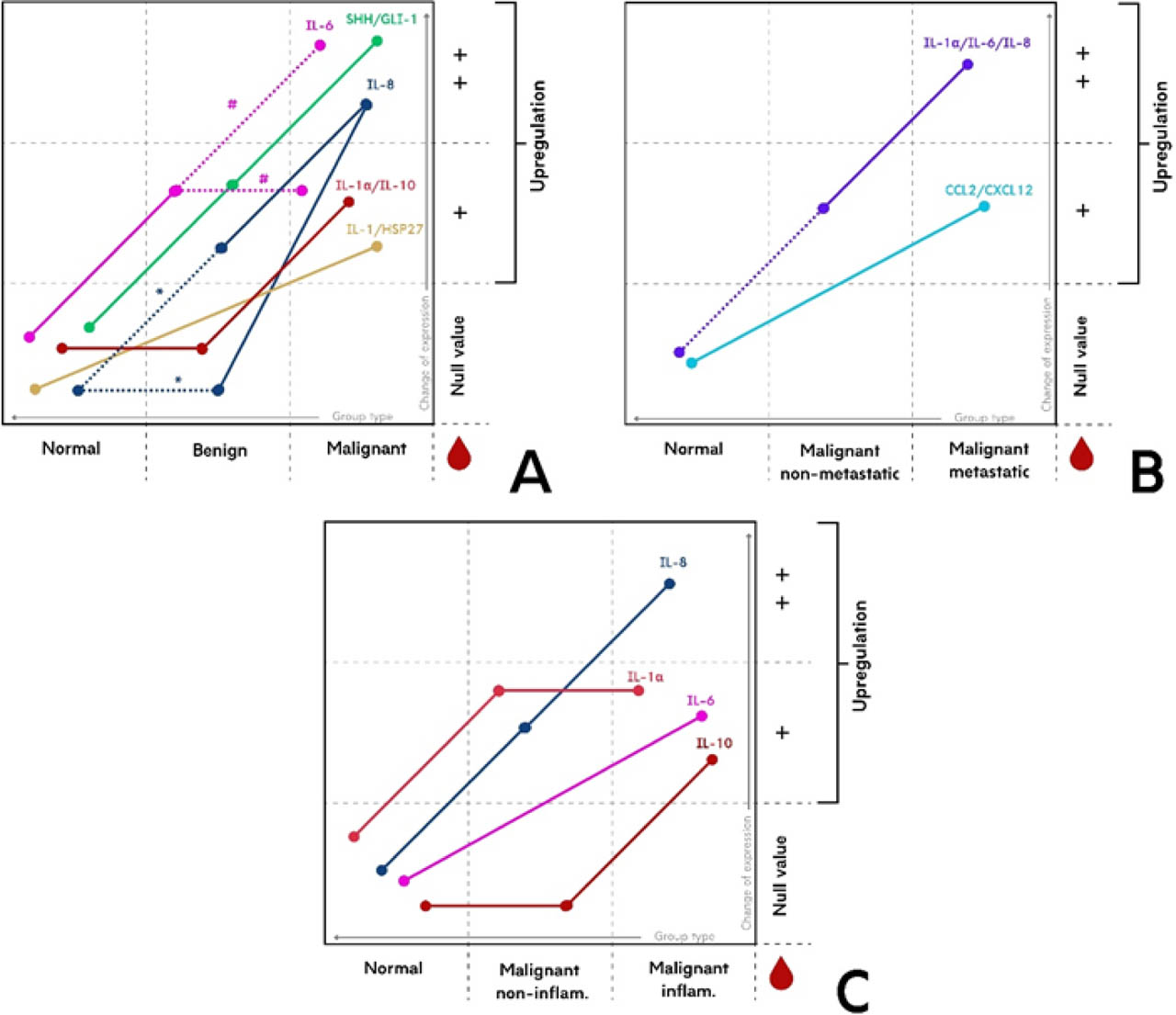
Figure 2.
Schematic representation of differences in concentrations of potential blood CMT bio-markers between groups. Colours denote expression patterns for individual proteins or grouped proteins that, in this review, showed a consistent upward regulation. The Y-axis reflects qualitative levels of change: + indicates overexpression relative to the reference level (Null value), and ++ indicates a higher-order overexpression relative to +. The X-axis presents the comparator groups between which expression differences were observed. Points mark the position of each group within a given pattern, and the connecting lines depict the trend in change across groups. Protein abbreviations are explained in Table 1. (A) Groups: normal (control), benign and malignant CMTs. Dashed lines indicate cases where concentrations are conflicting between groups; * denotes discrepancies between “Normal” and “Benign”, and # denotes discrepancies between “Benign” and “Malignant”. (B) Groups: normal (control), non-analysed. (C) Groups: normal (control), non-inflammatory and inflammatory CMTs.
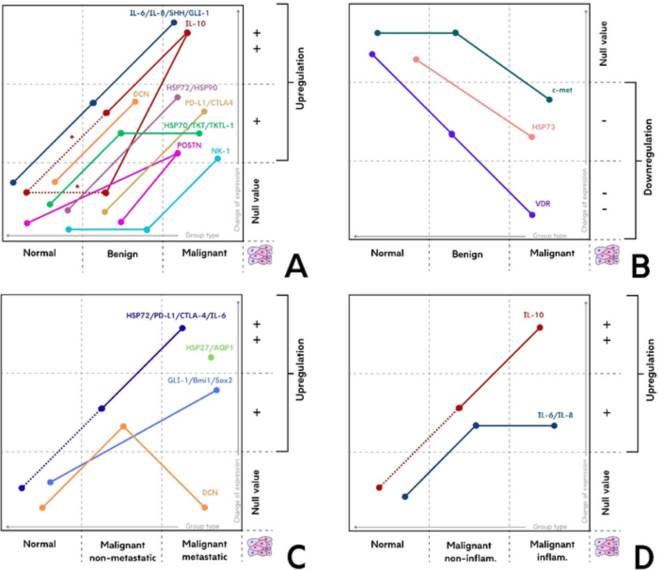
Figure 3.
Schematic representation of differences in expressions of potential tissue CMT biomarkers between groups. Colours denote expression patterns for individual proteins or grouped proteins that, in this review, showed a consistent upward regulation. The Y-axis reflects qualitative levels of change: +/− indicates up-/down-regulation relative to the reference level (Null value), and ++/−− indicates a higher-order up-/down-regulation relative to +/−. The X-axis presents the comparator groups between which expression differences were observed. Points mark the position of each group within a given pattern, and the connecting lines depict the trend in change across groups. The presence of a dot without a line means that the protein was identified only in this group, where it was not found in others. Protein abbreviations are explained in Table 2. (A) and (B) Groups: normal (control), benign and malignant CMTs. Dashed lines indicate cases where concentrations are conflicting between groups; * denotes discrepancies between “Normal” and “Benign” (C) Groups: normal (control), non-metastatic and metastatic CMTs. Dashed lines indicate that there was a difference in concentrations between normal and malignant (overall pool of malignant tumours) groups analysed. (D) Groups: normal (control), non-inflammatory and inflammatory CMTs. Dashed lines indicate that there was a difference in concentrations between normal and malignant (overall pool of malignant tumours) groups analysed.
In order to fully present the pattern of all the proteins discussed, Figure 2, Figure 3 and Figure 4 were created.
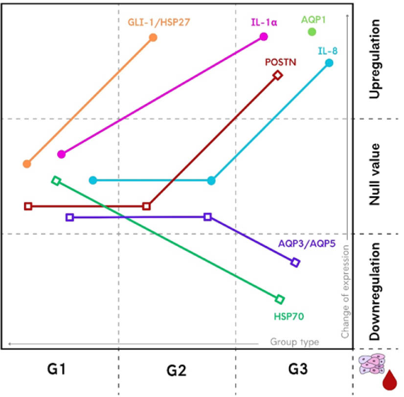
Figure 4.
Differences in concentration/expression of potential blood/tissue CMT biomarkers between tumours with different histological grade (G1, G2, G3). • – potential in blood; □ – potential in tissue. Colours denote expression patterns for individual proteins or grouped proteins that, in this review, showed a consistent change. The Y-axis reflects qualitative levels of change: up- or down-regulation relative to the reference level (Null value). The X-axis presents the comparator groups between which expression differences were observed. Points mark the position of each group within a given pattern, and the connecting lines depict the trend in change across groups. The presence of a dot without a line means that the protein was identified only in this group, where it was not found in others. Protein abbreviations are explained in Table 1 and Table 2.
Despite the promising potential of the proteins and research approaches discussed in the present article, significant gaps remain in the current literature. Most studies have been conducted on small study groups with inconsistent group sizes, and many lack a control group comprising healthy dogs or healthy tissues, substantially limiting the validity of their conclusions. Furthermore, critical subgroups, such as inflammatory, invasive, and meta-static tumours are often absent. These limitations underscore the need for additional studies to verify existing findings.