
In 2022, aquaculture surpassed capture fisheries as the primary source of aquatic animal production worldwide, with farmed species making up 51% of total production. Inland aquaculture accounted for 62.6% of the total, underscoring its increasing importance in world food systems (FAO, 2024). Additionally, inland fisheries were crucial, especially in Asia and Africa, which contributed 11.3 million tons of vital food for regional food security, accounting for 63.4 and 29.4 percent of the total production, respectively (FAO, 2024). China, Myanmar, Bangladesh, Indonesia, and India were among the top producers. Concerns about feed quality and safety have increased due to the quick growth of aquaculture and inland fishing output (FAO, 2024). This is especially true when it comes to mycotoxin contamination of aquafeeds, which poses serious dangers to fish health, productivity, and ultimately food safety (Paulino et al., 2018). The incorporation of technology advancements and mechanization in key production sectors has propelled aquaculture’s progress. Significant advancements in feed formulation techniques, with an emphasis on adjusting diets to the unique nutritional needs of cultured species at various phases of their production cycle, are among the major achievements (Paulino et al., 2018). About 70–80% of total production costs are attributed to feed, making it one of the most important cost factors in aquaculture operations (Paulino et al., 2018). Usually, a combination of components from plant and animal sources is used in its composition (Dawood, 2021). However, plant-based components are very vulnerable to microbial colonization, especially cereal grains. These substrates may encourage the growth of toxic fungus that can produce dangerous mycotoxins in response to environmental stress or nutritional imbalance (Hossain et al., 2024), which might endanger fish health and food safety.
Numerous mycotoxins, mostly generated by fungus belonging to the genera Aspergillus, Fusarium, and Penicillium, have been repeatedly detected in aquafeeds by quantitative evaluations (Nogueira et al., 2024). Fish performance and health may suffer both direct and indirect consequences from consuming feed tainted with mycotoxins. Growth and feed efficiency may be hampered by these harmful compounds’ direct injury to vital organs involved in digesting and nutrient absorption. According to Koletsi et al. (2021), they may indirectly impair immunological function, making fish more vulnerable to opportunistic infections. Furthermore, a number of mycotoxins have been linked to serious systemic toxicities, such as cytotoxic, neurotoxic, carcinogenic, estrogenic, and nephrotoxic effects (Oliveira and Vasconcelos, 2020). These effects may have an influence on fish welfare and aquaculture production.
In light of these concerns, this review aims to investigate the plant sources of mycotoxin contamination and their prevalence in aquafeeds, evaluate the toxicological effects of these toxins on fish species, and discuss both established and new methods for mitigation and control in aquaculture systems.
Sources and prevalence of mycotoxin contamination in aquafeeds
In the formulation of fish diets, nutrient sources are generally classified into two primary categories: protein-rich ingredients of both animal and plant origin and energy-providing feedstuffs. Fishmeal continues to be one of the most popular components among animal-based protein sources because of its high concentration of vital amino acids, accessible minerals and vitamins (Koletsi et al., 2021; Marijani et al., 2019). Notwithstanding its higher nutritional value, fishmeal’s growing demand across a number of businesses, notably the terrestrial cattle and poultry sectors, has led to increased prices and a shortage for use in aquaculture. The hunt for sustainable and affordable alternative protein sources that may partially or completely replace fishmeal in aquafeeds without sacrificing growth performance or health outcomes in cultured species has accelerated due to this resource- and economic-based limitation (Koletsi et al., 2021).
Soybean meal, corn, rice, and wheat are the most commonly utilized plant-derived protein sources in aquafeed formulation due to their worldwide availability, reduced cost when compared to fishmeal, and comparatively good digestibility (Gonçalves et al., 2017). Because of these qualities, they might be a good option to substitute fish-meal entirely or in part in commercial aquaculture diets. Because of its high starch content (Rokvić et al., 2020), favorable palatability, and accessibility, maize stands out as a primary carbohydrate input in terms of energy sources. However, strict quality control procedures are required when incorporating these plant elements that are high in protein and energy into industrial-scale feed production, especially because of the possibility of mycotoxin contamination (Table 1).
Table 1.
Mycotoxin contamination in plant ingredients used for formulating fish diets
| Ingredients | Mycotoxins (MQL µg/kg)* | Place | References |
|---|---|---|---|
| Rice | AFB1 (40.0), AFB2 (9.15), NIV (116.0), DAS (12.53), FB1 (75.1), ZEN (114.0), HT2 (32.0), DON (115.0), OTA (24.0) | Pakistan | Majeed et al., 2018 |
| ZEN (126.31) | Brazil | Savi et al., 2018 | |
| Oats | NIV (500.0), T-2 (132.0) | Switzerland | Martin et al., 2018 |
| Corn | AFB1 (24.0) | Japan | Nomura et al., 2018 |
| DON (725) | Turkey | Ünüsan, 2019 | |
| DON (826.0), FB1 (2234.0), FB2 (262.0), FB3 (189.0), FA (266.0), FX (96.0), ZEN (165.0), AFB1 (12.0), AFB2 (19.0), AFG1 (12.0), AFG2 (7.0), OTA (24.0), T-2 (81.0), HT-2 (190.0), NIV (661.0) | Europe | Koletsi et al., 2021 | |
| Soybean | AF (3430.0), OTA (125.0) and FB (4286.0) | Nigeria | Egbuta et al., 2016 |
| DON (2.265) and ZEN (853.0) | Croatia | Pleadin et al., 2017 | |
| DON (85.0), FB1 (371.0), FB2 (83.0), FB3 (50.0), FA (89.0), FX (65.0), ZEN (81.0), AFB1 (1.0), AFB2 (4.0), AFG1 (19.0), AFG2 (2.0), OTA (3.0), T-2 (49.0), HT-2 (155.0), NIV (231.0) | Europe | Koletsi et al., 2021 | |
| Wheat | OTA (34.75) | Algeria | Zebiri et al., 2019 |
| DON (470.0), FB1 (561.0), FB2 (59.0), FB3 (67.0), FA (54.0), FX (91.0), ZEN (64.0), AFB1 (2.0), AFB2 (9.0), AFG1 (3.0), AFG2 (3.0), OTA (6.0), T-2 (46.0), HT-2 (44.0), NIV (275.0) | Europe | Koletsi et al., 2021 |
* AFB1: aflatoxin B1, AFB2: aflatoxin B2, AFG1: aflatoxin G1, AFG2: aflatoxin G2, AF: aflatoxins, NIV: nivalenol, DAS: diacetoxyscirpenol, FB1: fumonisin B1, FB2: fumonisin B2, FB3: fumonisin B3, FB: fumonisins, ZEN: zearalenone, HT2: HT-2 toxin, DON: deoxynivalenol, OTA: ochratoxin A, T-2: T-2 toxin, FA: fusaric acid, FX: fusarenon X.
Animal health is seriously threatened by harmful secondary metabolites, which are mostly generated by fungus including Aspergillus, Fusarium, and Penicillium (Nogueira et al., 2020). Climate, crop genetics, and geographic location are some of the factors that affect the presence and concentration of mycotoxins in plant-based raw materials (Nogueira et al., 2020, 2024). Additionally, the risk of contamination can be increased by using less-than-ideal methods for cultivation, harvesting, post-harvest handling, and storage, both before and after industrial processing (Marijani et al., 2019). Therefore, it is essential to put preventative and monitoring measures into place to guarantee the security and nutritional value of aquafeeds.
Mycotoxin contamination exhibits marked geographical and ingredient-specific variability (Table 1). Tropical and subtropical regions including Nigeria, Pakistan and Brazil consistently show the highest contamination levels, a trend likely linked to warm, humid climates and limited cold storage facilities that favor fungal proliferation during both pre- and post-harvest strategies. Among the ingredients presented, soybean emerges as particularly vulnerable, with Nigerian soybean samples containing AF exceeding limits recommended by feed safety authorities. This raises concern regarding fish health, performance, international trade compliance and export-oriented economy. Another striking pattern is frequent co-occurrence of multiple mycotoxins within single feed batches hinting at additive or synergistic toxic effects. Yet there is no regulatory framework to address mycotoxins individually. For aquaculture species, where chronic low-dose exposure is more likely than acute poisoning, this gap in understanding multi-toxin interactions represents a major research and regulatory challenge. Likewise, the need for region-specific surveillance and tailored mitigation strategies is crucial. Wheat is highly susceptible due to its carbohydrate-rich composition, whereas oilseeds like soybean provide both high moisture retention and extended storage durations, creating ideal conditions for fungal colonization. These insights highlight why cost-effective detection methods and preventive management practices are urgently needed for the betterment of aquafeeds.
According to long-term global assessments of food safety, over 70% of food commodities may include at least one kind of mycotoxin, and it is not uncommon for multiple mycotoxins to co-occur in a single raw material (Kosicki et al., 2016). Since the manufacturing process sometimes entails blending many batches of materials from diverse sources, this poses a substantial barrier in the creation of aquafeed. A novel feed matrix with a distinct and perhaps unpredictable mycotoxin risk profile is created as a result of such combination (Pietsch et al., 2013). Therefore, it is not only possible but also likely that numerous mycotoxins might be present in aquafeeds at the same time and have cumulative harmful effects. The effects of these interactions on fish health and performance might be exacerbated if they are additive or synergistic (Gonçalves et al., 2020). The consequences of this kind of co-contamination highlight the necessity of thorough monitoring and risk assessment techniques in feed formulation and safety control. In this context, reliable quantification techniques are essential to accurately detect and measure multiple mycotoxins, assess their combined toxicity and support informed decisions in feed management.
Occurrence and methods of quantitative determination of mycotoxins in aquafeeds
Using herbal ingredients in feed formulation has led to a greater introduction of mycotoxins into crop systems because plant matrices have higher degrees of contamination (Modra et al., 2018). Contamination by mycotoxin is common, especially in underdeveloped and tropical areas (Matejova et al., 2017 a). All of the research points to mycotoxin contamination in feed rations as a major and widespread problem in aqua-culture that poses health and financial hazards in several nations (Gruber-Dorninger et al., 2019).
The presence and monitoring of mycotoxins in the diets of different animal species around the world have been documented in numerous studies (Arroyo-Manzanares et al., 2015; Kotinagu et al., 2015; Kim et al., 2017). However, there is still a lack of information regarding the presence of these contaminants in fish feed (Table 2). A key insight is the trade-off between sensitivity, cost and field applicability. Advanced chromatographic methods offer unparalleled sensitivity and multi-toxin detection capability, making them gold standards for regulatory laboratories. However, their high price and requirement of technical expertise and laboratory infrastructure limits widespread adoption in resource limited regions. On the contrary, ELISA-based kits provide a rapid, cost-effective tool but may lack precision for co-contamination scenarios. Novel biosensor platforms and portable immunoassays show promise in bridging this gap (Vardali et al., 2023). Mycotoxin contamination in feed is difficult to prevent, and their concentrations are often low within this matrix, so accurate detection requires the use of sensitive, precise, robust, selective, and reliable analytical methods (Greco et al., 2015). The most commonly used detection techniques for mycotoxins are gas chromatography, liquid chromatography, thin-layer chromatography, and ELISA (Vardali et al., 2023). Detectors made to strengthen the analyte signal are frequently used in conjunction with these chromatographic techniques. For example, gas chromatography is usually connected with flame ionization detection or mass spectrometry, whereas high-performance liquid chromatography can be paired with UV, diode array detection, fluorescence, or mass spectrometry (Vardali et al., 2023). For identifying mycotoxins both qualitatively and quantitatively, LC and GC combined with MS are among the most effective analytical techniques. They offer high selectivity and can detect multiple mycotoxins at once, making them ideal for confirmatory analysis (Albero et al., 2022; Vardali et al., 2023).
Table 2.
Prevalence of analytical approaches for mycotoxin detection in aquafeeds
| Method of quantification* | Mycotoxins obtained | References |
|---|---|---|
| 1 | 2 | 3 |
| Aflatest™ and Ochratest ™ | AFB1, OTA | Salama, 2007 |
| TLC and ELISA | AFB, AFB1 | Altuğ and Özyurt, 2003 |
| UPLC | AFB1, B2, G1, G2, M1, T2, HT-2, VCG, CTN, SMC, OTA, 3-ADON, 15-ADON, ZEN, FX, NIV, DON, ZEN+IS, ZEN−IS | Ren et al., 2007 |
| UPLC | AFB1, B2, G1, G2, OTA | Lopez Grio et al., 2010 |
| UPLC and mass spectrometry (MS) | AFB1, B2, G1, G2, OTA, FB1, FB2, HT-2, T-2, ZEN | Nácher-Mestre et al., 2013 |
| ELISA-based analytical test kits | AFs, DON, FUM, OTA, ZEN, T-2 | Greco et al., 2015 |
| LC-DAD AND LC-FL | DON, ZEN | Pietsch et al., 2013 |
| LC-FL | ZEN | Woźny et al., 2013 |
| LC-MS/MS | ENA, ENA1, ENB, ENB1, BEA | Tolosa et al., 2014 |
| ELISA and TLC | AFB1, FB1, OTA | Barbosa et al., 2013 |
| ELISA | AFB1 | Gonçalves-Nunes et al., 2015 |
| LC-MS/MS | AFB1, AFB2, AFG1 AFG2, FB1, FB2, FB3, DON, 3-ADON, 15-ADON, DAS, AOH, T-2, NIV | Marijani et al., 2017 |
| LC-MS/MS | 3-ADON, 15-ADON, AFB1, AFB2, AFG1 AFG2, BEA, DON, DAS, FB1, HT-2, NIV, OTA, T2, ZEN | Modra et al., 2018 |
| LC-MS/MS | AFB1, AFB2, AFG1 AFG2, ZEN, DON, NIV, 3- ADON, 15-ADON, FB1, FB2, FB3, T-2, HT-2, DAS, NEO, OTA | Gonçalves et al., 2018 a |
| ELISA and TLC | AFB1 | Jangampalli Adi and Matcha, 2018 |
| LC-HRMS/MS | AFB1, G1 | Mwihia et al., 2018 |
| HPLC | AFB1, B2, G1, G2, M1 | Michelin et al., 2017 |
| ELISA | AFB1, OTA | Osibona et al., 2018 |
| Multiplexed capillary microfluidic immunoassay | AFB1, OTA, DON | Machado et al., 2018 |
| LC-HRMS/MS | DON, T-2, HT-2, ZEN, ENN | Johny et al., 2019 |
| HPLC-FLD | AFB1, OTA, ZEN | Nogueira et al., 2020 |
| LC-MS/MS | 15-acetyl-DON, 3-acetyl-DON, AFB1, B2, G1, G2, alternariol, BEA, citreoviridin, citrinin, cyclopiazonic acid, DON, diacetoxy-scirpenol, ergometrin(in)e, FB1, FB2, FB3, fusarenon X, T-2 toxin, moniliformin, methylergonovine, NEO, NIV, OTB, OTA, patulin, T-2 toxin, verruculogen, ZEN | Koletsi et al., 2021 |
| HPLC-MS/MS | AFB1, AFM1, T-2 toxin, HT-2 toxin, DON, OTA, ZEN | Bi et al., 2022 |
| HPLC-MS/MS | NIV, DON, AFB1, B2, FB1, FB2, OTA, ZEN, ENNB, BEA, ENNB1, ENNA1, ENNA | Albero et al., 2022 |
| ELISA | AFs, OTA | Bashorun et al., 2023 |
| LC–MS/MS | AFB1, B2, G1, FB (B1+B2), DON, OTA | Terada-Nascimento et al., 2023 |
* UPLC: ultra-performance liquid chromatography; ELISA: enzyme linked immunosorbent assay; TLC: thin layer chromatography; LC-DAD: liquid chromatography with diode array detection; LC-FL: liquid chromatography with fluorescence detection; LC-MS/MS: liquid chromatography–tandem mass spectrometry; LC-HRMS/MS: liquid chromatography–high-resolution tandem mass spectrometry; HPLC: high-performance liquid chromatography; UHPLC-QTOF-MS/MS: ultra-high-performance liquid chromatography–quadrupole time-of-flight–tandem mass spec-trometry; HPLC-MS/MS: high-performance liquid chromatography–tandem mass spectrometry; AFM1: aflatoxin M1; OTB: ochratoxin B; CTN/CIT: citrinin; α-ZEL: alpha-zearalenol; β-ZEL: beta-zearalenol; D3G: deoxynivalenol-3-glucoside; 3-ADON: 3-acetyl-deoxynivalenol; 15-ADON: 15-acetyl-deoxynivalenol; ZEN+IS/ZEN-IS: zearalanone with/without internal standard; BEA: beauvericin; VCG: verrucogemefuranone (verruculogen); AOH: alternariol; SMC: sterigmatocystin; NEO: neosolaniol; ENNA: enniatin A; ENNA1: enniatin A1; ENNB: enniatin B; ENNB1: enniatin B1; MON: moniliformin.
Adverse toxicological effects of mycotoxins on fish
It is crucial to comprehend the toxicological consequences of mycotoxins in fish because prior research has indicated that these chemical compounds might have a variety of negative impacts (Baldiserra et al., 2020 a; He et al., 2024). Damage to cells and organs, abnormalities in morphological and functional development, and, in extreme situations, higher mortality rates in exposed fish populations have all been connected to mycotoxins (Anater et al., 2016). While several studies reported reduced growth performance in O. niloticus and C. carpio following AFB1 exposure (Ayyat et al., 2018; Deng et al., 2010), others noted paradoxical growth promotion at low doses (Imani et al., 2017). These discrepancies may reflect differences in exposure duration, fish developmental stage and baseline nutritional status, suggesting that dose-response relationship for AFB1 toxicity are non-linear and species specific. Similarly, evidence of OTA toxicity is heterogenous. Hepatotoxic and nephrotoxic effects dominate reports in C. carpio yet immune suppression appears inconsistent across studies at comparable doses (Hassan et al., 2018). Environmental stressors and dietary protein levels may explain why immune endpoints differ even when growth and organ pathology align across trials. Similar contradictory effects are found in case of DON, suggesting species-specific detoxification or microbial degradation capacities. Likewise, ZEN induces feminization and gonadal alterations in carp (Schwartz et al., 2010) but fails to trigger comparable endocrine effects in tilapia at similar doses (Pietsch et al., 2015), reflecting differences in hormone receptor sensitivity and endocrine baselines. Exhaustive literature review points out such contradictions across species-specific sensitivity, experimental viability and neglected microbiome interactions. Future research should prioritize standardized dosing protocols, incorporate microbiome analyses and explore interactive effects throughout the system which would enhance the predictive power of toxicological assessments and improve the relevance of findings for aquaculture management.
Aflatoxins and fumonisins consistently impair growth performance and liver function in O. niloticus and C. carpio yet similar exposure levels produce negligible effects in S. salar or C. gariepinus (Table 3), hinting at species-specific difference in detoxification metabolism, gut microbiome composition and immune competence. Also, there is a paucity of standardized dosing protocols across studies which hampers cross-trial comparisons and meta-analytical synthesis. Numerous factors influence the lethality linked to consuming mycotoxin-contaminated feed, such as the species involved (Ji et al., 2019), the type and concentration of the mycotoxin (Bitsayah et al., 2018), the animal’s age (Gonçalves et al., 2018 a; Khezri et al., 2018), and most significantly, the length of exposure, which is crucial in describing the possible effects and outcomes in affected animals (Zhou et al., 2017). The non-specific symptoms and delayed commencement of toxic effects from different mycotoxin exposures make it difficult to diagnose mycotoxin-induced disorders in fish (Oliveira and Vasconcelos, 2020). Furthermore, current research is extremely limited, frequently concentrating on a small number of mycotoxins in a few different fish species worldwide (Oliveira and Vasconcelos, 2020). There have been conflicting reports, though. For instance, Imani et al. (2017) examined the toxicity of aflatoxin B1 at concentrations of 0, 25, and 50 ng/kg in rainbow trout (Oncorhynchus mykiss) and found that fish fed with contaminated diets gradually increased their digestive enzyme activities, such as lipase, amylase, and alkaline protease, while also showing significantly higher weight gain (P<0.05). In a similar vein, Pietsch et al. (2014) evaluated the effects of DON-supplemented meals at dosages of 352 µg/kg, 619 µg/kg, and 953 µg/kg in common carp (Cyprinus carpio L.) and discovered no discernible changes in growth performance throughout the course of the trial. After being exposed to the greatest concentration of mycotoxin, the only negative impact seen was an increase in lipid peroxidation in the liver, kidney, and spleen. According to these results, estimation of weight change by itself is not a sensitive enough metric to identify mycotoxin contamination in aquafeed.
Table 3.
Impact of mycotoxins on fish health across species
| Mycotoxin | Species | Dose and administration | Toxic effects seen | References |
|---|---|---|---|---|
| 1 | 2 | 3 | 4 | 5 |
| OTA | Ictalurus punctatus (juvenile) | 0.5, 1.0, 2.0, 4.0 and 8.0 mg/kg for 8 weeks | Mortality, inadequate feed conversion, decreased haematocrit, and decreased weight growth | Manning et al., 2003 a |
| 2 and 4 mg/kg for 6 weeks | Reduced weight gain | Manning et al., 2005 | ||
| 2, 4 and 8 mg/kg for 8 weeks | Reduced weight gain and growth, low feed conversion | Zahran et al., 2016 | ||
| Colossoma macropomum (juvenile) | 0.8, 1.6 and 2.4 mg/kg for 14 days | Decreased growth performance, hiked muscle oxidative stress (ROS and LPO), decreased antioxidant enzyme activity (SOD and GPx), and deterioration of fillet fatty acid profile. | Baldiserra et al., 2020 a | |
| Colossoma macropomum (juvenile) | 1.6 and 2.4 mg/kg for 14 days | Impaired growth, reduced liver and body weight, and altered liver energy metabolism. Hepatic ATP and adenosine levels increased, while NTPDase (ATP) activity rose and ADA activity declined | Baldissera et al., 2020 b | |
| STG | Oreochromis niloticus (fingerling) | 1.6 µg/kg for 4 weeks | Clastogenic, weight reduction, chromosomal aberrations in the kidneys | Abdel-Wahhab et al., 2005 |
| Aflatoxins | Cyprinus carpio (juvenile) | 0.5, 0.7 and 1.4 mg/kg for 3 weeks | Decrease in lysozyme activity and reduction of immunoglobulin in blood plasma | Bitsayah et al., 2018 |
| AFB1 | Oreochromis niloticus and Oreochromis aureus (juvenile) | 19, 85, 245, 638, 793 and 1641 µg/kg for 20 weeks | Induction of hepatic disorder, resulting in decreased lipid content, hepatosomatic index, cytochrome P450 A1 activity, and elevated plasma alanine aminotransferase activity | Deng et al., 2010 |
| Oreochromis niloticus (fingerling) | 2 mg/kg for 14 weeks | Reduction in weight gain, low feed conversion; decreased hemoglobin concentrations in the blood, total erythrocytes, total proteins, albumin and globulins | Ayyat et al., 2018 | |
| Rhamdia quelen (juvenile) | 1177 µg/kg for 3 weeks | Affects neurotransmitters, hyperlocomotion, increased acetylcholinesterase activity; reduced sodium-potassium pump activity | Baldissera et al., 2018 a | |
| Changes in the cerebral phosphotransferase network | Baldissera et al., 2018 b | |||
| Cyprinus carpio (juvenile) | 50 µg/kg for 8 weeks | Reduced growth and low feed conversion | Fan et al., 2018 | |
| Pangasius hypophthalmus (fingerling) | 50, 100, 250, 500 and 1000 µg/kg for 8 weeks | Reduction in weight gain, increase in hepatosomatic rate, increase in adipose somatic index, elevated levels of alanine aminotransferase and aspartate aminotransferase | Gonçalves et al., 2018 c | |
| Ctenopharyngodon idella (juvenile) | 0, 29, 59, 86, 110 and 147 μg/kg for 60 days | Produced abnormalities and stunted development in young grass carp, with residues building up in the spleen and kidney of the head. Through apoptosis, it altered the structure of immunological organs, decreased antioxidant capability, and weakened intercellular integrity by damaging tight junctions. | Zeng et al., 2019 | |
| Danio rerio (larvae) | 0.01, 0.05, 0.1 mg/kg and 0.1, 0.5, 1 mg/kg for 3 days post fertilization | Developmental abnormalities, disrupted thyroid hormone and vitellogenin levels, induced oxidative stress and lipid peroxidation, and triggered the expression of pro-apoptotic genes in fish larvae | Di Paola et al., 2021 | |
| Ctenopharyngodon idella (juvenile) | 0.04, 29.48, 58.66, 85.94, 110.43 and 146.92 μg/kg for 60 days | Reduced myofiber density, collagen content, and antioxidant enzyme levels while increasing myofiber diameter, oxidative stress, and MMP activity, impairing grass carp muscle growth and meat quality. It suppressed myogenic regulatory pathways and TGF-β/Smads. | He et al., 2024 | |
| DON | Cyprinus carpio L. (juvenile) | 352, 619 and 953 µg/kg for 4 weeks | Cytotoxic effects on immune cells | Pietsch et al., 2014 |
| 352, 619 and 953 µg/kg for 6 weeks | Increased lipid peroxidation in the liver, kidney and spleen | Pietsch et al., 2014 | ||
| Oncorhynchus mykiss (juvenile) | 0.3, 0.8, 1.4, 2.0 and 2.6 ppm/kg for 8 weeks | Reduction in weight gain, low growth rate and feed conversion, morphological changes in the liver | Hooft and Bureau, 2017 | |
| Oncorhynchus mykiss (fingerling) | 4.5 and 10.5 mg/kg for 8 weeks | Reduction in weight gain, low feed conversion, altered proteolytic enzyme activity, elevated messenger RNA levels and nutrient retention | Gonçalves et al., 2018 b | |
| Cyprinus carpio L. (juvenile) | 332, 621 and 797 µg/kg for 4 weeks | Cytotoxic effects on immune cells | Pietsch et al., 2015 | |
| DON and ZEN | Oreochromis niloticus and Oreochromis mossambicus (fingerling) | 0.07, 0.31, 0.50, 0.92, 1.15 mg/kg and 0.01, 0.09, 0.21, 0.37, 0.98 mg/kg for 8 weeks | Reduction in weight gain and growth, low feed conversion | Tola et al., 2015 |
| T-2 | Cyprinus carpio L. | 5.3 mg/kg for 4 weeks | Reduction in weight gain, low feed conversion, anemia, lymphopenia, oxidative stress | Matejova et al., 2017 b |
| I. punctatus | 0.625, 1.25 and 5 mg/kg for 8 weeks | Dose-dependent deficits in survival, hematocrit levels, feed conversion efficiency, and body weight gain. Gastric lesions and a decrease in hematopoietic cells in the head kidney were among the histopathological alterations. There were also more melanomacrophage centers with hemosiderin and lipopigments. | Manning et al., 2003 b | |
| 1 and 2 mg/kg for 6 weeks | Reduction in weight gain | Manning et al., 2005 | ||
| Oncorhynchus mykiss (adult) | 1.0 and 1.8 mg/kg for 4 weeks | Oxidative stress, increased lipid peroxidation, increased ceruloplasmin activity | Modra et al., 2018, 2020 | |
| FB1 and FB2 | Oreochromis niloticus (fingerling) | 20, 40 and 60 mg/kg for 4 weeks | Reduction of messenger RNA levels, reduced expression of the CASP7 gene | Claudino-Silva et al., 2018 a, b |
| AFB1 and FB1 | Clarias gariepinus (juvenile) | 2.0, 7.3, 17.6, 48.0, 93.0 µg/kg and 3.0, 15.0, 24.5, 43.0, 83.0 mg/kg for 8 weeks | Reduction in weight gain and length, low feed conversion | Adeyemo et al., 2018 |
| Mixed mycotoxins (low levels) | Oreochromis niloticus (juvenile) | 40 µg/kg AFB1, 600 µg/kg FB, 50 µg/kg ZEN, and 150 µg/kg DON | Liver degeneration, intestinal and renal tissue damage, splenic lymphoid depletion, and clinical signs like fin rot and ascites; impaired immune responses and disrupted normal growth and biochemical parameters | Hussein et al., 2024 |
As reported by Schwartz et al. (2010), adult zebrafish’s gonadal morphology, hatching, survival, and fertility were all unaffected by ZEN exposure (100–3200 ng/L). However, Bakos et al. (2013) found that ZEN caused anomalies in the development of the heart and eyes in embryos and produced estrogenic effects in adults, indicating broader ontogenetic disruption. A heat map (Figure 1) shows reported organ damage or changes following mycotoxin exposure in fish.
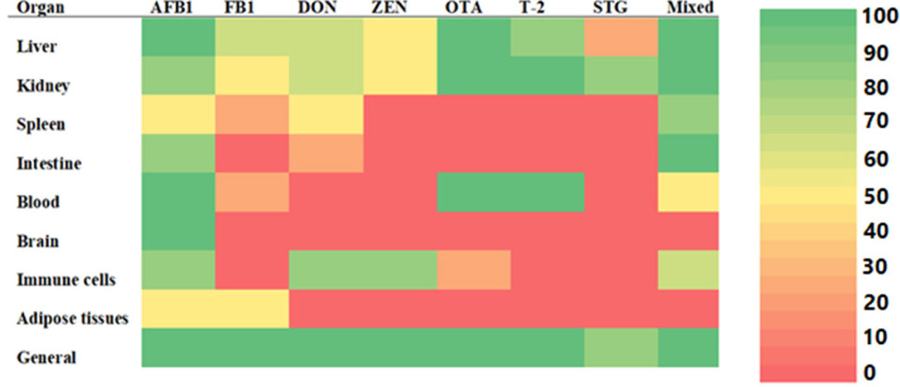
Figure 1.
Heat map summarizing the frequency of reported organ specific effects induced by individual and mixed mycotoxins based on a systematic literature review. The color scale represents the percentage frequency of studies reporting effects for a given organ mycotoxin combination, normalized to a 0 to 100 scale. Red (0 percent) indicates no available data or no reports of effects. Light orange (1 to 25 percent) denotes very low frequency of reported effects. Yellow (26 to 50 percent) indicates low to moderate frequency. Light green (51 to 75 percent) represents moderate to high frequency, while dark green (76 to 100 percent) indicates a high frequency of reported effects across studies. “Mixed” refers to studies evaluating combinations of two or more mycotoxins. “General” denotes systemic or non-organ specific alterations reported in the literature rather than changes confined to a single organ.
These results highlight how important it is to standardize toxicological evaluations of mycotoxins in fish, especially with regard to exposure time, doses given, and experimental setups (Anater et al., 2016). In addition to the direct harm to aquatic life, there is a serious health risk to humans when fish are contaminated by eating feed that contains mycotoxin. This danger results from the possibility of mycotoxin bioaccumulation and toxic synergism in the food chain (FAO, 2024). Supporting this concern, Michelin et al. (2017) demonstrated the transfer of aflatoxin B1 at concentrations of 10, 20, and 50 µg/kg from lambari feed (Astyanax altiparanae) to fish muscle tissue, with mycotoxin levels in muscle closely mirroring those in the ration after 120 days of exposure. This evidence partly accounts for the predominance of studies addressing the toxicity of aflatoxins (Table 3), given their well-documented carcinogenic, mutagenic, and teratogenic properties, which pose substantial threats to human health and incur significant economic losses.
Regulatory limits for mycotoxins in fish feed
Regulations have been put in place in a number of nations to limit the amount of mycotoxins that may be present in food items meant for human and animal consumption. About 101 nations have passed laws addressing mycotoxin contamination (FAO, 2024). The regulatory thresholds, termed as maximum tolerable limits, were mostly determined using toxicological data from a variety of terrestrial animal species, such as cattle, pigs, and birds. There is still a substantial knowledge gap regarding fish species-specific guideline values (Anater et al., 2016; Gonçalves et al., 2020). The majority of current rules are derived from laws governing terrestrial cattle feed; there are presently no legal maximum values for mycotoxins in fish feed that are particular to aquaculture (Gonçalves et al., 2020). For instance, advisory levels for a number of important mycotoxins are outlined in the European Commission’s Recommendation 2006/576/EC. The maximum amount of aflatoxin B1 in whole feedstuffs is 20 µg/kg (Koletsi et al., 2021). Ochratoxin A (OTA), fumonisins, ZEN, and deoxynivalenol (DON) have comparable indicative values (Oliveira and Vasconcelos, 2020; Koletsi et al., 2021). Certain samples occasionally may be found above the limitations, especially for OTA and DON, but the majority of commercial aquafeeds adhere to these regulatory benchmarks (Sodeeq et al., 2023). Fish may react to mycotoxins differently than terrestrial animals owing to their diverse metabolisms and physiologies. The growing use of plant-based components in aquafeeds, which are more vulnerable to fungal contamination, exacerbates this worry (Hossain et al., 2024). In order to safeguard fish health and guarantee food safety, current evaluations advocate for specific regulatory criteria for aquaculture feeds (Pietsch et al., 2013; Gonçalves et al., 2020). Additionally it is advised that better feed management techniques should be followed and routine monitoring to be exercised (Sodeeq et al., 2023; Koletsi et al., 2021).
Control of mycotoxins in aquafeed rations
Preventive and physical detoxification approaches
A widespread problem impacting markets worldwide is mycotoxin contamination of agricultural commodities (Luo et al., 2021). The best way to reduce the presence of mycotoxin in raw materials and processed goods like animal feed is still to avoid fungal growth and contamination, although these methods are sometimes insufficient for total removal. Detoxification methods are therefore commonly used in control attempts (Bashorun et al., 2023). It has been shown that mycotoxin contamination may be successfully reduced or eliminated by implementing good agricultural practices along the whole grain production and feed manufacturing chain (Bashorun et al., 2023). Furthermore, developments in the creation of genetically engineered crops that are intended to reduce fungal growth and the synthesis of mycotoxin as a result have been explored (Sissener et al., 2011). However, the possibility of such genetically modified crops being widely adopted is limited by the significant geographical and climatic heterogeneity among countries (Medina et al., 2015). In order to reduce mycotoxin concentrations to levels that meet regulatory requirements, detoxification procedures are used as supplemental treatments (Manning et al., 2003 a). The most common of these techniques in feed detoxification procedures is the use of adsorbent compounds (Puvača et al., 2023). The nutritional inertness and capacity to bind mycotoxins in the gastrointestinal system, therefore minimizing or stopping their absorption during digestion, are characteristics of effective adsorbents. This adsorption process reduces the spread of pollutants throughout the food chain and lessens their harmful effects on animals (Avantaggiato et al., 2021; Puvača et al., 2023). An ideal adsorbent should be economical, safe for the environment, leave no residues, maintain product quality, and not alter palatability (Puvača et al., 2023). But none of the detoxification plans in use today satisfy all of these requirements. In addition to mycotoxin type, concentration, feed matrix, and environmental factors, adsorbent effectiveness is also influenced by pore size, charge, polarity, and structure (Kabak et al., 2006; Avantaggiato et al., 2021). Since aflatoxins are polar, aluminosilicate-based adsorbents have shown promise in binding them. Their wider usefulness is limited, nevertheless, because their binding affinity is greatly diminished when they target non-polar mycotoxins such OTA, ZEN, and trichothecenes (Avantaggiato et al., 2021). Scaglioni and Badiale-Furlong (2016) examined the effectiveness of rice husk as a natural adsorbent for aflatoxin B1 (AFB1) and aflatoxin M1 (AFM1) in milk as part of an inquiry into non-traditional adsorbents. Adsorption rates for AFB1 and AFM1 were from 77.2% to 122% and 73.1% to 125%, respectively, according to the study, with recoveries surpassing the European Commission’s standards. The results highlight the potential use of agro-industrial byproducts in mycotoxin reduction, notwithstanding the study’s exclusive emphasis on the aflatoxin group. Alternative techniques, such as chemical and biological detoxification procedures, are being assessed for their efficacy in mycotoxin management in addition to physical adsorption.
Chemical and biological detoxification approaches
Chemical detoxification techniques work by changing the mycotoxin’s molecular makeup or binding properties. The effectiveness of ozone gas treatment was highlighted by Hassan et al. (2018), who showed that ozonation considerably (P<0.05) decreased AFB1 concentrations in animal feed. Alcántar-Barrios and Méndez-Albores (2017) also assessed the combined impact of tartaric acid and nonionizing radiation on B-group aflatoxins in maize flour (particle size <420 µm) and found that, after only 3 minutes of treatment (P<0.01), aflatoxin levels significantly decreased (43–87%). Ascorbic acid significantly (P<0.05) increased mycotoxin degradation at all tested temperatures, according to Kokkinidou et al. (2014), who examined the thermal degradation of patulin with and without its presence. This is corroborated by Su et al. (2018), who found that ascorbic acid supplementation reduces mycotoxin-induced immunosuppression and toxicity by improving immunological function and antibody generation in addition to aiding in mycotoxin breakdown. Ammonization is another chemical method that is approved in some countries and is well known for its efficacy in detoxifying foods infected with aflatoxin (Doğan and Hayırlı, 2022). Its effectiveness, however, is restricted to aflatoxins and does not extend to other kinds of mycotoxin. Furthermore, too much residual ammonia in treated feed might be harmful to animals’ health (Doğan and Hayırlı, 2022), which limits its further use.
The use of biological techniques for mycotoxin detoxification has grown in popularity as an efficient and ecologically friendly approach. These strategies are appealing substitutes for chemical and physical procedures because of their comparatively low environmental effect and need for just mild operating conditions. Mycotoxin binding by biological adsorbents or enzymatic inactivation by certain bacteria or their byproducts are the two main methods of biological detoxification (Liu et al., 2022). Rhizopus oryzae and Trichoderma reesei, two non-toxic bacteria, have been shown by Kupski et al. (2018) to have the ability to biodegrade OTA. According to their results, T. reesei broke down 57.7% of OTA in 72 hours, but R. oryzae reduced OTA concentration by 63.5% after 48 hours of treatment. These findings demonstrate how microbial biotransformation may be used as a practical method to reduce mycotoxin contamination in food and feed systems. The use of plant-derived extracts as natural agents to regulate fungal growth and mycotoxin production has been investigated in a number of research. These extracts have shown promise as a safe and environmentally friendly substitute for synthetic antifungal medicines by suppressing the production of mycotoxins in addition to inhibiting fungal growth (Qi et al., 2023). Gandomi et al. (2009) looked at how essential oil from Zataria multiflora affected Aspergillus flavus in terms of its antifungal and anti-mycotoxigenic properties. According to their research, fungal growth was suppressed by 92.5% at a concentration of 200 ppm of the essential oil, and total suppression was noted at doses higher than 400 ppm. Additionally, aflatoxin production was considerably (P<0.05) decreased at all measured doses, with a maximum reduction of 99.4% at 150 ppm. Table 4 provides a thorough inventory of all the different control techniques or tactics. Phenolic extracts from Spirulina sp. and Nannochloropsis sp. microalgae were tested by Scaglioni et al. (2019) against Fusarium graminearum and its related mycotoxin DON. Spirulina and Nannochloropsis extracts decreased DON contamination by 62% and 97%, respectively, and inhibited fungal biomass by 34% and 25%. Phenolic chemicals originating from plants have attracted a lot of interest lately due to their ability to reduce the production of mycotoxin and regulate fungal growth. These naturally occurring substances, which were isolated from different matrices, have proven to be effective substitutes for traditional chemical fungicides due to their broad-spectrum antifungal and anti-mycotoxigenic properties (Table 4). Despite the paucity of research on aquafeed mycotoxin management, these results combined imply that plant-based extracts have great potential as safe and efficient ways to reduce mycotoxin contamination in aquafeed systems. Table 4 highlights the cost-efficacy tradeoffs in current detoxification strategies available worldwide. While adsorbents like bentonite offer affordability and ease of use, they lack toxin specificity and may reduce nutrient bioavailability. Biological approaches emerge as ecofriendly and promote gut health, but their large-scale validation and economic feasibility studies remain meagre. Herbal extracts present a promising frontier yet challenges in standardization and stability limit their commercial adoption, underscoring the need for integrated, field-ready solutions.
Table 4.
Comprehensive control strategies for mycotoxins in food and aquafeed
| Control method | Remarks/Mode of action | Reference |
|---|---|---|
| 1 | 2 | 3 |
| Tri-octahedral bentonite | Clay mineral with high adsorption capacity for multiple mycotoxins; suitable for inclusion in fish feed | Pascari et al., 2025 |
| Calcium and sodium bentonite | Clays effectively bind ochratoxin A both in vitro and in vivo, reducing its bioavailability and potential toxicity | Hamad et al., 2022 |
| Pulse electric field (PEF) | Electrical pulses permeabilize cell membranes, leading to fungal inactivation and partial mycotoxin degradation | Gavahian et al., 2022 |
| Supercritical CO2 (SC-CO2) | SC-CO2 penetrates fungal cells and mycotoxins, causing structural damage and facilitating toxin extraction | Gavahian et al., 2022 |
| Ultrasound (USN)/Sonication | Cavitation generates microbubbles that physically disrupt fungal cells and degrade mycotoxins via shear forces and ROS | Gavahian et al., 2022 |
| Water-assisted microwave treatment (WMT) | Effectively reduces aflatoxin B1 in corn by combining thermal and moisture-driven degradation while minimizing heat damage to kernels | Zhang et al., 2025 |
| High-pressure processing (HPP) | High hydrostatic pressure disrupts fungal cell structures and can inactivate mycotoxins without heating | Gavahian et al., 2022 |
| Tri-octahedral smectite + lignocellulose additive | Dual-action: smectite adsorbs toxins; lignocellulose supports gut health | Greco et al., 2022 |
| Yeast cell wall extract (e.g., β-glucans) | Effectively adsorb aflatoxin B1, ochratoxin A, and fumonisins – demonstrated in vitro and in animal studies | Xu et al., 2023 |
| Saccharomyces cerevisiae cell wall | Reduced aflatoxin B1 absorption in rodent model; considered for broader feed applications | Imani et al., 2017 |
| Postbiotic yeast blends | Mitigated multi-mycotoxins and improved gut health and performance in livestock | Lázaro et al., 2024 |
| Mixed-origin biocontrol agents (LAB, yeasts) | Bind and degrade mycotoxins via enzymatic activity or cell wall adsorption | Bashorun et al., 2023 |
| Bacillus cereus VOCs | VOCs suppress fungal growth and sporulation; reduce mycotoxin biosynthesis | Bashorun et al., 2023 |
| Proper feed storage and moisture control | Reduces fungal proliferation by controlling humidity and temperature | Pietsch et al., 2020 |
| Botanical phenolic extracts | Inhibit mycotoxin synthesis via antioxidant action and downregulation of toxin genes | Ahmed et al., 2022 |
| Cold atmospheric plasma and gamma irradiation | Effectively degrade aflatoxins while preserving food quality | Misra et al., 2019 |
| Natural essential oils (NEOs) | Disrupt fungal membranes; antifungal and antimycotoxigenic effects | Mirza Alizadeh et al., 2022 |
| Natural phenolics against Fusarium | Inhibit trichothecene biosynthesis; eco-friendly method to control food contamination | Chtioui et al., 2022 |
| Thiol compounds (–SH) | Efficient detoxification of patulin in juice via nucleophile binding (–SH, –OH, –NH2) | Diao et al., 2022 |
| Fatty acids and oxylipins | Broad antifungal effect targeting fungal enzymes and membranes; eco-friendly with low resistance potential | Qiu et al., 2021 |
| Active food packaging (biopolymer-based) | Inhibits fungal growth via polysaccharide/lipid/protein matrices; biodegradable and sustainable | Ghobish et al., 2025 |
| Lactic acid bacteria (LAB) as biodetox agents | Reduce mycotoxin biosynthesis and promote degradation in food matrices | Nasrollahzadeh et al., 2022 |
| Biogenic Trichoderma harzianum-derived selenium nanoparticles | Stronger antifungal and anti-mycotoxigenic effect compared to nanoparticles alone | Hu et al., 2019 |
| Cold plasma treatment of jujube juice (for Alternaria toxins) | Effectively degrades mycotoxins while preserving juice quality and physical characteristics | Wang et al., 2022 |
Plant microbiome and mycotoxin contamination in aquafeed crops
Recognizing the plant microbiome as a determinant of mycotoxin risk strengthens the continuum between crop ecology and feed safety, providing a scientific basis for targeted detection and control strategies discussed in this review. The crops used in aquafeed production harbor a diverse plant-associated microbiome, encompassing both epiphytic and endophytic microbial communities. These microbial consortia significantly influence pre-harvest mycotoxin risk by either suppressing or facilitating the colonization of mycotoxigenic fungi (Medina et al., 2015; Pascari et al., 2025). Beneficial endophytes can act as biological control agents producing antifungal metabolites or competing for ecological niches, thereby reducing toxin accumulation in host plants (Nasrollahzadeh et al., 2022). However, abiotic stressors such as water shortage, temperature rise and nutrient imbalance often disrupt microbial load, creating optimal conditions favorable for toxin producing fungi. Similarly, soil management practices and pesticide applications can shift microbial diversity indirectly influencing mycotoxin biosynthesis (Hossain et al., 2024). Pascari et al. (2025) highlighted that microbiome engineering through seed treatments with beneficial endophytes or crop rotation practices effectively lowered mycotoxin contamination before harvest. Nasrollahzadeh et al. (2022) showed that Trichoderma harzianum inoculation in maize decreased Fusarium colonization and fumonisin levels. Given the aquafeed industry’s increasing dependence on plant-based ingredients, integrating crop microbiome management into feed safety protocols represents a proactive intervention point. Understanding these microbial dynamics will not only advance risk prediction models for mycotoxin outbreaks but also inform the development of eco-friendly mitigation strategies linking agricultural practices with aquafeed safety.
Fish gut microbiome as modulator of mycotoxin toxicity
The bioavailability of ingested mycotoxins and the host’s immunological competence are both impacted by the gut microbiota of fish, which is crucial in controlling host–toxin interactions. This intricate ecology, which includes bacteria, fungus, archaea, and protozoa, aids in immune modulation, mucosal barrier maintenance, and nutrition metabolism (Dawood, 2021). According to recent research, depending on the host’s genetics, exposure circumstances, and microbial makeup, gut-associated microorganisms can either lessen or increase mycotoxin toxicity (Xu et al., 2023). Enzymatic mechanisms found in a number of commensal microorganisms can biotransform mycotoxins into less harmful metabolites. Lactic acid bacteria (LAB) and derivatives of Saccharomyces cerevisiae may adsorb aflatoxins and break down trichothecenes, lowering toxicity and systemic absorption in aquatic species (Bashorun et al., 2023). Likewise, microbial enzymes like dehydrogenases and esterases aid in detoxification processes and may reduce tissue levels of toxins such as deoxynivalenol and ZEN (Liu et al., 2022). On the other hand, mycotoxin initiated dysbiosis can weaken the intestinal barrier, set off inflammatory reactions, and lower immunological homeostasis, making the host more vulnerable to metabolic stress and secondary infections (Pietsch et al., 2015). Long-term exposure to fumonisin has been associated with changes in intestinal microbiota diversity, which can impair tilapia’s ability to absorb nutrients and their immune system (Claudino-Silva et al., 2018 a). These results imply that, in addition to direct cellular effects, microbiome disturbance is a crucial amplification mechanism for mycotoxin toxicity. Furthermore, the formation and control of mucosal immunity are influenced by the gut microbiota, which serves as an immune training center. According to Xu et al. (2023), microbial metabolites including short-chain fatty acids (SCFAs) have anti-inflammatory properties and may mitigate the oxidative stress brought on by prolonged exposure to mycotoxin. Despite these realizations, there are very few studies that focus specifically on fish, and the majority of the data is extrapolated from livestock systems on land. To understand the fish–microbiome–toxin axis, find indicators of susceptibility, and create microbiome-targeted therapies for aquaculture feed safety, future studies should include metagenomic and metabolomic analysis.
Comparative evaluation of detection methods and detoxification strategies
Though LC-MS/MS remains the gold standard for sensitivity and multi-mycotoxin detection, it is only restricted to well-equipped laboratories due to expertise and cost factors. ELISA kits and lateral flow devices offer quick and affordable screening choices that are appropriate for regular farm-level monitoring but are less sensitive. A possible future path for aquafeed safety surveillance is the combination of enhanced sensitivity and on-site application offered by emerging biosensor systems (Figure 2).
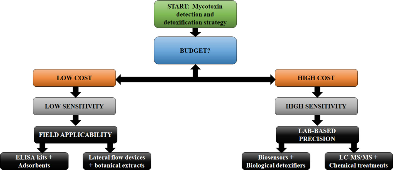
Figure 2.
A decision tree figure showing how users can choose appropriate detection and detoxification strategies.
Despite their lack of toxin selectivity and potential to lower feed nutrient quality, physical adsorbents are nonetheless appealing for extensive, low-cost treatments. Although there are few commercially available research on the economic viability of biological detoxification techniques, such as probiotics and yeast derivatives, they provide environmentally friendly alternatives with extra gut health advantages (Liu et al., 2022). Chemical detoxification is quite effective, yet there are issues with residual safety and regulations with no standardized protocols being available. Botanical extracts still need to be further validated for consistency, stability, and dose optimization (Liu et al., 2022). In a variety of aquaculture contexts, this comparison methodology supports evidence-based decision-making for mycotoxin control.
Despite their lack of toxin selectivity and potential to lower feed nutrient quality, physical adsorbents are nonetheless appealing for extensive, low-cost treatments. Although there are few commercially available research on the economic viability of biological detoxification techniques, such as probiotics and yeast derivatives, they provide environmentally friendly alternatives with extra gut health advantages (Liu et al., 2023). Chemical detoxification is quite effective, yet there are issues with residual safety and regulations with no standardized protocols being available. Botanical extracts still need to be further validated for consistency, stability, and dose optimization (Liu et al., 2023). In a variety of aquaculture contexts, this comparison methodology supports evidence-based decision-making for mycotoxin control.
Conclusion
The many issues raised by mycotoxin contamination in aquafeeds are highlighted in this study, including the sources of contamination, detection techniques, toxicological consequences, and mitigation measures. A thorough review of the literature identifies a number of open research questions that need to be addressed in order to protect feed safety and aquaculture output. The first is that species-specific regulatory thresholds for mycotoxins in aquafeeds are desperately needed since existing standards frequently extrapolate limitations from terrestrial livestock without taking into account the physiological and metabolic variations across fish species. Second, the co-occurrence of many mycotoxins is still not well understood, particularly in relation to low-level, long-term exposures where synergistic or additive effects could happen but are not well understood. Third, detection techniques need to be more sensitive and applicable in the field, especially in environments with limited resources where quick and affordable screening technologies might allow for early intervention and avert major contamination problems. Pre-harvest prevention, host resilience, and feed safety may all be linked into a single framework by integrating microbiome-based insights at the crop and fish gut levels. Lastly, there is still insufficient attention paid to how climate change affects mycotoxin epidemiology. Fungal ecology, crop vulnerability, and mycotoxin prevalence are expected to change due to more unpredictable weather patterns, necessitating the development of feed formulation and storage techniques that are climate robust. Future research should prioritize developing standardized toxicological protocols for different fish species, designing of hybrid detection systems combining affordability with high sensitivity and investigate microbiome-mycotoxin interactions for both crops and fish. The aquafeed industry may move toward a proactive, systems-level approach to mycotoxin control by filling in these gaps, guaranteeing safer, more sustainable aquaculture in the face of issues with global food security.
Acknowledgement
The authors sincerely thank the Hon’ble Vice-Chancellor of The Neotia University for his continuous support, encouragement, and facilitation of research activities that enabled the successful preparation and submission of this manuscript.