
Table 1.
Role of TLRs in mastitis pathogenesis
| TLRs | Polymorphisms/ Expression | Mastitis phenotypic traits | Experimental model | References |
|---|---|---|---|---|
| TLR2 | High expression | Associated with mastitis susceptibility | Mammary gland tissues from both healthy and infected cows | (Zhang B. et al., 2025) |
| TLR2 | High expression | Associated with inflammation, apoptosis, and angiogenesis in mammary epithelial cells of dairy cows with bacterial-induced mastitis | Mammary epithelial cells (MECs) | (Bai et al., 2022) |
| TLR2 | High expression | Elevated level of TLR2/MyD88/PI3K/AKT followed by high level of TNF-α and IL-6 with inflammatory changes. | Bovine mammary epithelial cells (BMECs) | (Cai et al., 2021) |
| TLR4, TLR2 | High expression | Linked with enhancement of mastitis pathogenesis in response to Bacillus cereus and Bacillus subtilis | Murine model | (Yanthi et al., 2025) |
| TLR4 | g.107062971A>B | Correlated with mastitis resistance | Hardhenu cattle | (Sindhu et al., 2025) |
| TLR4 | rs8193069T >C | Linked to mastitis resistance | Holstein cows | (Chen H. et al., 2022) |
| TLR4 | High expression | Upregulated the expression IL-1β, IL-8, NF-κB and NLR family pyrin domain containing 3 (NLRP3) in response to LPS-stimulation. The inflammation changes were associated with regulating TLR4/SYK/NF-κB by LPS. | BMECs | (Yang F. et al., 2022) |
| TLR4 | High expression | Increased levels of lipid peroxidation (MDA), TNF-α and IL-1β and NF-κB p65 with inflammatory changes in mammary glands. | BMECs | (Lebda et al., 2022) |
| TLR4 | High expression | Increased levels of TNF-α, IL-1β, IL-6, CXCL2, CXCL5, CCL5, CXCL8 in LPS-treated cells. This showed that TLR4, MyD88 and NF-κB p65 higher expression led to mammary gland infection. | BMECs | (Jiang et al., 2022) |
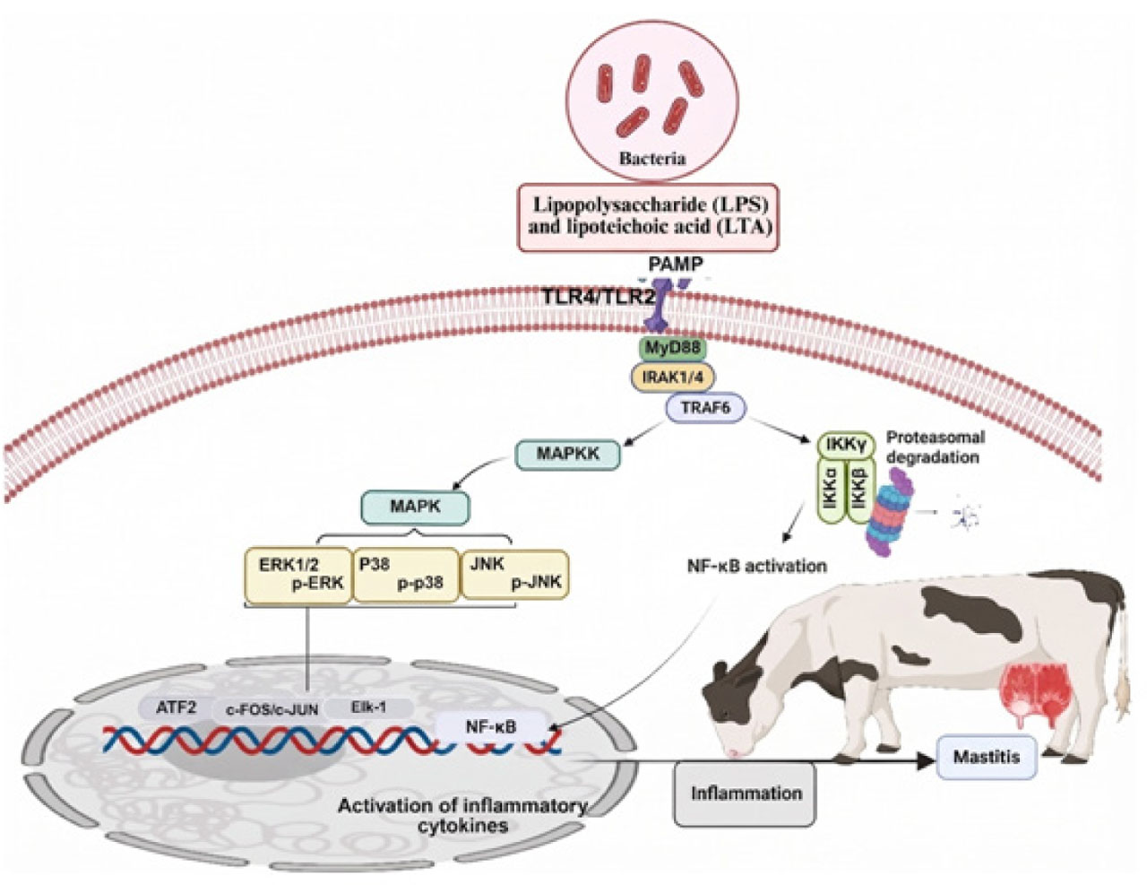
Figure 1.
Bacterial-induced inflammatory signaling pathway leading to mastitis in dairy cattle. Bacterial pathogen-associated molecular patterns activated TLR4/TLR2-MyD88 signaling, which diverged into IRAK1/4-TRAF6-IKKγ/IKKα-NF-κB and MAPKK-MAPK (ERK1/2, p38, JNK) cascades. These pathways converged on nuclear transcription factors (ATF2, c-FOS/c-JUN, Elk-1, NF-κB) that induced proinflammatory cytokine expression, culminating in mammary gland inflammation. PAMP (pathogen-associated molecular pattern).
Table 2.
Summary of studies targeting TLRs/NF-κB/MAPK signaling pathways for mastitis pathogenesis
| Causative agent | Targeted pathway | Biological effect | Experimental model | References |
|---|---|---|---|---|
| 1 | 2 | 3 | 4 | 5 |
| S. aureus | TLR2/NF-κB/MAPK signaling pathway | Enhanced TLR2-MyD88-TRAF6 components followed by upregulation of protein phosphorylation associated with the MAPK and NF-κB signaling pathways Increased inflammatory changes in BMECs | BMECs | (Guo et al., 2025) |
| S. aureus | NF-κB signaling pathway | mRNA expressions of IL-1β, NF-κB, MPO, nitric oxide and TNF-α in mammary tissues with moderate fibrosis and infiltration of macrophages and neutrophils in mammary tissue sections | Mouse model | (Soni et al., 2025) |
| S. aureus | TLR2/Nrf2-Keap-1/MAPK signaling pathway | Increased expression of MPO, MDA, caspase-3 with elevating and suppressed Nrf2, Keap-1 levels The expressions of TLR2, NOD2, and MAPK, NF-κB P65 and p-ERK were increased | BMECs | (Edres et al., 2025) |
| S. aureus | TLR4/MyD88/NF-κB signaling pathway | Showed high expression of TLR4, MyD88 and NF-κB with significant inflammatory changes in BMECs | BMECs | (Arsaute et al., 2024) |
| S. aureus and E. coli | MAPK signaling pathway | Upregulated the expressions of MAPK components including ERK1/2, P38, and JNK and consequent inflammatory changes in BMECs | BMECs | (Zhang Y. et al., 2025) |
| E. coli | NF-κB/MAPK signaling pathway | Elevated level of inflammatory cytokines (TNF-α, IL-1β, IL-6, and IL-8) in bovine bone marrow-derived macrophages (BMDMs) | BMDMs | (Yang X. et al., 2025) |
| LPS | TLR4/NF-κB signaling pathway | Increased mRNA and protein levels of TNF-α, IL-1β, IL-6, TLR4 and NF-κB with elevated inflammatory changes | Mouse MECs | (Hu et al., 2025) |
| LPS | Nrf2/MAPK signaling pathway | Induced oxidative stress and inflammation in BMECs | BMECs | (Yang M. et al., 2025) |
| LPS | TRAF1/ NF-κB signaling pathway | Enhanced inflammation in BMECs | BMECs | (Gao et al., 2025) |
| LPS | TLR4/NF-κB/MAPK signaling pathway | LPS significantly elevated the phosphorylation of key NF-κB (IκBα and p65) and MAPK (JNK, ERK, and p38) Upregulated of the expressions of Myd88 and Traf6 and consequent inflammatory changes in porcine mammary epithelial cells (PMECs) | PMECs | (He et al., 2025) |
| LPS | TLR4/NF-kB signaling pathway | Induced inflammatory changes with high expression of TNF-α, IL-1β, IL-6, and IL-8 in BMECs. Suppressed the level of total antioxidant capacity (T-AOC), glutathione (GSH), superoxide dismutase (SOD), and elevated the expression of MDA, nitric oxide (NO) content assay and ROS | BMECs | (Wang et al., 2024) |
| LPS | TLR4/MAPK/NF-κB signaling pathway | Induced inflammatory changes in mouse MECs | Mouse model | (Xie et al., 2024) |
| LPS | MAPK/NF-κB signaling pathway | Elevated levels of IL-1β, IL-6, IL-8, TNF-α, and IL-17A with reduced phosphorylation levels of ERK and p38 proteins Inflammatory changes in cow MECs | Cow mammary epithelial cells (CMECs) | (Zhang et al., 2024) |
| LPS | MAPK/NF-κB signaling pathway | Elevated levels of IL-6 and IL-1β with significant increased changes in mouse MECs | Mouse mammary epithelial cells (MMECs) | (Cai et al., 2024) |
| LPS | NF-κB/MAPK signaling pathway | Increased the expression of Mas1 gene with reduced blood-milk barrier proteins including zonula occludens 1 (ZO-1), occludin, and claudin-3 Enhanced inflammatory changes in BMECs | BMECs | (Yan et al., 2024) |
| LPS | NF-κB/MAPK signaling pathway | Reduced blood-milk barrier proteins expression and elevated the levels of inflammatory cytokines (TNF-α, IL-6, and IL-1β) and inhibited the activation of MPO to induce inflammation | MMECs | (Luo et al., 2024) |
| LPS | p38-MAPK/NF-κB↔TNF-α signaling pathway | Inhibited the phosphorylation of p65, p50, p38, IκB, and MKK3/6 proteins and enhanced the expression of downstream inflammatory factors TNF-α, IL-1β, IL-6, IL-8, and COX-2 to promote inflammatory changes in MMECs | MMECs | (Qin et al., 2024) |
| Lactococcus garvieae | TLR2/NLRP3/NF-kB signaling pathway | Elevated inflammatory changes in mouse mammary gland tissue followed by mastitis | Mouse mammary gland cells | (Wu et al., 2024) |
| Streptococcus agalactiae | NF-κB/NLRP3 signaling pathway | Inhibited CDK5 regulatory sub-unit-associated protein 3 (CD-K5RAP3) followed by activation of NF-κB /NLRP3 signaling pathway which may lead to inflammatory changes in BMECs | BMECs | (Yan et al., 2023) |
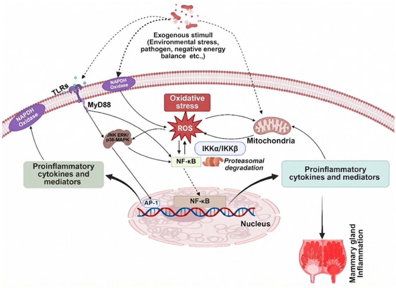
Figure 2.
Molecular pathways of stress-induced inflammatory response in mammary epithelial cells. Exogenous stressors activated TLRs and NADPH oxidase, initiating MyD88-dependent JNK/ERK/p38-MAPK signaling. ROS generation served as a central hub linking oxidative stress to IKKα/IKKβ-mediated NF-κB and AP-1 activation. Nuclear translocation of these transcription factors upregulated proinflammatory mediators, establishing a mechanistic connection between oxidative stress and mammary gland inflammation.
Table 3.
Summary of studies targeting TLRs/NF-κB/MAPK signaling pathways via therapeutic agents for mastitis treatment
| Therapeutic agent | Biological effect | Experimental model | References |
|---|---|---|---|
| 1 | 2 | 3 | 4 |
| Maslinic acid | Inhibited the production of pro-inflammatory cytokines (IL-6, IL-1β, TNF-α, iNOS, and COX2) in LPS-treated MMECs Decreased the expression of NLRP3 inflammasome, AKT/NF-κB, and MAPK signaling pathways Enhanced intestinal microflora | MMECs | (Li K. et al., 2023 a) |
| Apigenin | Apigenin blocked the activation of TLR4/NF-κB followed by suppression of myeloperoxidase (MPO), TNF-α, IL-1β, and IL-6 induced by LPS in rat’s mammary gland Prevents inflammatory changes in vitro and mastitis in vivo | Rats model | (Hu et al., 2025) |
| Taraxacum mongolicum | Protected MMECs from S. aureus- induced inflammatory changes by inhibiting TLR2-NF-κB/MAPKs pathways Alleviated inflammation in MECs with significant decrease in TNF-α, IL-6 and IL-1β levels, and MPO levels Inhibited the expression of TLR2, and the phosphorylations of inhibitor κBα (IκBα), p65, p38, extracellular signal-regulated kinase 1/2 (ERK1/2) and c- JNK proteins | MMECs | (Ge et al., 2021) |
| Berberine and Cyperus rotundus rhizomes extract | Reduced the mRNA expression levels of MPO, NF-κB P65, p-ERK, TLR2, NOD2, Keap-1, MDA and MAPK induced by S. aureus in a rat mammary gland | Rats model | (Edres et al., 2025) |
| Luteolin | Suppressed the S. aureus-induced inflammatory changes by targeting TLR2-MyD88-TRAF6 components following inhibition of MAPK and NF-κB signaling pathways in BMECs | BMECs | (Guo et al., 2025) |
| Diflunisal | Ameliorated inflammatory changes caused by S. aureus in BMECs Diflunisal inhibited the NF-κB signaling pathway, resulting in suppression of mRNA expression of IL-1β and TNF-α in BMECs | BMECs | (Soni et al., 2025) |
| Danggui Buxue decoction (DBD) | Attenuated the inflammation and oxidative stress in MMECs induced by E. coli The DBD suppressed the expression of TLR4/NF-κB and Nrf2/HO-1 signaling pathways to alleviate oxidative stress and inflammation | MMECs | (Wang et al., 2025 a) |
| Bacillus subtilis Lactobacillus casei Lactobacillus plantarum 17–5 | Reduced E. coli-induced inflammatory changes in BMECs and MMECs by suppressing proinflammatory cytokines (TNF-α, IL-1β, and IL-6) via inhibiting TLR4/NF-κB/MAPK signaling | BMECs MMECs | (Li et al., 2024; Hu et al., 2023; Li K. et al., 2023 b; Li K. et al., 2022) |
| Jiawei Yanghe decoction | Reduced TLR4/MyD88/NF-κB signaling pathway activation which is correlated with prevention of inflammatory changes in MMECs | MMECs | (Zhao et al., 2023) |
| Paeoniflorin | Suppressed expression of TLR2/NF-κB pathway and related genes (NF-κB unit p65, NF-κB unit p50, and MyD88) in Staphylococcus haemolyticus-treated BMECs | BMECs | (Shen et al., 2023) |
| Taurine | Prevented S. uberis-induced inflammatory changes by suppressing TLR2/TAK1/MAPK/NF-κB signaling activation in RAW264.7 | BMECs | (Li et al., 2023) |
| Quyu Xiaozhong recipe | Inhibited activation of TLR4/NF-κB pathway following prevention of acute mastitis caused by S. aureus in rat mammary glands | Rat MECs | (Wang and Jin, 2023) |
| Chlorogenic acid from Taraxacum officinale | Downregulated the expression of TNF-α, IL-6, and IL-1β and TLR2/NF-κB signaling pathway in BMECs treated with lipoteichoic acid (LTA). | BMECs | (Xu et al., 2023) |
| Hexadecanamide | Ameliorated the inflammatory changes in MMECs caused by S. aureus Inhibited the activation of the NF-κB pathway and improved barrier integrity in MMECs | MMECs | (Bao et al., 2023) |
| Medulla Tetrapanacis | LPS-induced MAPK activation followed by elevated inflammatory cytokines (TNF-α and IL-6) and nitric oxide levels were significantly suppressed in mouse monocyte macrophages (RAW 264.7) by Medulla Tetrapanacis treatment | Mouse monocyte macrophages (RAW 264.7) | (Kwok et al., 2024) |
| Rhapontici Radix ethanol extract (RRE) (isochlorogenic acid B), oligomeric proanthocyanidins and isoliquiritigenin | RRE suppressed LPS-induced inflammatory changes (decreased levels of TNF-α, IL-6, and IL-1β, c-Jun N-terminal kinase 1/2, p38, and extra-cellular regulated protein kinase 1/2) by targeting NF-κB/MAPK pathways in ovine mammary alveolar (MAC-T) cells. The inhibition of NF-κB/MAPK was upregulated by G protein-coupled receptor 161 (GPR161) and transmembrane protein 59 (TMEM59) | Bovine mammary alveolar (MAC-T) cells | (Lv et al., 2024; Ma et al., 2020; Li M. et al., 2022) |
| Quinic acid (QA) and isochlorogenic acid B (ICAB) | Suppressed levels of NOD-like receptor 3 (NLRP3) inflammasome, caspase-11, gasdermin D (GSDMD), and p65 of nuclear factor κB (NF-κB) in MAC-T cells Reduced the inflammatory factors (TNF-α, IL-1β, and IL-6) and oxidative stress factors (COX-2 and iNOS) in both MAC-T cells and mouse mammary glands Target with NF-κB/NLRP3 to suppress inflammatory changes | MAC-T cells | (Lv et al., 2025) |