
Mastitis, the inflammatory condition affecting mammary gland tissue, represents one of the most significant and widespread infectious diseases in global dairy production (Rasmussen et al., 2024; Khan et al., 2024 a). This multifaceted condition poses substantial challenges to the dairy industry through its diverse impacts on animal health and farm operations, including reduced milk yield, increased veterinary interventions, reproductive complications, and premature culling of affected animals (Khan et al., 2023; Corrêa et al., 2024; Kour et al., 2023; Richardet et al., 2023; Samaraweera et al., 2022; Hogeveen et al., 2019; Arikan et al., 2024). The global burden of mastitis is considerable, affecting dairy operations across all major milk-producing regions (Puerto et al., 2021; Rollin et al., 2015). The condition presents persistent challenges worldwide, with significant impacts documented in major dairy-producing countries including the United States, Canada, and China, where the scale of the problem affects numerous farms and herds (Puerto et al., 2021; Rollin et al., 2015; Wang et al., 2019).
Beyond its economic implications, mastitis raises critical concerns for animal welfare and food safety. The inflammatory process fundamentally compromises mammary gland function and milk quality, creating a complex disease syndrome that affects multiple aspects of dairy production (De Vliegher et al., 2018). The pathogenesis of mastitis involves intricate interactions between invading pathogens – predominantly bacterial species including Escherichia coli (E. coli) and Staphylococcus aureus (S. aureus), and Streptococcus species – and the host’s innate and adaptive immune responses (Tong et al., 2025; Li Y. et al., 2025; Srithanasuwan et al., 2024; Tommasoni et al., 2023; Sadam et al., 2024; Arbab et al., 2022).
Clinically, mastitis manifests in two primary forms with distinct characteristics and diagnostic challenges. Clinical mastitis presents with pronounced pathological symptoms, including redness, pain, and fever, accompanied by physical changes such as swelling and increased temperature in mammary gland tissues. In contrast, sub-clinical mastitis, particularly that caused by S. aureus, often remains undetected due to the absence of obvious clinical signs, with elevated somatic cell counts and decreased milk yield serving as the primary indicators of infection (Khan et al., 2022 a, b, 2020 a; Cobirka et al., 2020). Multiple factors influence mastitis susceptibility, including anatomical udder positioning, lactation stage, animal age, and pregnancy-related stress during the periparturient period (Tommasoni et al., 2023; Khan et al., 2022 c).
The mammary gland’s defense against pathogenic invasion relies heavily on sophisticated molecular recognition and signaling mechanisms that detect microbial threats and orchestrate appropriate inflammatory responses (Khan et al., 2025, 2022). Central to this defense system are toll-like receptors (TLRs), a family of pattern recognition receptors that serve as the first line of detection for pathogen-associated molecular patterns (PAMPs) and damage-associated molecular patterns (DAMPs) (Rainard et al., 2022). Upon activation, TLRs initiate cascading signaling pathways that ultimately lead to the production of inflammatory mediators, antimicrobial peptides, and the recruitment of immune cells to the site of infection (Khan et al., 2020 b).
Two critical downstream signaling pathways activated by TLRs – nuclear factor kappa B (NF-κB) and mitogen-activated protein kinase (MAPK) – play pivotal roles in orchestrating the inflammatory response during mastitis (Li K. et al., 2023 a; Khan et al., 2020 a). The NF-κB pathway serves as a master regulator of inflammatory gene expression, controlling the transcription of numerous cytokines, chemokines, and adhesion molecules essential for mounting an effective immune response (Khan et al., 2020 a, 2024 a). The role of the NF-κB pathway has been well reported in mastitis (Wang et al., 2025 b). Simultaneously, MAPK signaling pathways, including p38, extracellular signal regulated protein kinase (ERK)1/2, and Jun N-terminal kinase (JNK), modulate cellular responses ranging from cytokine production to cell survival and proliferation, fine-tuning the intensity and duration of the inflammatory response. Understanding the intricate interplay between TLRs, NF-κB, and MAPK signaling in mastitis pathogenesis has emerged as a critical area of research, offering insights into both protective immunity and pathological inflammation (Khan et al., 2022 b). While these pathways are essential for pathogen clearance and tissue protection, their dysregulation can lead to excessive inflammation, tissue damage, and chronic mastitis conditions that compromise mammary gland function and animal health.
This review synthesizes current knowledge regarding the activation and regulation of TLRs/NF-κB/MAPK signaling pathways in mastitis, examining their roles in pathogen recognition, inflammatory response initiation, and disease resolution. We discuss the molecular mechanisms underlying pathway activation, cross-talk between signaling networks, and their implications for mastitis susceptibility and severity. Furthermore, we explore how understanding these pathways may inform the development of novel therapeutic strategies and management approaches for preventing and treating mastitis in dairy animals.
Literature search methodology
A comprehensive literature search was conducted across multiple electronic databases to identify relevant studies published between January 2020 and June 30, 2025. The databases systematically searched included PubMed, Web of Science Core Collection, Scopus, ScienceDirect, and Google Scholar for additional grey literature and recent publications. The search strategy employed free-text keywords related to mastitis, TLRs, NF-κB, and MAPK signaling pathways. Primary terms included “mastitis” OR “mammary gland inflammation” OR “bovine mastitis” OR “dairy cow mastitis”. Molecular pathway terms encompassed “toll-like receptor” OR “TLR” OR “pattern recognition receptor”, “NF-κB” OR “nuclear factor kappa B” and “MAPK” OR “mitogen-activated protein kinase” OR “p38” OR “ERK” OR “JNK”. Additional search terms included “innate immunity” OR “inflammatory response” OR “cytokine” OR “pathogen recognition” and “signaling pathway” OR “signal transduction” OR “molecular mechanism”. Studies were included if they were published between January 1, 2020, and December 31, 2025, written in the English language, and focused on mastitis in dairy animals (primarily bovine). Additional inclusion criteria required that studies investigated TLRs, NF-κB, and/or MAPK signaling pathways in the context of mastitis, included original research articles, review articles, meta-analyses, and systematic reviews, involved both clinical mastitis and subclinical mastitis, utilized in vitro, in vivo, and ex vivo experimental studies, and examined both pathogen-induced and non-pathogen-induced mastitis. Studies were excluded if they were published before 2020, were non-English publications, consisted of conference abstracts, case reports, and editorials without substantial data, focused solely on treatment outcomes without molecular pathway analysis, were conducted exclusively in non-mammary tissues or cell lines not relevant to mastitis, or investigated mastitis in dairy cattle.
Role of TLR2/4 in mastitis pathogenesis
The mammary epithelium constitutes a critical primary barrier against pathogenic invasion, with TLRs serving as essential mediators of innate immune recognition (Rainard et al., 2022; Duan et al., 2022; Bhattarai et al., 2018). These phylogenetically conserved pattern recognition receptors detect distinct PAMPs characteristic of diverse microbial threats. In bovine mastitis pathogenesis, TLR2 functions as a key sensor for gram-positive bacterial components, specifically recognizing peptidoglycan and lipoteichoic acid derived from S. aureus, a predominant etiological agent of intramammary infection (Bai et al., 2022). Conversely, TLR4 demonstrates specificity for lipopolysaccharide (LPS), the characteristic endotoxin of gram-negative bacteria such as E. coli, which represents another significant mastitis pathogen (Hu et al., 2025). The binding of these cognate ligands to their respective TLRs triggers the activation of downstream signaling cascades that orchestrate the innate immune response. Upon pathogen recognition, TLRs undergo conformational changes that facilitate the recruitment of adaptor proteins, primarily myeloid differentiation primary response 88 (MyD88) and TIR-domain-containing adapter-inducing interferon-β (TRIF) (Guo et al., 2025; Li B. et al., 2020; Yang J. et al., 2022). This recruitment event triggers the activation of interleukin-1 receptor-associated kinases (IRAKs) and TNF receptor-associated factor 6 (TRAF6), leading to the activation of downstream signaling pathways (Guo et al., 2025; Khan et al., 2024 a).
A study investigated immune homeostasis disruption in mammary glands of dairy cows with clinical mastitis caused by S. aureus compared to healthy controls (Qudratullah et al., 2022; Zhang B. et al., 2025). Proteomics analysis identified coronin1A (CORO1A) as a key protein involved in immune response regulation through phagosome formation in monocytes, macrophages, and neutrophils. The molecular mechanism showed that TLR2 recognizes pathogens and recruits CORO1A to enhance phagocytic activity, with both proteins showing increased expression correlated with mastitis severity. CORO1A upregulation appears to disrupt mammary gland immune homeostasis during mastitis, providing new insights into disease pathogenesis and potential therapeutic targets (Zhang B. et al., 2025). Consistently, a study investigated the temporal gene expression patterns of TLRs and chemokine receptors (CXCRs) in murine mammary epithelial HC11 cells exposed to Bacillus cereus and B. subtilis to understand their roles in mastitis pathogenesis (Yanthi et al., 2025). HC11 cells were infected with both bacterial species, and expression levels of interleukin (IL)-6, IL-8, TLR2, TLR4, IL-1α, and CXCR1 were evaluated by quantitative real-time polymerase chain reaction (PCR) at multiple time points over 72 hours post-infection. Their findings showed that TLR2 exhibited a biphasic expression pattern with early upregulation followed by suppression, TLR4 demonstrated higher expression in response to B. subtilis than B. cereus, IL-6 displayed prolonged expression under B. subtilis challenge but transient expression under B. cereus, IL-1α showed consistent expression across both bacterial challenges suggesting its potential as a stable biomarker, and CXCR1 exhibited delayed but sustained expression indicative of secondary neutrophil recruitment. The findings demonstrate that immune responses are gene- and pathogen-specific, with TLR and CXCR genes showing distinct temporal profiles, identifying IL-1α, IL-6, and CXCR1 as promising targets for genetic selection and therapeutic intervention in mastitis management (Yanthi et al., 2025). It is well established that MYD88 acts as an adapter protein for TLRs to mediate inflammatory changes during mastitis pathogenesis (Lin et al., 2024). Consistently, it has been reported that MYD88-deficient mice in the mammary gland showed reduced inflammation along with inflammatory cytokines including interleukin-1β (IL-1β), tumor necrosis factor-α (TNF-α), and IL-6, CXCL1, CXCL2, and CCL2 (Lin et al., 2024). Similarly, studies have shown that polymorphisms in TLR4 were associated with mastitis susceptibility with higher milk SCC in dairy cattle (Sindhu et al., 2025; Bhat et al., 2024). Similarly, another study reported that TLR4 polymorphism (rs8193069T >C) in the TLR4 gene is associated with mastitis resistance in Holstein cows (Chen H. et al., 2022). The summary of research development on TLRs role in mastitis pathogenesis has been provided in Table 1.
Table 1.
Role of TLRs in mastitis pathogenesis
| TLRs | Polymorphisms/ Expression | Mastitis phenotypic traits | Experimental model | References |
|---|---|---|---|---|
| TLR2 | High expression | Associated with mastitis susceptibility | Mammary gland tissues from both healthy and infected cows | (Zhang B. et al., 2025) |
| TLR2 | High expression | Associated with inflammation, apoptosis, and angiogenesis in mammary epithelial cells of dairy cows with bacterial-induced mastitis | Mammary epithelial cells (MECs) | (Bai et al., 2022) |
| TLR2 | High expression | Elevated level of TLR2/MyD88/PI3K/AKT followed by high level of TNF-α and IL-6 with inflammatory changes. | Bovine mammary epithelial cells (BMECs) | (Cai et al., 2021) |
| TLR4, TLR2 | High expression | Linked with enhancement of mastitis pathogenesis in response to Bacillus cereus and Bacillus subtilis | Murine model | (Yanthi et al., 2025) |
| TLR4 | g.107062971A>B | Correlated with mastitis resistance | Hardhenu cattle | (Sindhu et al., 2025) |
| TLR4 | rs8193069T >C | Linked to mastitis resistance | Holstein cows | (Chen H. et al., 2022) |
| TLR4 | High expression | Upregulated the expression IL-1β, IL-8, NF-κB and NLR family pyrin domain containing 3 (NLRP3) in response to LPS-stimulation. The inflammation changes were associated with regulating TLR4/SYK/NF-κB by LPS. | BMECs | (Yang F. et al., 2022) |
| TLR4 | High expression | Increased levels of lipid peroxidation (MDA), TNF-α and IL-1β and NF-κB p65 with inflammatory changes in mammary glands. | BMECs | (Lebda et al., 2022) |
| TLR4 | High expression | Increased levels of TNF-α, IL-1β, IL-6, CXCL2, CXCL5, CCL5, CXCL8 in LPS-treated cells. This showed that TLR4, MyD88 and NF-κB p65 higher expression led to mammary gland infection. | BMECs | (Jiang et al., 2022) |
The NF-κB /MAPK signaling role in mastitis pathogenesis
The recognition of PAMPs by TLRs initiates a sophisticated cascade of intracellular signaling events that culminate in the coordinated inflammatory response characteristic of mastitis. Upon PAMP binding, TLRs primarily activate MyD88-dependent pathways, leading to rapid NF-κB activation and subsequent transcriptional upregulation of pro-inflammatory mediators, including IL-1β, TNF-α, and IL-6, alongside chemokines that orchestrate neutrophil recruitment to the infection site (Khan et al., 2020 a, 2024, 2025). These kinases undergo autophosphorylation and subsequently recruit TRAF6, which functions as an E3 ubiquitin ligase. TRAF6 then activates the transforming growth factor-β-activated kinase 1 (TAK1) complex, leading to the phosphorylation and activation of the IκB kinase (IKK) complex. The activated IKK complex phosphorylates IκBα, targeting it for proteasomal degradation and allowing NF-κB to translocate to the nucleus (Zhang J. et al., 2025; Khan et al., 2020 a).
Rather than operating in isolation, the NF-κB pathway demonstrates extensive cross-talk with complementary signaling cascades, creating a sophisticated regulatory network that ensures appropriate inflammatory responses during mastitis. The MAPK pathways, particularly p38 MAPK and JNK, modulate NF-κB activity through multiple convergent mechanisms (Che et al., 2023; He et al., 2023). Simultaneously, integration with phosphoinositide 3-kinase (PI3K)/Akt signaling influences both IKK complex activation and nuclear retention of NF-κB (He et al., 2023). This intricate signaling network extends beyond transcription factor activation to encompass various inflammatory mediator pathways, influencing prostaglandin synthesis, nitric oxide production, and complement cascade activation. The parallel activation of MAPK pathways, including ERK, JNK, and p38, through TLR-mediated signaling creates a dual regulatory system that ensures robust and sustained inflammatory gene expression (Qiu et al., 2022). This coordinated response provides multiple therapeutic intervention points while maintaining the specificity required for effective pathogen clearance.
Recent investigations have revealed the temporal specificity of anti-inflammatory interventions, highlighting the critical importance of treatment timing in modulating NF-κB/MAPK signaling. Lactoferrin (LF) demonstrates striking timing-dependent effects on inflammatory responses in BMECs (Zhang K. et al., 2025). When administered prophylactically before LPS-induced inflammation, LF significantly attenuates inflammatory cytokine production (IL-1β, IL-6, IL-8, TNF-α) and suppresses key signaling pathways (NF-κB/p65 and MAPK/p38/ERK) while downregulating TLR4 receptor expression (Figure 1). However, post-inflammatory administration fails to reduce cytokine levels, indicating that LF exerts protective anti-inflammatory effects primarily through preventive mechanisms targeting TLR4 receptors and upstream signaling cascades (Zhang K. et al., 2025).
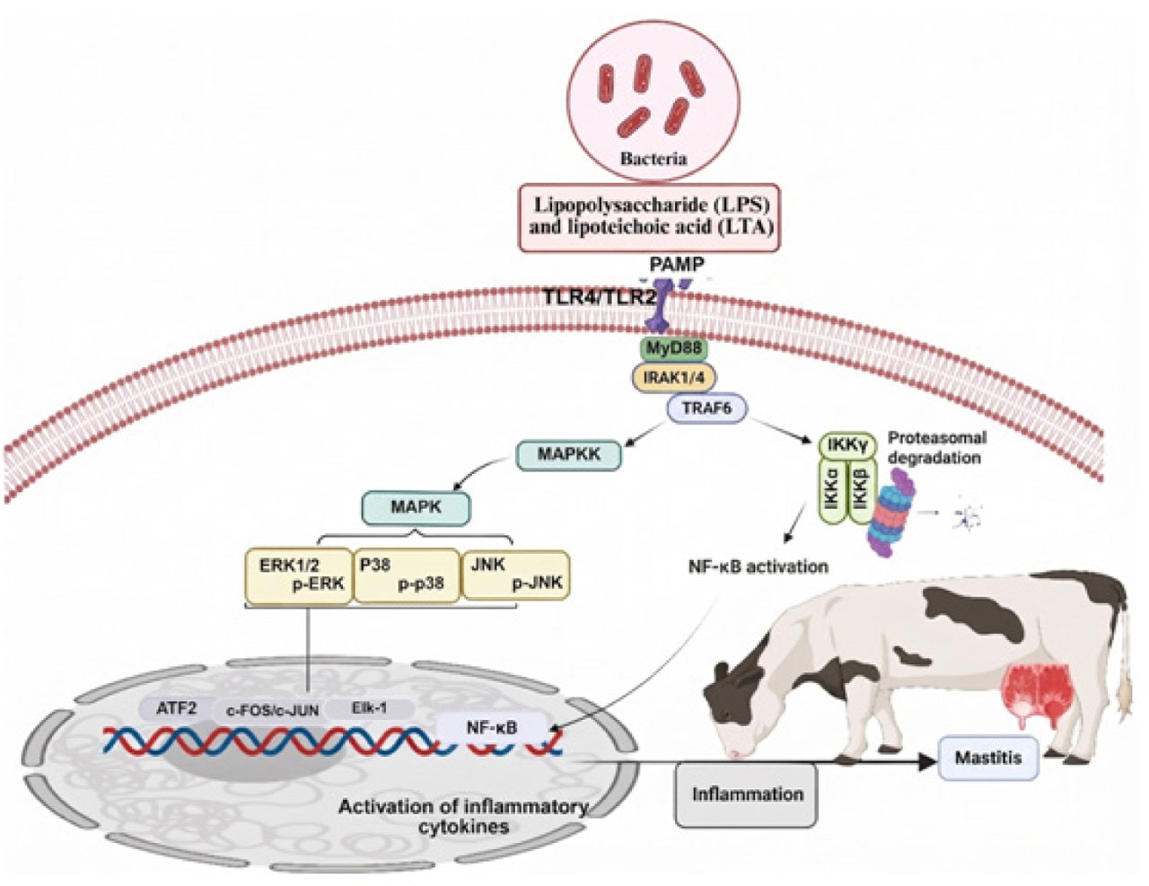
Figure 1.
Bacterial-induced inflammatory signaling pathway leading to mastitis in dairy cattle. Bacterial pathogen-associated molecular patterns activated TLR4/TLR2-MyD88 signaling, which diverged into IRAK1/4-TRAF6-IKKγ/IKKα-NF-κB and MAPKK-MAPK (ERK1/2, p38, JNK) cascades. These pathways converged on nuclear transcription factors (ATF2, c-FOS/c-JUN, Elk-1, NF-κB) that induced proinflammatory cytokine expression, culminating in mammary gland inflammation. PAMP (pathogen-associated molecular pattern).
The pathophysiological relevance of NF-κB signaling in naturally occurring mastitis has been confirmed through clinical observations demonstrating elevated NF-κB expression in inflamed bovine udders. Furthermore, mastitis significantly increases cathelicidin-3 (CATHL3) expression through NF-κB signaling, particularly via p65 phosphorylation, with blood glucose levels also influencing CATHL3 expression independent of inflammatory status (Zhang J. et al., 2025). These findings underscore the complex metabolic-inflammatory interactions that characterize mastitis pathogenesis. Consistently, different mastitis pathogens activate distinct but overlapping signaling pathways, reflecting the specificity of host-pathogen interactions. Mycoplasma bovis infection activates both TLR1/2/6/9/MyD88/NF-κB and TLR3/TRIF/IRF signaling cascades, demonstrating the multifaceted nature of immune recognition during mycoplasmal mastitis (Yang J. et al., 2022). This pathogen-specific activation pattern suggests that therapeutic strategies may need to be tailored based on the causative organism and its unique inflammatory signature.
Emerging therapeutic strategies targeting the NF-κB/MAPK network demonstrate promising anti-inflammatory efficacy. Nanocurcumin treatment significantly reduces lipid peroxidation, oxidized glutathione, and pro-inflammatory cytokines (TNF-α, IL-1β) while decreasing expression of TLR4, NF-κB p65, and high mobility group box 1 (HMGB1) in LPS-induced mastitis models (Lebda et al., 2022). Simultaneously, nanocurcumin enhances reduced glutathione levels and nuclear factor erythroid 2-related factor 2 (Nrf2) activity, preserving normal mammary architecture and suggesting a dual antioxidant-anti-inflammatory mechanism. Furthermore, a study documented that Taraxacum mongolicum extract significantly reduced pro-inflammatory cytokines (TNF-α, IL-6, IL-1β) and myeloperoxidase activity in S. aureus-infected mastitis, while effectively attenuating histopathological damage and cell necrosis in mammary gland tissues (Ge et al., 2021). Traditional medicinal approaches have also yielded mechanistic insights into NF-κB/MAPK modulation. Taraxacum mongolicum extract effectively reduces pro-inflammatory cytokines (TNF-α, IL-6, IL-1β) and myeloperoxidase activity in S. aureus-induced mastitis while attenuating histopathological damage through TLR2 expression inhibition and suppression of key signaling protein phosphorylation (IκBα, p65, p38, ERK1/2, JNK) (Ge et al., 2021). Consistently a study has shown that LPS-induced inflammation in bovine mammary epithelial cells showed a higher expression of TNF-α, IL-6, IL-1β, P38 and ERK1/2 protein phosphorylation which is associated with inflammatory changes (Kan et al., 2021). This provides scientific validation for traditional therapeutic approaches while elucidating their molecular mechanisms of action. Recent advances in understanding epigenetic regulation of mastitis inflammation have revealed novel regulatory mechanisms involving long non-coding RNAs (lncRNAs). The lncRNA CA12-AS1 functions as a pro-inflammatory regulator by targeting miR-133a, with its silencing significantly reducing LPS-induced inflammatory factors (IL-6, IL-8, IL-1β) and decreasing NF-κB and apoptosis-related gene expression (BAX, caspase3, caspase9) in BMECs (Feng et al., 2024). Importantly, CA12-AS1 silencing promotes tight junction signaling genes (Claudin-1, Occludin, ZO-1), anti-apoptotic gene BCL2, and proliferation genes (CDK2, CDK4, PCNA), suggesting that epigenetic modulation may offer therapeutic potential through simultaneous reduction of inflammation and enhancement of mammary epithelial cell function. The NF-κB/MAPK signaling network represents a central regulatory hub in mastitis pathogenesis, integrating diverse pathogen recognition signals into coordinated inflammatory responses. The temporal specificity of therapeutic interventions, pathogen-specific activation patterns, and emerging epigenetic regulatory mechanisms highlight the complexity of this system while providing multiple avenues for therapeutic development. For ease of review, the summary of research development on role of TLRs/NF-κB/MAPK in mastitis pathogenesis is provided in Table 2.
Table 2.
Summary of studies targeting TLRs/NF-κB/MAPK signaling pathways for mastitis pathogenesis
| Causative agent | Targeted pathway | Biological effect | Experimental model | References |
|---|---|---|---|---|
| 1 | 2 | 3 | 4 | 5 |
| S. aureus | TLR2/NF-κB/MAPK signaling pathway | Enhanced TLR2-MyD88-TRAF6 components followed by upregulation of protein phosphorylation associated with the MAPK and NF-κB signaling pathways Increased inflammatory changes in BMECs | BMECs | (Guo et al., 2025) |
| S. aureus | NF-κB signaling pathway | mRNA expressions of IL-1β, NF-κB, MPO, nitric oxide and TNF-α in mammary tissues with moderate fibrosis and infiltration of macrophages and neutrophils in mammary tissue sections | Mouse model | (Soni et al., 2025) |
| S. aureus | TLR2/Nrf2-Keap-1/MAPK signaling pathway | Increased expression of MPO, MDA, caspase-3 with elevating and suppressed Nrf2, Keap-1 levels The expressions of TLR2, NOD2, and MAPK, NF-κB P65 and p-ERK were increased | BMECs | (Edres et al., 2025) |
| S. aureus | TLR4/MyD88/NF-κB signaling pathway | Showed high expression of TLR4, MyD88 and NF-κB with significant inflammatory changes in BMECs | BMECs | (Arsaute et al., 2024) |
| S. aureus and E. coli | MAPK signaling pathway | Upregulated the expressions of MAPK components including ERK1/2, P38, and JNK and consequent inflammatory changes in BMECs | BMECs | (Zhang Y. et al., 2025) |
| E. coli | NF-κB/MAPK signaling pathway | Elevated level of inflammatory cytokines (TNF-α, IL-1β, IL-6, and IL-8) in bovine bone marrow-derived macrophages (BMDMs) | BMDMs | (Yang X. et al., 2025) |
| LPS | TLR4/NF-κB signaling pathway | Increased mRNA and protein levels of TNF-α, IL-1β, IL-6, TLR4 and NF-κB with elevated inflammatory changes | Mouse MECs | (Hu et al., 2025) |
| LPS | Nrf2/MAPK signaling pathway | Induced oxidative stress and inflammation in BMECs | BMECs | (Yang M. et al., 2025) |
| LPS | TRAF1/ NF-κB signaling pathway | Enhanced inflammation in BMECs | BMECs | (Gao et al., 2025) |
| LPS | TLR4/NF-κB/MAPK signaling pathway | LPS significantly elevated the phosphorylation of key NF-κB (IκBα and p65) and MAPK (JNK, ERK, and p38) Upregulated of the expressions of Myd88 and Traf6 and consequent inflammatory changes in porcine mammary epithelial cells (PMECs) | PMECs | (He et al., 2025) |
| LPS | TLR4/NF-kB signaling pathway | Induced inflammatory changes with high expression of TNF-α, IL-1β, IL-6, and IL-8 in BMECs. Suppressed the level of total antioxidant capacity (T-AOC), glutathione (GSH), superoxide dismutase (SOD), and elevated the expression of MDA, nitric oxide (NO) content assay and ROS | BMECs | (Wang et al., 2024) |
| LPS | TLR4/MAPK/NF-κB signaling pathway | Induced inflammatory changes in mouse MECs | Mouse model | (Xie et al., 2024) |
| LPS | MAPK/NF-κB signaling pathway | Elevated levels of IL-1β, IL-6, IL-8, TNF-α, and IL-17A with reduced phosphorylation levels of ERK and p38 proteins Inflammatory changes in cow MECs | Cow mammary epithelial cells (CMECs) | (Zhang et al., 2024) |
| LPS | MAPK/NF-κB signaling pathway | Elevated levels of IL-6 and IL-1β with significant increased changes in mouse MECs | Mouse mammary epithelial cells (MMECs) | (Cai et al., 2024) |
| LPS | NF-κB/MAPK signaling pathway | Increased the expression of Mas1 gene with reduced blood-milk barrier proteins including zonula occludens 1 (ZO-1), occludin, and claudin-3 Enhanced inflammatory changes in BMECs | BMECs | (Yan et al., 2024) |
| LPS | NF-κB/MAPK signaling pathway | Reduced blood-milk barrier proteins expression and elevated the levels of inflammatory cytokines (TNF-α, IL-6, and IL-1β) and inhibited the activation of MPO to induce inflammation | MMECs | (Luo et al., 2024) |
| LPS | p38-MAPK/NF-κB↔TNF-α signaling pathway | Inhibited the phosphorylation of p65, p50, p38, IκB, and MKK3/6 proteins and enhanced the expression of downstream inflammatory factors TNF-α, IL-1β, IL-6, IL-8, and COX-2 to promote inflammatory changes in MMECs | MMECs | (Qin et al., 2024) |
| Lactococcus garvieae | TLR2/NLRP3/NF-kB signaling pathway | Elevated inflammatory changes in mouse mammary gland tissue followed by mastitis | Mouse mammary gland cells | (Wu et al., 2024) |
| Streptococcus agalactiae | NF-κB/NLRP3 signaling pathway | Inhibited CDK5 regulatory sub-unit-associated protein 3 (CD-K5RAP3) followed by activation of NF-κB /NLRP3 signaling pathway which may lead to inflammatory changes in BMECs | BMECs | (Yan et al., 2023) |
Oxidative stress-mediated inflammatory pathways in bovine mastitis
Environmental stress and bacterial pathogens synergistically contribute to oxidative stress in mastitis through multiple interconnected mechanisms that disrupt the physiological equilibrium between reactive oxygen species (ROS) production and antioxidant defense systems in mammary tissue (Khan et al., 2025; Huang et al., 2024; Senthamilan et al., 2023; Zhu et al., 2023; Rakib et al., 2020). Environmental stressors, including heat stress, nutritional deficiencies, and suboptimal housing conditions, compromise antioxidant capacity by reducing the intake and absorption of essential micronutrients such as selenium, vitamin E, and zinc (Xiao et al., 2021). This reduction diminishes the activity of critical antioxidant enzymes, including glutathione peroxidase and superoxide dismutase, while simultaneously elevating cortisol levels that suppress immune function and increase metabolic oxygen consumption (Khan et al., 2025, 2022; Tao et al., 2022).
Concomitantly, mastitis-causing bacteria, particularly S. aureus, E. coli, and Streptococcus species, directly generate free radicals through toxin production and metabolite release. Moreover, bacterial lipopolysaccha-rides and pathogen-associated molecular patterns trigger massive neutrophil infiltration and inflammatory cascades, resulting in excessive ROS production (superoxide, hydrogen peroxide, hypochlorous acid) and elevated pro-inflammatory cytokines (TNF-α, IL-1β, IL-6), which further amplify oxidative stress and suppress antioxidant enzyme expression (Biswas et al., 2023; Cai et al., 2024; Ayemele et al., 2021). Consequently, the resultant oxidative damage manifests as lipid peroxidation of cellular membranes, protein oxidation that impairs enzymatic function, and DNA damage leading to mammary epithelial cell death, tight junction disruption, increased vascular permeability, and compromised milk synthesis, thereby establishing a self-perpetuating cycle wherein oxidative tissue damage facilitates bacterial invasion and sustains inflammatory responses that exacerbate mastitis pathogenesis.
Building upon this oxidative cascade, ROS-induced activation of redox-sensitive signaling pathways further perpetuates inflammatory responses within the mammary gland. Specifically, oxidative stress activates NF-κB and MAPK signaling pathways by increasing the activity of redox-sensitive kinases, such as ASK1, and promoting IKK complex activation (Xie et al., 2024; Lyu et al., 2024; Yu et al., 2024; Jiang et al., 2020). This activation occurs through direct oxidative modifications, whereby ROS oxidize cysteine residues in IκB kinase, leading to IκBα phosphorylation and degradation, subsequently facilitating NF-κB dimer translocation to the nucleus (Luo et al., 2024; Malik et al., 2023; Chen Y. et al., 2022). The sustained nuclear translocation of NF-κB, coupled with the phosphorylation of p38, JNK, and ERK MAPK pathways, culminates in amplified and prolonged transcription of pro-inflammatory genes, including TNF-α, IL-1β, and IL-6, which regulate apoptosis and inflammatory responses (Figure 2) (Ma F. et al., 2022).
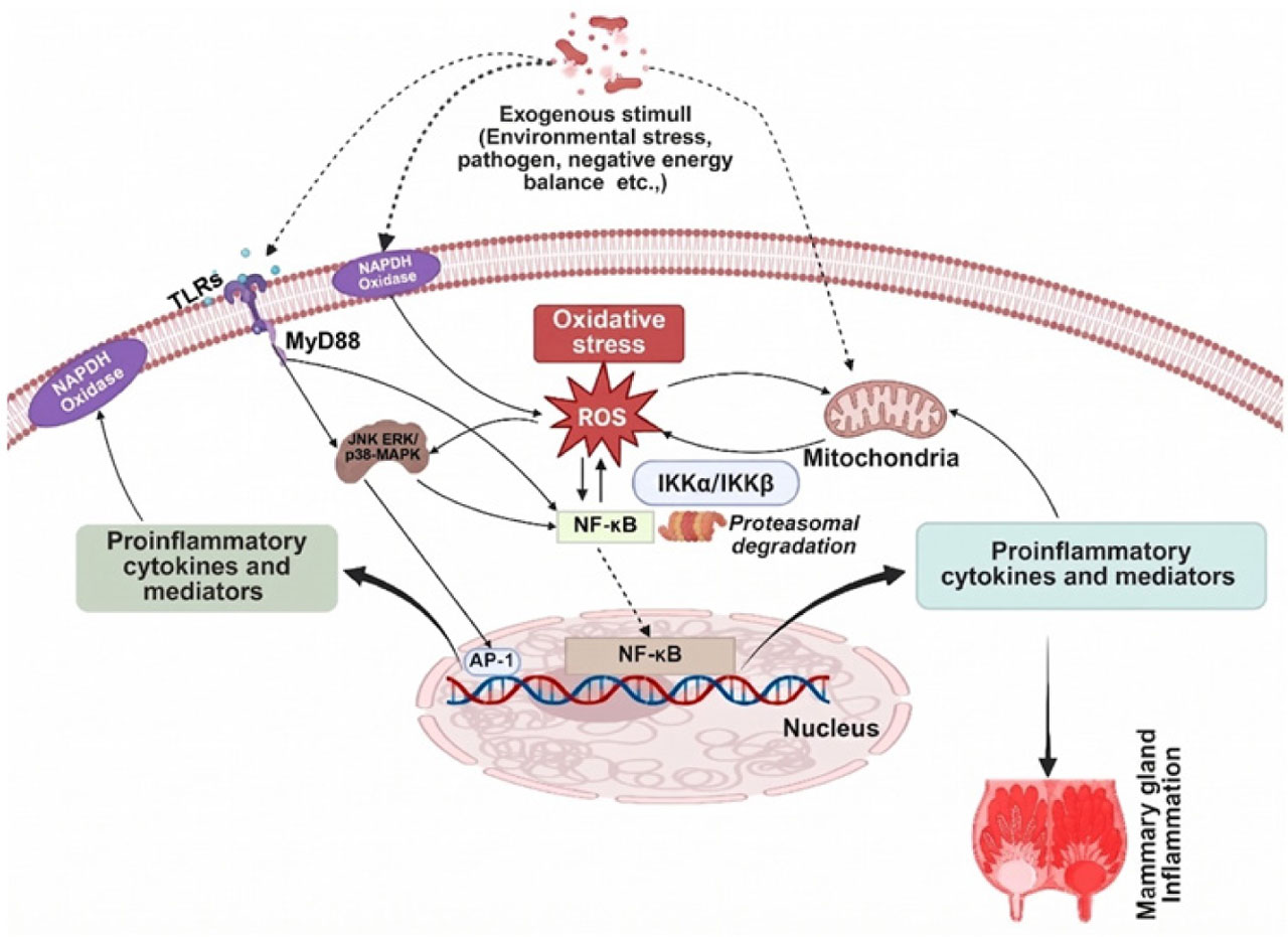
Figure 2.
Molecular pathways of stress-induced inflammatory response in mammary epithelial cells. Exogenous stressors activated TLRs and NADPH oxidase, initiating MyD88-dependent JNK/ERK/p38-MAPK signaling. ROS generation served as a central hub linking oxidative stress to IKKα/IKKβ-mediated NF-κB and AP-1 activation. Nuclear translocation of these transcription factors upregulated proinflammatory mediators, establishing a mechanistic connection between oxidative stress and mammary gland inflammation.
Furthermore, mitochondrial dysfunction represents a critical component of oxidative stress-mediated pathology in mastitis. Mitochondria serve as both major sources and primary targets of ROS during bacterial-induced oxidative stress (Li et al., 2020). Excessive ROS production overwhelms mitochondrial antioxidant defenses, precipitating lipid peroxidation of mitochondrial membranes, oxidation of mitochondrial DNA, and damage to respiratory chain complexes (Figure 2). This mitochondrial injury establishes a detrimental feedback loop wherein damaged mitochondria generate additional ROS, further exacerbating oxidative stress. Additionally, mitochondrial dysfunction severely compromises ATP production through impaired oxidative phosphorylation, thereby limiting cellular energy availability and constraining the mammary epithelium’s capacity for milk synthesis, immune defense, and tissue repair.
Drugs target TLRs/NF-κB /MAPK to prevent/treat bovine mastitis
The strategic targeting of TLR, NF-κB, and MAPK signaling pathways has emerged as a promising therapeutic approach for both the prevention and treatment of bovine mastitis (Fan et al., 2025; Khan et al., 2024 a; Sun et al., 2022; Li S. et al., 2022). These interconnected pathways form the molecular backbone of the innate immune response, orchestrating the inflammatory cascade that characterizes mastitic infections. While the activation of TLR-mediated signaling through NF-κB and MAPK pathways is essential for pathogen recognition and initial immune defense, excessive or prolonged activation can lead to tissue damage, compromised milk production, and chronic inflammation (Khan et al., 2020 a). Consequently, pharmacological interventions that modulate these pathways offer the potential to maintain protective immunity while mitigating harmful inflammatory responses, representing a paradigm shift from traditional antimicrobial approaches toward precision immunomodulatory therapy in bovine mastitis management (Tao et al., 2022; Cao et al., 2023; Xu et al., 2022; Ali et al., 2022; Ma Q. et al., 2022; Liu R. et al., 2022).
Several therapeutic agents, including curcumin, Lycium barbarum polysaccha-ride, hederacoside-C, sodium butyrate, resveratrol, meloxicam, palmatine, epicatechin and gambogic acid etc., have been documented to target the TLRs/NF-κB/MAPK signaling pathway both in vitro and in vivo to prevent inflammatory changes in mammary epithelial cells and mitigate mastitis development (Malik et al., 2023; Liu R. et al., 2022; Liu K. et al., 2024; Ma H. et al., 2021; Ma X. et al., 2021; Muhammad et al., 2020; Sun et al., 2020; Tang et al., 2020; Xu et al., 2020). Table 3 summarizes the research progress on therapeutic agents evaluated for their efficacy in preventing inflammatory responses and bovine mastitis through modulation of the TLRs/NF-κB/MAPK signaling cascade.
Table 3.
Summary of studies targeting TLRs/NF-κB/MAPK signaling pathways via therapeutic agents for mastitis treatment
| Therapeutic agent | Biological effect | Experimental model | References |
|---|---|---|---|
| 1 | 2 | 3 | 4 |
| Maslinic acid | Inhibited the production of pro-inflammatory cytokines (IL-6, IL-1β, TNF-α, iNOS, and COX2) in LPS-treated MMECs Decreased the expression of NLRP3 inflammasome, AKT/NF-κB, and MAPK signaling pathways Enhanced intestinal microflora | MMECs | (Li K. et al., 2023 a) |
| Apigenin | Apigenin blocked the activation of TLR4/NF-κB followed by suppression of myeloperoxidase (MPO), TNF-α, IL-1β, and IL-6 induced by LPS in rat’s mammary gland Prevents inflammatory changes in vitro and mastitis in vivo | Rats model | (Hu et al., 2025) |
| Taraxacum mongolicum | Protected MMECs from S. aureus- induced inflammatory changes by inhibiting TLR2-NF-κB/MAPKs pathways Alleviated inflammation in MECs with significant decrease in TNF-α, IL-6 and IL-1β levels, and MPO levels Inhibited the expression of TLR2, and the phosphorylations of inhibitor κBα (IκBα), p65, p38, extracellular signal-regulated kinase 1/2 (ERK1/2) and c- JNK proteins | MMECs | (Ge et al., 2021) |
| Berberine and Cyperus rotundus rhizomes extract | Reduced the mRNA expression levels of MPO, NF-κB P65, p-ERK, TLR2, NOD2, Keap-1, MDA and MAPK induced by S. aureus in a rat mammary gland | Rats model | (Edres et al., 2025) |
| Luteolin | Suppressed the S. aureus-induced inflammatory changes by targeting TLR2-MyD88-TRAF6 components following inhibition of MAPK and NF-κB signaling pathways in BMECs | BMECs | (Guo et al., 2025) |
| Diflunisal | Ameliorated inflammatory changes caused by S. aureus in BMECs Diflunisal inhibited the NF-κB signaling pathway, resulting in suppression of mRNA expression of IL-1β and TNF-α in BMECs | BMECs | (Soni et al., 2025) |
| Danggui Buxue decoction (DBD) | Attenuated the inflammation and oxidative stress in MMECs induced by E. coli The DBD suppressed the expression of TLR4/NF-κB and Nrf2/HO-1 signaling pathways to alleviate oxidative stress and inflammation | MMECs | (Wang et al., 2025 a) |
| Bacillus subtilis Lactobacillus casei Lactobacillus plantarum 17–5 | Reduced E. coli-induced inflammatory changes in BMECs and MMECs by suppressing proinflammatory cytokines (TNF-α, IL-1β, and IL-6) via inhibiting TLR4/NF-κB/MAPK signaling | BMECs MMECs | (Li et al., 2024; Hu et al., 2023; Li K. et al., 2023 b; Li K. et al., 2022) |
| Jiawei Yanghe decoction | Reduced TLR4/MyD88/NF-κB signaling pathway activation which is correlated with prevention of inflammatory changes in MMECs | MMECs | (Zhao et al., 2023) |
| Paeoniflorin | Suppressed expression of TLR2/NF-κB pathway and related genes (NF-κB unit p65, NF-κB unit p50, and MyD88) in Staphylococcus haemolyticus-treated BMECs | BMECs | (Shen et al., 2023) |
| Taurine | Prevented S. uberis-induced inflammatory changes by suppressing TLR2/TAK1/MAPK/NF-κB signaling activation in RAW264.7 | BMECs | (Li et al., 2023) |
| Quyu Xiaozhong recipe | Inhibited activation of TLR4/NF-κB pathway following prevention of acute mastitis caused by S. aureus in rat mammary glands | Rat MECs | (Wang and Jin, 2023) |
| Chlorogenic acid from Taraxacum officinale | Downregulated the expression of TNF-α, IL-6, and IL-1β and TLR2/NF-κB signaling pathway in BMECs treated with lipoteichoic acid (LTA). | BMECs | (Xu et al., 2023) |
| Hexadecanamide | Ameliorated the inflammatory changes in MMECs caused by S. aureus Inhibited the activation of the NF-κB pathway and improved barrier integrity in MMECs | MMECs | (Bao et al., 2023) |
| Medulla Tetrapanacis | LPS-induced MAPK activation followed by elevated inflammatory cytokines (TNF-α and IL-6) and nitric oxide levels were significantly suppressed in mouse monocyte macrophages (RAW 264.7) by Medulla Tetrapanacis treatment | Mouse monocyte macrophages (RAW 264.7) | (Kwok et al., 2024) |
| Rhapontici Radix ethanol extract (RRE) (isochlorogenic acid B), oligomeric proanthocyanidins and isoliquiritigenin | RRE suppressed LPS-induced inflammatory changes (decreased levels of TNF-α, IL-6, and IL-1β, c-Jun N-terminal kinase 1/2, p38, and extra-cellular regulated protein kinase 1/2) by targeting NF-κB/MAPK pathways in ovine mammary alveolar (MAC-T) cells. The inhibition of NF-κB/MAPK was upregulated by G protein-coupled receptor 161 (GPR161) and transmembrane protein 59 (TMEM59) | Bovine mammary alveolar (MAC-T) cells | (Lv et al., 2024; Ma et al., 2020; Li M. et al., 2022) |
| Quinic acid (QA) and isochlorogenic acid B (ICAB) | Suppressed levels of NOD-like receptor 3 (NLRP3) inflammasome, caspase-11, gasdermin D (GSDMD), and p65 of nuclear factor κB (NF-κB) in MAC-T cells Reduced the inflammatory factors (TNF-α, IL-1β, and IL-6) and oxidative stress factors (COX-2 and iNOS) in both MAC-T cells and mouse mammary glands Target with NF-κB/NLRP3 to suppress inflammatory changes | MAC-T cells | (Lv et al., 2025) |
Conclusions
The TLRs/NF-κB/MAPK signaling networks represent fundamental regulatory hubs orchestrating both protective immunity and pathological inflammation in bovine mastitis. While essential for pathogen clearance, dysregulation of these pathways leads to excessive inflammation, tissue damage, and compromised mammary function. This review demonstrates that strategic modulation of these interconnected pathways offers promising therapeutic approaches beyond traditional antimicrobial treatments. The temporal specificity of interventions, pathogen-specific activation patterns, and emerging epigenetic regulatory mechanisms highlight the complexity of these systems. Understanding cross-talk between signaling networks and oxidative stress responses provides multiple therapeutic intervention points while maintaining protective immunity, representing a paradigm shift toward precision immunomodulatory therapy in mastitis management.
Notes
[1] Contributed by Author contributions:
Conceptualization, writing – original draft: X.F., M.Z.K, and Q.U.; writing – review & editing, data collection and data curation: X.F., Q.U., X.J., M.Z.K, X.J., S.O.R., F.M.A., K.J.A and K.F.A.; visualization, funding sources and supervision: M.Z.K., Q.U., and X.F. All authors have read and agreed to the published version of the manuscript.
[2] Financial disclosure Funding
This work was funded by the Liaocheng Municipal Bureau of Science and Technology, High-talented Foreign Expert Introduction Program (GDWZ202401) and the Dean-ship of Research and Graduate Studies, King Khalid University, Abha, Saudi Arabia under grant number (R.G.P.2/136/47).