
Increasing numbers of neoplasms are observed among domestic animals, especially dogs. It has been estimated that the development of cancer disease is a major health problem in pet dogs and is responsible for ~30% of deaths across breeds (Gustafson et al., 2018).
Spleen tumours (STs) may be categorised as malignant diseases, e.g. haemangiosarcoma (HSA) and non-haemangiosarcoma (non-HSA) such as histiocytic sarcoma, liposarcoma, leiomyosarcoma, and lymphoma, and benign diseases, e.g. abscess, haemangioma, leiomyoma, hematoma, and splenic fibrosis (Magas et al., 2018; O’Byrne and Hosgood, 2019).
HSAs are neoplasms of mesenchymal origin. They can be classified into non-visceral or visceral forms. Spleen tumours (visceral form) are more common in dogs; they are very aggressive neoplasms that can metastasise to distant sites and, therefore, have a poorer prognosis than non-visceral HSA (De Nardi et al., 2023; Kim et al., 2015).
Currently, it has been proposed that, in terms of the cellular origin, HSA is derived from bone marrow pluripotent cells in the pre-differentiation phase with high potential of metastasis formation. These cells are able to migrate to sites of vascularisation, such as the spleen, where they undergo neoplastic transformation. This may explain the high prevalence of HSA in this canine organ (De Nardi et al., 2023; Kapturska and Pawlak, 2023).
Vascular haemangiosarcomas arising from malignant endothelial cells may develop in any tissue or organ containing vascular structures. The most common primary sites for canine HSAs are the spleen and the right atrium of the heart (Griffin et al., 2021; Kim et al., 2015). As shown in various studies, the prevalence of canine haemangiosarcomas in the haemoabdomen varies from 50% to 70% (Davies and Taylor, 2020).
Visceral hemangiosarcoma is a life-threatening disease because its development is unnoticeable at early stages. The clinical manifestation of splenic HSA is based on nonspecific signs, such as anorexia, cachexia, dyspnoea, syncope, emesis, diarrhoea, and hyperthermia, as well as local signs like splenomegaly, abdominal pain on palpation, and haemoperitoneum (De Nardi et al., 2023).
Metastases spread occurs via haematogenous routes or seeding throughout a body cavity commonly to the liver, omentum, mesentery, and lungs. Dogs may show no clinical signs until critical condition usually related to concealed haemorrhage and tumour rupture resulting in rapid death (Griffin et al., 2021). Currently, canine HSA in the spleen is considered as the primary cause of nontraumatic haemoperitoneum (Aronsohn et al., 2009).
There are many reports describing the occurrence of canine spleen tumours; majority of them are histopathological evaluations (Brigandi et al., 2024; Hamid et al., 2010; Kim et al., 2015; Magas et al., 2018; Sobczynska-Rak et al., 2009). At present, little is known about the molecular background of STs, albeit a few papers focused on nuclear DNA analyses have been published in recent years and mutations in genes NRAS, PTEN, PIK3CA, PIK3R1, PLCG1, and TP53 have been reported (Megquier et al., 2019; Wang et al., 2020, 2017).
Despite these results, the complex character and aggressive profile of malignant spleen tumour remain unknown. Tumourigenesis as an energy-dependent condition is related to mitochondria and mitochondrial DNA. Thus, abnormalities within mtDNA may lead to genomic instability and predispose to development of neoplasms in specific localisations (Kopinski et al., 2021). Currently, more reports of mtDNA alterations are available for many different human than canine cancers. However, changes in mtDNA in canine mammary tumours (Bertagnolli et al., 2009; Kowal et al., 2022), mast cell tumours (Śmiech et al., 2019), and transmissible venereal tumours (Strakova et al., 2016) have been confirmed.
To the best authors’ knowledge, this is the first study involving dogs diagnosed with STs wherein detailed mtDNA analyses based on whole mitochondrial genome sequencing were performed. The aim of the research was to identify mtDNA markers that could be useful in molecular diagnostics of canine STs. To achieve this goal, analyses of entire mitochondrial genomes in combination with histopathological evaluations were performed to identify mtDNA polymorphisms, mutations, indels, and heteroplasmic sites related to STs.
Material and methods
The study included 19 dogs diagnosed with spleen tumour (Table 1). Blood, tumour tissue, and healthy tissue were collected from each animal for molecular analyses. In total, 57 samples were collected by veterinarians during a routine procedure. The animals did not undergo chemotherapy or radiotherapy.
Table 1.
List of dogs diagnosed with spleen tumours (STs), histopathological assessment, and characteristics of individuals
| Number | Breed/Crossbreed | Sex | Age of dog | Size* | Tumour profile | Tumour type | Malignant/Benign |
|---|---|---|---|---|---|---|---|
| ST002 | Crossbreed | F | 10 | M | one | haemangiosarcoma | M |
| ST008 | Fox Terrier Smooth | M | 10 | S | one | liposarcoma | |
| ST010 | English Cocker Spaniel | F | 15 | M | two | haemangiosarcoma | |
| ST017 | Crossbreed | M | 12 | L | one | histiocytic sarcoma | |
| ST030 | Boxer | M | 12 | L | one | haemangiosarcoma | |
| ST056 | German Shepherd | M | 10 | L | one, disintegrative | ||
| ST073 | Crossbreed | F | 10 | M | two | haemangioma | B |
| ST074 | Dachshund | M | 12 | S/M | one | haemangiosarcoma | M |
| ST076 | Crossbreed | F | 12 | S | one | haemangioma | B |
| ST088 | Crossbreed | M | 13 | M | one, disintegrative | lymphoma | M |
| ST089 | English Pointer | F | 12 | M/L | one | haemangiosarcoma | |
| ST094 | Crossbreed | M | 15 | M | one | ||
| ST126 | German Shepherd | F | 9 | L | many, disintegrative | ||
| ST145 | Miniature Schnauzer | F | 11 | S | one | ||
| ST148 | Crossbreed | M | 12 | M | one, disintegrative | ||
| ST164 | Jack Russell Terrier | M | 5 | S | one | ||
| ST174 | Miniature Schnauzer | F | 10 | S | one | ||
| ST177 | Crossbreed | M | 8 | L | one | ||
| ST197 | German Shepherd | M | 11 | L | one, disintegrative |
At the beginning, the post-operative tumour tissue was evaluated histopathologically. The collected samples were routinely fixed with 10% buffered formalin (pH 7.2), passed through increasing concentrations of alcoholic solutions to acetone and xylene, and embedded in paraffin blocks. For detailed histopathological evaluation, the preparations were stained with haematoxylin and eosin (H&E staining) and examined under a light microscope coupled with a digital camera (Olympus BX43, Olympus SC100, Tokyo, Japan) (Figure 1).
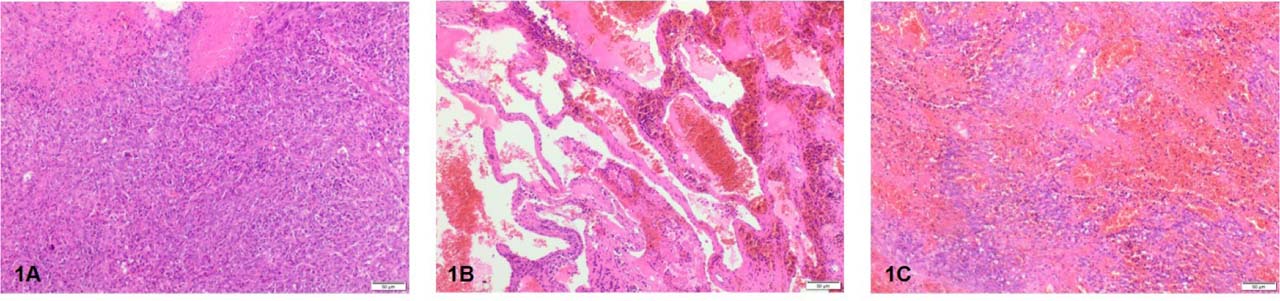
Figure 1.
Histopathological evaluations of spleen tumours in dogs: 1A. Histiocytic sarcoma – visible infiltration of polymorphic round to spindyloid multinucleate cells and foci of necrosis, 1B. Haemangioma – visible irregularly dilated vascular channels in stromal collagen, 1C. Haemangiosarcoma – visible bundles of spindle-shaped cells and slit-like vascular spaces. All tissues were H&E stained × 100
The histopathological evaluations revealed: haemangiosarcomas (14), haemangiomas (2), histiocytic sarcoma (1), liposarcoma (1), and lymphoma (1) among 19 STs. Seventeen out of the 19 neoplasms were malignant tumours (89.5%), with the exception of haemangiomas (Table 1). The histopathological evaluations of selected spleen tumours are presented in Figure 1.
After histopathological evaluations, molecular analyses were performed using the DNeasy Blood & Tissue Kit (Qiagen, Hilden, Germany) to extract DNA from blood and tissue according to the manufacturer’s instructions. The concentration of obtained DNA samples was measured using a NanoDrop™ One/OneC Microvolume UV-Vis spectrophotometer (Thermo Scientific, Waltham, MA, USA), and quality was assessed by electrophoretic separation in 1.5% agarose gel (multiSUB™ Maxi, Cleaver Scientific Ltd, UK).
To obtain the entire mitochondrial genome sequence for each sample, selective amplification was performed using two pairs of primers as in Imes et al. (2012): F1418 and R11041, ~9.5kb PCR product, F9190 and R2382, ~9.8kb PCR product. Next, for each sample, full coverage of mtDNA was obtained after amplification of two long range PCR products obtained using KAPA HiFi PCR Kit reagents (KAPA Biosystems, Wilmington, USA). The PCR DNA template mix (~1 ng) was used as an input and an Illumina shotgun library was constructed using the Nextera XT Kit (Illumina, San Diego, CA, USA) following the manufacturer’s instructions. The library sample was sequenced on an Illumina MiSeq sequencer (Illumina, San Diego, CA, USA) using a 600-cycle kit (v3) in a paired-end mode targeting at least 600x coverage. The detailed information about sequencing data generated using the Illumina MiSeq sequencer is presented in Table S8.
The obtained mtDNA sequences were analysed in comparison with the reference sequence (GenBank accession No. U96639) (Kim et al., 1998) using the bioinformatic program Unipro UGENE (v.34.0) (Okonechnikov et al., 2012) to identify changes, such as heteroplasmic sites, mutations, and polymorphisms, in the tested samples. A mutation was identified when mtDNA alteration was found only in blood, tumour tissue or healthy tissue, and a polymorphism was noted when a nucleotide change was observed in all three tested samples compared to the reference sequence (Ślaska et al., 2014). Next, bioinformatics tools such as ExPASy Server (Gasteiger et al., 2003), SIFT (sorting intolerant from tolerant) (Vaser et al., 2016), and SOPMA (Self-Optimized Prediction Method with Alignment) (Combet et al., 2000; Geourjon and Deleage, 1995) were used for detailed physicochemical and structural protein analyses. The identified variants in DNA and proteins were described according to the guidelines of the Human Genome Variation Society (HGVS) (den Dunnen et al., 2016).
Results
Study includes 19 animals diagnosed with splenic tumours from which blood, healthy and tumour tissue were collected. For each sample whole mitochondrial genome sequencing was performed. The molecular analyses on 57 mtDNA genomes revealed a total of 1123 nucleotide changes: single nucleotide polymorphisms (SNPs, 1011), SNP mutations (20), indels (45), indel mutations (10), and heteroplasmic sites (37) (Table S1, Table S2, Table S3).
The vast majority of identified mtDNA variations viz. 778 out of 1123 (69.3%), including 730 SNPs, 42 indels, 2 indel mutations, and 4 heteroplasmic sites were found in protein-coding mitochondrial genes. Among 37 genes (13 proteins, 22 tRNAs, and 2 rRNAs) encoded by mtDNA, the highest number of polymorphisms was found in genes encoding the respiratory chain and oxidative phosphorylation (OXPHOS). Numerous SNPs were observed in OXPHOS complexes: IV-cytochrome oxidase: COX1 (105) and COX3 genes (71), I-NADH dehydrogenase: ND4 (95), ND5 (88), and ND1 (60) genes, and V-ATP synthase: ATP6 gene (72). In turn, the lowest number of changes occurred in the ND4L (4), ND3 (7), and ATP8 (1) genes (Table S3, Figure 2).
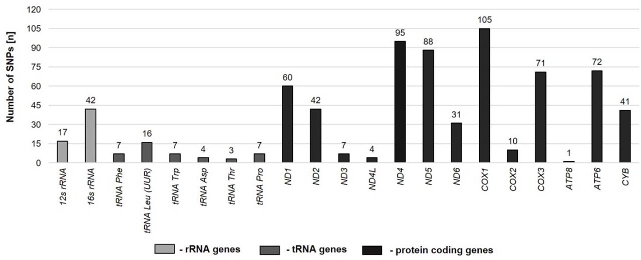
Figure 2.
Number of SNPs found in mtDNA genes
Also, 10 nucleotide alterations were observed in 8 out of the 22 tRNA genes: tRNA Phe, m.16T>C (7/19), tRNA Leu (UUR), m.2678_2679insG (19/19), m.2683G>A (15/19), tRNA Trp, m.5009C>T (7/19), tRNA Asn, m.5151A/T (1/19), m.5152A/T (1/19), tRNA Asp, m.7014T>C (4/19), tRNA Arg, m. 9865_9866insA (3/19), tRNA Thr, m.15372G>A (3/19), and tRNA Pro, m.15435G>A (7/19). These changes were found in tRNA regions: DHU arm (3), DHU loop (1), TΨC loop (2), TΨC arm (1), central loop (1), and anticodon arm (2) (Table S6). 17 SNPs were observed in the 12s rRNA gene and 42 SNPs were detected in the 16s rRNA gene (Table S3, Table S4). Also, two deletions: m.1486delA and m.2267delA were found in the 16s rRNA gene in spleen tumour from ST148 and ST094 (Table S5).
The analyses also showed 312 changes in the non-coding region of the displacement loop (D-loop), including 281 SNPs, 20 mutations, 3 indels, and 8 indel mutations. The highest number of SNP polymorphisms was found in HVI: 141 (50.1%), followed by VNTR: 89 (31.7%) and HVII: 51 (18.2%) (Table S2, Table S3).
In the VNTR localised between 16,130 and 16,430 bp of the D-loop, there is a 10-nucleotide tandem repeat motif (5’-GTACACGT(A/G)C-3’). In this region from 16,138 to 16,408 bp, 30 out of 36 (83.3%) heteroplasmic sites were observed (Table S2). Interestingly, in 5 out of 19 (26.3%) dogs: ST002, ST030, ST089, ST126, and ST148, a loss of the aforementioned motif was observed: m.16288delGTACACGTAC (ST030), m.16408delGTACACGTGC (ST002, ST030), and m.16418delGTACACGTAC (ST089, ST126, ST164) (Table S2). It is worth noting that, in the case of dog ST030, the VNTR motif differed from that in the other dogs, with T>C or T/C alteration in the last position of the repetitive element.
The whole mitochondrial genome sequencing analysis of all the samples from dogs diagnosed with STs showed 9 common polymorphisms across all the samples. Among them, 7 out of 11 occurred in the protein-coding genes of OXPHOS (COX1, COX3, ATP6, ND4L, ND5), 1 in tRNA-Leu (UUR) (m.2678_2679insG), and 1 in the HVI region (m.15814C>T) of the D-loop (Table 2). It is important to emphasise the impact of identified common SNPs in all the samples. These changes may be crucial in aetiopathogenesis because of their common presence in spleen tumours.
Table 2.
List of common polymorphisms present in all the dogs diagnosed with spleen tumours (STs)
| Gene/Region | Reference sequence | Sequence variant | Codon change/position in tRNA | Amino acid change/tRNA region | SIFT1 for nonsynonymous changes |
|---|---|---|---|---|---|
| tRNA Leu (UUR) | m.2678 | m.2678_2679insG | 8_9 | between acceptor stem and DHU stem | – |
| COX1 | m.5367C | m.5367C>T | CTG→TTG | p.Leu7= | |
| m.5444T | m.5444T>C | GCT→GCC | p.Ala32= | ||
| m.6065A | m.6065A>G | GGA→GGG | p.Gly239= | ||
| ATP6 | m.8368C | m.8368C>T | CTC→CTT | p.Leu135= | |
| COX3 | m.8807G | m.8807G>A | TGC→TAC | p.Cys55Tyr | tolerant |
| ND4L | m.9911_9912 | m.9911_9912insGT | ATG→GTG | p.Met1Val | intolerant |
| ND5 | m.13299T | m.13299T>A | TCA→ACA | p.Ser508Thr | tolerant |
| D-loop | m.15814C | m.15814C>T | – | – | – |
To assess the effects of the identified mtDNA variants on the protein level, the SIFT method was used and revealed that the variants noted in the COX1 (3) and ATP6 (1) genes resulted in no protein changes: p.Leu7=, p.Ala32=, p.Gly239=, and p.Leu135=, respectively. These synonymous changes had no deleterious effects on the amino acid sequences. However, variants noted in the COX3 gene: p.Cys55Tyr, the ND4L gene: p.Met1Val, and the ND5 gene: p.Ser508Thr caused non-synonymous changes in the protein level, but only the amino acid change in the ND4L gene was found intolerant (Table 2).
In addition to the 9 common SNPs, protein analyses were performed for all non-synonymous changes (32) identified in 10 protein-coding genes using the SIFT and SOPMA tools (Table S7). The obtained results showed that the majority of these changes (78.1%) were tolerant.
All the 20 SNP mutations were found in the D-loop, including HVI: m.16003A>G (1), VNTR: m.16148A>G (2), m.16168A>G (2), m.16178A>G (1), m.16188G>A (1), m.16228G>A (1), m.16338G>A (1), m.16358A>G (1), m.16368G>A (1), m.16378G>A (1), m.16388A>G (1), and HVII: m.16431C>T(1), m.16664T>C (1), m.16665T>C, (1), m.16672C>T (1), m.16675T>C (1). Among 10 indel mutations 2 were found in 16s rRNA gene: m.1486delA and m.2267delA and 8 changes were noted in D-loop: HVI (2), VNTR (2), and HVII (4). Detailed information of mutations and samples is presented in Table S5.
The changes occurring during tumourigenesis included complex and enormously diversified molecular variations which were visible in the number of mtDNA alterations per sample. In the case of malignant STs, i.e. histiocytic sarcoma (ST017), 93 SNPs and 18 heteroplasmic sites were observed (Table S4). Moreover, 8 mutations were found in the materials from this dog, including: m.15931delA (D-loop, HVI) in the tumour, m.16338G>A (D-loop, VNTR) in the blood, and m.16664T>C, m.16665T>C, m.16672C>T, m.16675T>C, m.16678insTC (D-loop, HVII) in the healthy tissue. Another individual example of malignant ST was lymphoma with 100 SNPs, m.16148A>G and 13 heteroplasmic sites (Table S4, Table S5). In turn, two dogs (ST073, ST076) with benign neoplasms: haemangiomas had 40 SNPs and 38 SNPs, respectively. Despite the commonly occurring SNP in the tRNA Leu (UUR) gene m.2678_2679insG in all the samples, other SNPs or mutations localised in tRNA genes were not detected in these two benign tumours in comparison to malignant STs. However, the following mutations were found: m.16358A>G (ST076B) and m.16378G>A (ST073B) in the D-loop, VNTR and m.16674delC (ST076H) in the D-loop, HVII (Table S5).
Haemangiosarcomas constituted the most numerous groups of STs in this study. The number of mtDNA variations in these tumours ranged from 12 to 104 SNPs and from 5 to 19 heteroplasmic sites. It is worth noting that, in the dogs diagnosed with HSA, three groups (I, II, III) of mtDNA alterations were identified in protein-coding genes. Group I – if no changes were observed in 12s rRNA and 16s rRNA, the total number of observed SNPs varied from 12 to 22 (ST002, ST010, ST094, ST174, ST177). Group II – when polymorphisms were observed in 16s rRNA, from 38 to 42 SNPs in total were found in dogs ST056, ST074, and ST197 (Table S4). Two of these animals had spleens in poor condition with disintegrative tumour (Table 1). Group III – if at least 1 SNP was observed in both 12s rRNA and 16s rRNA, a higher number of SNPs (from 47 to 104) were identified (ST030, ST089, ST126, ST145, ST148, ST164). Also, SNPs (from 2 to 5) always occurred in the CYB gene in these animals (Table S4, Figure 3).
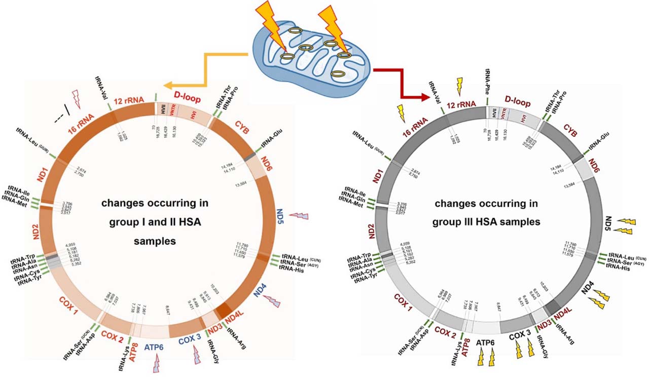
Figure 3.
Association between the HSA sample group (I, II, III) and the increasing number of SNPs in the ND4, ND5, COX3, and ATP6 genes
The detailed analysis of the number of SNPs found in the HSA samples showed that there was a visible tendency for accumulation of polymorphisms. Haemangiosarcomas samples belonging to group I and II had 1–2, 1–3, 1, and 1–2 SNPs in comparison to the animals from group III: 4–13, 4–13, 2–10, and 3–8 SNPs found in the ND4, ND5, COX3, and ATP6 genes, respectively (Table S4). Moreover, also in the other malignant tumour: histiocytic sarcoma (HS) and lymphoma (LP), a higher number of SNPs (12–13, 11–13, 7, 6) were found in the ND4, ND5, COX3, and ATP6 genes (Table S4).
Among the 19 analysed samples, the greatest diversity in the number of mtDNA variants was detected in samples from the following animals with HSA: ST148 (123), ST145 (110), ST169 (106) as well as ST126 (104) and ST089 (103) followed by the other malignant STs: lymphoma (ST088; 113) and histiocytic sarcoma (ST0017; 111) (Table S4).
Discussion
Spleen tumours pose a serious health problem in domestic animals, especially in dogs (Hendrick, 2016). Among these tumours, the most common life-threatening type is haemangiosarcomas (HSA), which have a poor prognosis due to the long period of imperceptible tumour development and simultaneous metastases to distant locations (De Nardi et al., 2023; Megquier et al., 2019).
Currently, many cases of canine HSA have been reported and its aggressive character is known (Sobczynska-Rak et al., 2009; Varela et al., 2022; Wang et al., 2020, 2017). Moreover, in recent years, various molecular analyses of nDNA were performed and mutations in genes NRAS, PTEN, PIK3CA, PIK3R1, PLCG1, and TP53 were found (Megquier et al., 2019; Wang et al., 2020, 2017). However, even these molecular results did not explain the complex character and common occurrence of spleen tumours.
Beyond nDNA, the genetic information is also localised in mitochondria in the form of mtDNA. These organelles are crucial in cells as principal generators of cellular energy in the form of ATP (adenosine triphosphate) via oxidative phosphorylation (OXPHOS), incorporating the electron-transferring respiratory chain (complexes I–IV) and ATP synthase (complex V). Mitochondria are under the dual genetic control of both mtDNA and nDNA (Hahn and Zuryn, 2019; Kopinski et al., 2021). Alterations in mtDNA may lead to severe metabolic diseases. Moreover, mtDNA disturbances in cellular energy metabolism were found to result in exacerbation of reactive oxygen species (ROS) production. To date, changes in canine mtDNA have been associated with the development of encephalomyelopathies (Baranowska et al., 2009; Li et al., 2006), myopathies (Paciello et al., 2003; Shelton et al., 2024), and cancer disease, including mammary gland tumours (Bertagnolli et al., 2009; Kowal et al., 2022), mast cell tumours (Śmiech et al., 2019), and canine transmissible venereal tumours (Strakova et al., 2016).
The whole mitochondrial genome sequencing performed in the present study showed a tremendous diversity in the analysed sequences of canine STs and revealed a total of 1123 mtDNA changes, including 1011 SNPs, 20 mutations, 45 indels, 10 indel mutations and 37 heteroplasmic sites (Table S1, Table S2, Table S3). The majority of the identified SNP variants (730/1011; 72.2%) were found in protein-coding genes, especially in OXPHOS complexes: IV – COX1 (105), COX3 (71), I – ND4 (95), ND5 (88), and ND1 (60), and V – ATP6 (72) (Table S3, Figure 2).
Mitochondrial genomes undergo mutagenesis in different types of tumours. Accumulation of replicative errors during replication process and generation of ROS lead to changes in mtDNA which dysregulate OXPHOS (Hahn and Zuryn, 2019; Kim et al., 2022). In our study the highest number of mtDNA variants was found in the OXPHOS IV complex. These molecular changes may be responsible for the disturbances in correct assembly of complex IV and may enhance the production of ROS (Hahn and Zuryn, 2019). In this study, the common SNPs in the COX1 gene: m.5367C>T, m.5444T>C, m.6065A>G were synonymous (Table 2), albeit additional variants m.6711T>A and m.6882A>G were intolerant (Table S7). Also, results obtained by de Oliveira et al. (2023) showed mutations in genes encoding mitochondrial complex IV may have pathogenic potential in breast cancers. Moreover, alterations in complex I genes are strongly linked with the development and growth of different types of cancer due to increased ROS production (Hahn and Zuryn, 2019). For instance, substitution of m.4776G>A in the ND2 gene contributed to HIF1α accumulation and upregulated PDK2 in head and neck squamous cell carcinoma (Sun et al., 2009). Also, Gorelick et al. (2021) described an m.3380G>A change in the ND1 gene, which was related to amino acid residue Arg25, usually resulting in synonymous mutations in colorectal patients. However, detailed modelling of the observed amino acid replacement p.Arg25Gln revealed serious changes resulting from the local charge environment due to loss of the relatively bulky, positively charged arginine side chain. As a result, it was suggested that the ND1 gene p.Arg25Gln promotes a transcriptional phenotype characterised by increased mitochondrial metabolism and suppressed expression of immune genes (Gorelick et al., 2021).
In this study, the multiplicity of mtDNA variants in the OXPHOS complex I genes indicates their high diversity (Table 2, Table S4, Table S7). All were nonsynonymous at the protein level, although tolerated, as revealed by the SIFT tool. As described by Gorelick et al. (2021), the issue may be related to the altered local environment during tumourigenesis and may have an impact on the constitution of selected proteins.
In this study, changes m.2678_2679insG (tRNA Leu (UUR)), m.5367C>T, m.5444T>C, m.6065A>G (COX1), m.8368C>T (ATP6), m.8807G>A (COX3), m.9911_9912insGT (ND4L), m.13299T>A (ND5), and m.15814C>T (D-loop) were found in each dog diagnosed with spleen tumour regardless of the histopathological type. Notably, other authors also found such polymorphisms in tRNA Leu (UUR), COX1, COX3, ND5 (Kowal et al., 2022), and the D-loop (Bertagnolli et al., 2009; Kowal et al., 2022) in mammary gland tumours and in testicular neoplasms (Tkaczyk-Wlizlo et al., 2024). Also, non-synonymous changes identified in protein-coding genes (Table S7) found in individual animals were compared with available literature data. Selected mtDNA variants in genes CYTB (m.14474G>A) (Śmiech et al., 2019) and ND4 (m.11402T>C) (Ślaska et al., 2016) have been previously described in canine mast cell tumour and adenocarcinoma, respectively. This observation may indicate that this mtDNA alteration could be related to tumourigenesis as a biological process. This is consistent with the fact that some mtDNA mutations are commonly identified in different types of tumours or are tumour-specific (Kopinski et al., 2021). Therefore, the above-mentioned mitochondrial genome aberrations should be monitored in future research, as some of them may be selected as tumour-related mtDNA markers.
Interestingly, it was observed in malignant STs that in protein-coding genes ND4, ND5, COX3, and ATP6, the number of mtDNA variants was increased when changes were concurrently found in 12s rRNA and/or 16s rRNA (Table S4). 12s rRNA and 16s rRNA form small and large subunits of mitochondrial ribosome. These genes are important for the proper synthesis of mitochondrially encoded proteins; hence, substitutions within the genes may result in distorted mitochondrial protein synthesis, lower stability of produced components, and loss of mitochondrial translation (Kabekkodu et al., 2014). Additionally, in patients with mutations in rRNA genes, reduced production of ATP and increased levels of mitochondrial superoxide were observed (Hahn and Zuryn, 2019). Alterations in mtDNA rRNAs were observed in human cancers as tumour-specific changes. Moreover, changes in 16s rRNA were higher in malignant samples (Kabekkodu et al., 2014). It is important in the future to elucidate the impact of changes in rRNAs on increased mtDNA variants in other protein-coding genes.
Also, detailed analyses of additional individual canine factors, such as age, breed, and sex, were conducted (Table 1). According to literature data, adult dogs between 7 to 15 years old are the most affected (De Nardi et al., 2023). In our study, the majority of animals were from 8 to 15 years old (94.7%), with one exception of sample ST164 (5 years old). The most common age of the animals at diagnosis was 12 years old (6 out of 19; 31.6%). The affected dogs (8 females and 11 males) represented different breeds of dogs: Boxer (1), Dachshund (1), English Cocker Spaniel (1), English Pointer (1), Fox Terrier Smooth (1), German Shepherd (3), Jack Russell Terrier (1), Miniature Schnauzer (2), and crossbreeds (8). This is consistent with other publications indicating that such canine breeds as Boxer, German Shepherd, and Miniature Schnauzer are predisposed to spleen tumour development (Brigandi et al., 2024; Story et al., 2020).
It is important to emphasise that for the first time mtDNA changes were described in canine STs. However, there is a need to confirm observed mtDNA variants in future research because the tested group was diversified. We acknowledge limitations to this study: (1) different breeds of dogs including crossbreed dogs; (2) affected animals were between 8 to 15 years old with one exception of 5 years old; (3) different histopathological type of splenic tumours. Also, additional analyses including statistics should be performed to identify functional effects of the observed mtDNA variants.
Conclusions
Spleen tumours, especially HSAs, are aggressive difficult-to-treat neoplasms. The present analyses based on whole mitochondrial genome sequencing revealed the complexity and diversity of these neoplasms supported by numerous mtDNA variants. Based on the obtained results, 9 common SNPs were found as well as many additional changes, i.e. polymorphisms, mutations, and heteroplasmic sites.
The majority of SNPs (71.2%) were identified in protein-coding genes, especially in COX1, COX3, ND4, ND5, ND1, and ATP6. It is worth highlighting that, in addition to the 9 common polymorphisms, additional changes in the ND5, COX1, COX3, and ATP6 genes were detected in samples from dogs diagnosed with malignant STs. The number of these SNPs could be related to the lack or occurrence of mtDNA variants in 12s rRNA and 16s rRNA. The changes in the D-loop were divided into SNPs that were more frequent in the HVI region and numerous heteroplasmic sites found in the VNTR region.
Notes
[3] Ethical statement
The study was approved by the II Local Ethical Commission for animal experiments in Lublin, Poland (resolution number 79/2014).
[4] Contributed by Authorship contribution statement
ATW: conceptualisation, formal analysis, data curation, funding acquisition, project administration, original draft; KK: formal analysis, software, visualisation, writing – review and editing; AS: investigation, writing – review and editing; BS: methodology, resources, supervision.
[5] Conflicts of interest Conflicts of interest
The authors declare that they have no known competing financial interests or personal relationships that could have appeared to influence the work reported in this paper.