
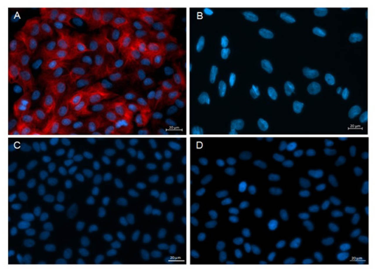
Figure 1.
The purity of endometrial epithelial cell culture confirmed by positive immunofluorescent staining for anti-cytokeratin (A) and negative anti-vimentin staining (B). Negative control without primary antibodies for cytokeratin (C) and vimentin (D). Scale bar: 20 μm.
Table 1.
Influence of noradrenaline (NA, 10−5 M) alone or agonists of α1-, α2- and β-adrenoreceptors (ARs, 10−4 M) alone or antagonists of β-ARs (10−4 M) alone on the prostaglandin-endoperoxidase synthase-2 (PTGS-2), microsomal PTGE synthase-1 (mPTGES-1) and mPTGES-2 protein abundances in the pig cultured endometrial epithelial cells. Enzyme abundances were determined by Western blotting, and values were normalized against glyceraldehyde-3-phosphate dehydrogenase (GAPDH) protein abundances. Treatments were conducted in triplicate for each of the four pigs. Data are presented as the mean ± SEM. Different letters (a, b, c) indicate statistical differences (P<0.01, P<0.001). CV: control value; agon.: agonist; anta.: antagonist
| Treatment | PTGS-2 | mPTGES-2 protein abundances (arbitrary units) | mPTGES-1 |
|---|---|---|---|
| CV | 0.19±0.03 | 0.03±0.01 | 0.29±0.03 a |
| NA | 0.18±0.08 | 0.04±0.02 | 0.54±0.04 b |
| α1 agon. | 0.22±0.04 | 0.04±0.02 | 0.18±0.05 a |
| α2 agon. | 0.16±0.05 | 0.03±0.01 | 0.21±0.02 a |
| β agon. | 0.22±0.03 | 0.03±0.02 | 0.71±0.06 b |
| β1 agon. | 0.23±0.02 | 0.07±0.03 | 0.58±0.02 b |
| β2 agon. | 015±0.04 | 0.05±0.03 | 1.23±0.04 c |
| β3 agon. | 0.25±0.03 | 0.03±0.01 | 0.59±0.03 b |
| β1 anta. | 0.21±0.05 | 0.06±0.02 | 0.22±0.04 a |
| β2 anta. | 0.15±0.03 | 0.04±0.01 | 0.25±0.05 a |
| β3 anta. | 0.18±0.06 | 0.04±0.02 | 0.21±0.07 a |
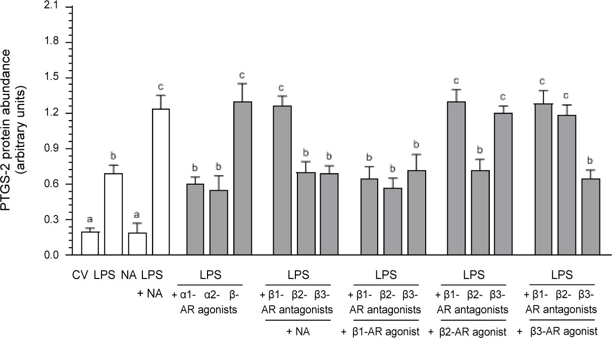
Figure 2.
Influence of noradrenaline (NA, 10−5 M) alone or agonists of α1-, α2- and β-adrenoreceptors (ARs, 10−4 M) or antagonists of β1-, β2- and β3-ARs (10−4 M) with NA or antagonists of β1-, β2- and β3-ARs (10−4 M) in combination with agonists of β1-, β2-, β3-ARs (10−4 M) on the prostaglandin-endoperoxidase synthase-2 (PTGS-2) protein abundance in the lipopolysaccharide (LPS, 10 ng/ml of medium)-exposed pig cultured endometrial epithelial cells, estimated by Western blotting. Treatments were conducted in triplicate for each of the four pigs. The values were normalized against glyceraldehyde-3-phosphate dehydrogenase (GAPDH) abundances. Data are presented as the mean (± SEM). Blots with representative bands for each treatment are given in Supplementary Figure 2 A. Different letters (a, b, c) indicate statistical differences (P<0.05–0.001). CV: control value.
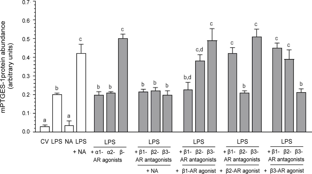
Figure 3.
Influence of noradrenaline (NA, 10−5 M) alone or agonists of α1-, α2- and β-adrenoreceptors (ARs, 10−4 M) or antagonists of β1-, β2-and β3-ARs (10−4 M) with NA or antagonists of β1-, β2- and β3-ARs (10−4 M) in combination with agonists of β1-, β2-, β3-ARs (10−4 M) on the microsomal prostaglandin E synthase-1 (mPTGES-1) protein abundance in the lipopolysaccharide (LPS, 10 ng/ml of medium)-exposed pig cultures endometrial epithelial cells, estimated by Western blotting. Treatments were conducted in triplicate for each of the four pigs. The values were normalized against glyceraldehyde-3-phosphate dehydrogenase (GAPDH) abundances. Data are presented as the mean (± SEM). Blots with representative bands for each treatment are given in Supplementary Figure 2 B. Different letters (a, b, c, d) indicate statistical differences (P<0.05–0.001). CV: control value.
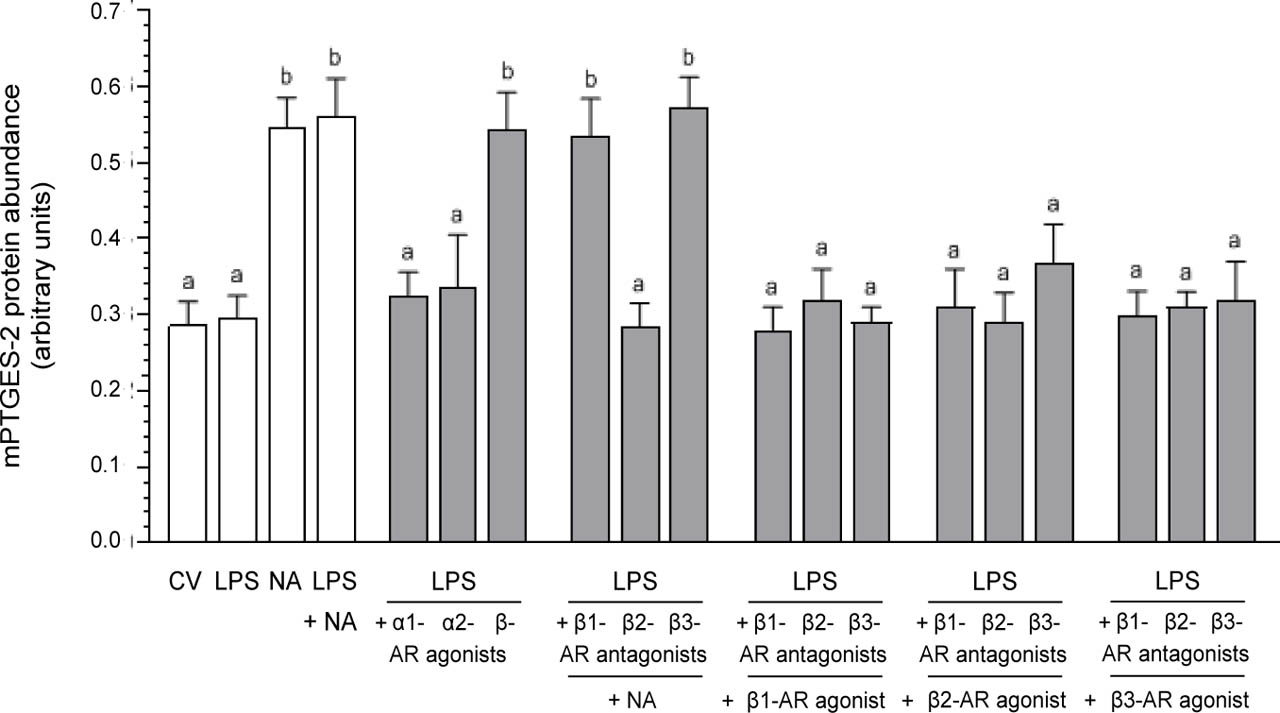
Figure 4.
Influence of noradrenaline (NA, 10−5 M) alone or agonists of α1-, α2- and β-adrenoreceptors (ARs, 10−4 M) or antagonists of β1-, β2-and β3-ARs (10−4 M) with NA or antagonists of β1-, β2- and β3-ARs (10−4 M) in combination with agonists of β1-, β2-, β3-ARs (10−4 M) on the microsomal prostaglandin E synthase-2 (mPTGES-2) protein abundance in the lipopolysaccharide (LPS, 10 ng/ml of medium)-exposed pig cultured endometrial epithelial cells, estimated by Western blotting. Treatments were conducted in triplicate for each of the four pigs. The values were normalized against glyceraldehyde-3-phosphate dehydrogenase (GAPDH) abundances. Data are presented as the mean (± SEM). Blots with representative bands for each treatment are given in Supplementary Figure 2 C. Different letters (a, b) indicate statistical differences (P<0.05, P<0.01). CV: control value.
Table 2.
Influence of noradrenaline (NA, 10−5 M) alone or agonists of α1-, α2- and β-adrenoreceptors (ARs, 10−4 M) alone or antagonists of β-ARs (10−4 M) alone on the prostaglandin E2 (PGE2) content in the medium of pig cultured endometrial epithelial cells. PGE2 concentrations were determined by ELISA. Treatments were conducted in triplicate for each of the four pigs. Data are presented as the mean ± SEM. Different letters (a, b, c, d) indicate statistical differences (P<0.05–0.001). CV: control value; agon.: agonist; anta.: antagonist
| Treatment | PGE2 content (ng/ml of medium) |
|---|---|
| CV | 0.71±0.15 a |
| NA | 1.52±0.13 b |
| α1 agon. | 0.59±0.17 a |
| α2 agon. | 0.68±0.14 a |
| β agon. | 1.91±0.15 cb |
| β1 agon. | 2.38±0.09 cd |
| β2 agon. | 2.74±0.11 d |
| β3 agon. | 2.65±0.13 d |
| β1 anta. | 0.62±0.14 a |
| β2 anta. | 0.53±0.09 a |
| β3 anta. | 0.83±0.14 a |
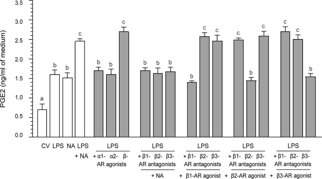
Figure 5.
Influence of noradrenaline (NA, 10−5 M) alone or agonists of α1-, α2- and β-adrenoreceptors (ARs, 10−4 M) or antagonists of β1-, β2-and β3-ARs (10−4 M) with NA or antagonists of β1-, β2- and β3-ARs (10−4 M) in combination with agonists of β1-, β2-, β3-ARs (10−4 M) on the prostaglandin E2 (PGE2) concentration in medium after incubation of the lipopolysaccharide (LPS, 10 ng/ml medium)-exposed pig cultured endometrial epithelial cells, estimated by ELISA. Treatments were conducted in triplicate for each of the four pigs. Data are presented as the mean (± SEM). Different letters (a, b, c) indicate statistical differences (P<0.001). CV: control value.