
Across the world, tilapia is the third most commonly cultivated finfish after grass carp, Ctenopharyngodon idela, and silver carp, Hypophthalmichthys molitrix, with 4.4 million metric tonnes produced worldwide (FAO, 2022). Because of its great commercial value, the Nile tilapia, Oreochromis niloticus, is particularly important and valuable throughout the world (Adeshina et al., 2021). Due to its quality flesh and economic gains, the technicalities deployed in its production are evolving with an intensive participation (FAO, 2021). This accomplishment is not unconnected to the advanced culturing practices such as high stocking density, supply of quality and adequate feed, among others but emergence of diseases and reoccurrence of old ones particularly parasite infestations, is impeding the tilapia operation (Adeshina et al., 2020).
The monogenean Dactylogyrus vastator is an ectoparasite that has a direct, single-host life cycle that multiplies easily in fish (Klinger and Floyd, 1998). Monogenean parasitic infestations usually occur on the skin and gills, resulting in severe disorders such as gill inflammation, excessive mucous secretions, de-colouration, descaling, dermatitis, accelerated respiration, oedema, lamellae fusion, branchial necrosis, and consequently high mortalities (Reed et al., 2009; Shamsi et al., 2009; Šimková et al., 2017; Thilakaratne et al., 2003). The presence of D. vastator on the gills of tambaqui, Colossoma macropomum, resulted in histopathological disorder and mortalities (Alves et al., 2019; Soares et al., 2016). Further, D. vastator causes disorders and huge mortality ranges from 29 to 68% in Nile tilapia, Carassius auratus (goldfish) (Reed et al., 2009), Cyprinus carpio (common carp) (Borji et al., 2012), and Carassius gibelio (Prussian carp) (Šimková et al., 2017). Therefore, it is important to adequately manage, control, and treat parasite infestations for profitable aquaculture ventures.
Chemotherapeutics are used more frequently as growth promoters and disease control agents, but their usage has become controversial as they contributed in increasing drug-resistant organisms, and environmental deterioration (Adeniyi et al., 2024; Adeshina et al., 2024; Eissa et al., 2024 a; Jahanbakhshi et al., 2023). That is why, there is a need of environmentally friendly alternatives for continuous, sustainable, and responsible aquaculture. There are several efforts to use plant-based feed additives, phytobiotics to ameliorate fish growth, health, and immunity (Fu et al., 2014, 2021; Lin et al., 2016). Plants and plant materials that have medicinal traditional usage are gaining popularity and have become promising feed additives and substitutes for antibiotics with low-side effects in aquaculture. They had high antiparasitic potentials due to the presence of high levels of active compounds and secondary metabolites. For instance, in Nile tilapia, Adeshina et al., (2021) showed that the use of Tridax procumbens extract had positive effect on the growth performance, antioxidant profile, innate immunity, and disease resistance against D. vastator infestation. Similarly, de Queiroz et al., (2022) reported that 60 mg L−1 Piper aduncum leaf extract protected tambaqui against monogenean infestation. Furthermore, studies have shown the stimulatory roles of some botanical herbs against parasites, i.e. Euphorbia fischeriana extract against D. vastator in gold fish (Zhang et al., 2014), acetone extract from Bixa orellana against Anacanthorus spathulatus in tambaqui (de Andrade et al., 2016), extracts of Mucuna pruriens, Carica papaya, Capsicum frutescens, or Galla chinensis against the Ichthyophthirius multifiliis (Ekanem et al., 2004; Ling et al., 2012; Zhang et al., 2013), and Mitracarpus scaber extract protected Nile tilapia against Gyrodactylus malalai infestation (Adeshina et al., 2021).
Polyherbal formulations, as widely used in traditional medicine, are made up of different plant constituents and bioactive compounds that have pharmacological benefits and are employed for the treatment of various ailments, as well as to maintain well-being (Eissa et al., 2024 b). This is based on the idea that multiple herbs with distinct bioactive components may complement one another using its synergistic potentiality and be more useful than a single extract. Based on the performance of each of these plants, thus, it appears that T. procumbens, M. scaber, Mucuna pruriens, and Carica papaya leaves extracts can be used to enhance growth performance, immune response, and reduction of pathogenic infestation (Adeshina et al., 2019 a). The combination of multiple herbs with individual importance could be what to look out for in this study in fish nutrition. The use of multiple herbs has been documented in human in treatment of immune disorders and other infections, nevertheless its usage in fish for the treatment of dactylogyrosis has yet to be fully clarified. Therefore, this study examined the effect of T. procumbens, M. scaber, M. pruriens, and C. papaya leaf extracts-based diets on the growth performance, intestinal histomorphometry, antioxidant status, and immunomodulation of Nile tilapia, along with its protection from D. vastator infestation.
Material and methods
Plant collection, identification, and extraction
The leaves of T. procumbens, M. scaber, M. pruriens, and C. papaya were obtained from a botanical garden and authenticated at the Herbarium of Forest Research Institute of Nigeria (FRIN), Ibadan, Nigeria, where the plants’ leaves were confirmed by a qualified plant taxonomist using microscopic and morphological identification of leaf shape, margin, venation, and trichomes, and then compared with floras, botanical keys, and monographs (Plant Resources of Tropical Africa). These leaves were allowed to dry in air at 35°C, ground in a hand-blender, and sieved into fine powder. The herbal leaves (HL) meals were mixed at 1:1:1:1 before extracted using 80% ethyl-acetate for 48 h to complete extraction in triplicates. Briefly, 20 g of the leaves’ mixture comprises 5 g of T. procumbens, M. scaber, M. pruriens, and C. papaya, each was added to 200 mL of ethyl-acetate. Then, No. 1 Whatman paper was used to filter the herbal leaves mixture extract (HLME) solution while the solvent was separated using a vacuum rotary evaporator (Zhang et al., 2014). The HMLE was then transferred to a container and stored at −20°C.
Phyto-chemical and phyto-constituents’ analyses
Several analytical procedures and reagents were used to quantitatively check the HLME for the presence of secondary metabolites. The following metabolites were analysed: tannins (Mayuri, 2012), terpenoids (Odebiyi and Sofowora, 1978), steroids (Marcano and Hasenawa, 1991), flavonoids (Harborne, 1998; Joshi et al., 2011), alkaloids (Joshi et al., 2011), and saponins (Harborne, 1998; Mayuri, 2012). The secondary metabolites were shown in Table 1.
Table 1.
Quantitative analysis of secondary metabolites in the HMLE1
| Parameter | Value (mg 100 g−1) |
|---|---|
| Alkaloids | 350.3 |
| Flavonoids | 72.4 |
| Saponins | 49.9 |
| Steroids | 114.3 |
| Tannins | 63.5 |
| Terpenoids | 702.6 |
Also, gas chromatography (GC, Agilent 5975, Avondale, PA USA)/mass spectrometry (MS, Agilent 7890A, Avondale, PA USA) containing a 5-meter-long HP column (0.25 cm internal diameter) was used to analyse the phyto-constituents of HLME. Briefly, an autosampler was used to inject one microliter of sample into the Agilent 190915-433HP-5M, containing 5% phenylmethylsilox (30 m × 250 µm × 0.25 µm), using a split-less injector (at 300°C) and a film thickness of 1.0 µm at an ionization energy of 70eV, while helium gas (flow rate = 1 mL per minute) was used as carrier. The oven temperature was designed to rise from 35°C to 300°C and then hold at 35°C for 5 minutes, before ramping up at 20°C per minute to 250°C for 5 minutes. An HP Chemostation System was used to record the results of the entire scan of the samples, which had a mass range of 50 to 750 units. The constituents of the extract were determined through comparison of their mass spectra and relative retention durations to those of real samples from the database’s analytical standards (Adeshina et al., 2018). Only structures that achieved 90% or greater probability were chosen for structural assignments by the library database (NIT XI) and shown in Table 2. The major active compounds in HMLE are β-sitosterol, p-cymene, quercetin, eicosanoic acid, octadecanoic acid, phytol, oleic acid, chlorogenic acid, p-coumaric acid, cadinene, kaempferol, caffeic acid, and protocatechuic acid (Table 2). They are mainly natural aromatic compounds with antioxidant, antimicrobial, and anti-inflammatory properties, and neuroprotective effects.
Table 2.
Selected phyto-constituents identified in HMLE1
| Name of compound | RT2 (min) | MF3 | MW (g mol−1)4 | Conc. (%)5 | QP (%)6 |
|---|---|---|---|---|---|
| Bifemelane | 23.17 | C18H23NO | 269.4 | 2.1 | 98 |
| α-pinene | 9.72 | C10H16 | 136.2 | 2.4 | 99 |
| β-Sitosterol | 20.13 | C29H50O | 414.7 | 1.1 | 96 |
| p-Cymere | 26.19 | C10H14 | 134.2 | 2.4 | 89 |
| Cadimene | 23.15 | C15H26 | 206.37 | 1.4 | 93 |
| Caryophyllene | 22.67 | C15H24 | 204.35 | 0.2 | 98 |
| Eicosanoic acid | 22.81 | C20H40O2 | 312.5 | 1.1 | 78 |
| Octadecanoic acid | 22.84 | C18H36O2 | 284.5 | 4.3 | 56 |
| Salicyclic acid | 1.87 | C7H6O3 | 138.12 | 5.7 | 98 |
| Phytol | 25.31 | C20H40O | 296.5 | 7.3 | 80 |
| Oleic acid | 27.71 | C18H34O2 | 282.5 | 1.1 | 94 |
| Tricosane | 23.76 | C23H48 | 324.6 | 2.7 | 88 |
| Protocatehuic acid | 11.12 | C7H6O4 | 154.12 | 2.3 | 76 |
| p-Coumaric acid | 11.89 | C9H8O3 | 164.16 | 0.7 | 99 |
| Caffeic acid | 13.03 | C9H8O4 | 180.16 | 4.1 | 97 |
| Kaempferol | 19.62 | C15H10O6 | 286.24 | 3.7 | 79 |
| Quercetin | 20.86 | C15H10O7 | 302.23 | 1.1 | 92 |
| Chlorogeric acid | 20.01 | C16H18O9 | 354.31 | 4.8 | 80 |
| Campesterol | 35.21 | C28H48O | 400.70 | 3.2 | 65 |
| Stigmasterol | 35.55 | C29H48O | 412.70 | 4.1 | 93 |
| 17-octadecynoic acid | 4.29 | C18H32O2 | 280.40 | 2.1 | 99 |
| Neophytadiene | 22.43 | C20H38 | 278.50 | 2.7 | 91 |
| n-Hexadecanoic acid | 23.57 | C16H32O2 | 256.42 | 1.3 | 68 |
| Undecane | 11.19 | C11H24 | 156.31 | 3.5 | 98 |
| Linoleic acid | 28.21 | C18H32O2 | 280.40 | 3.6 | 94 |
| Stearic acid | 28.47 | C18H36O2 | 284.50 | 1.2 | 99 |
| Trimethylsilane | 19.47 | C3H10Si | 74.20 | 2.3 | 85 |
| N-(Trimethylsilyl)acetamide | 8.35 | C5H13NOSi | 131.25 | 2.7 | 98 |
Experimental diet and culture technique
A basal diet (300 g kg−1 crude protein (CP)) using the ingredients as described by Furuya (2010) is presented in Table 3. The diet was then supplemented with HLME at varying concentrations of 0 g (control), 2 g, 4 g, or 6 g of HLME/kg of diet (Table 3). A 100 mL of sterile water was used to properly mix the 1 kg of diets to produce dough. A meat grinder was then used to pellet the diets into 1 mm diameter size. The pellets were allowed to dry at ambient temperature for 24 h and then stored at 4°C. In addition, the fresh diets were prepared after every 2 weeks to ensure the quality of nutrients and prevent HLME degeneration.
Table 3.
Components and proximate composition analysis (g kg−1 diet on dry matter basis) of diets containing different levels of HMLE
| Ingredients (g kg−1) | HMLE1 diets (g kg−1) | |||
|---|---|---|---|---|
| 0 | 2 | 4 | 6 | |
| Fish meal (55% CP) | 250 | 250 | 250 | 250 |
| Soybean meal (45% CP) | 250 | 250 | 250 | 250 |
| Maize flour | 430 | 428 | 426 | 424 |
| Vegetable oil | 10 | 10 | 10 | 10 |
| Vitamin premixes2 | 20 | 20 | 20 | 20 |
| Minerals premixes3 | 20 | 20 | 20 | 20 |
| Starch | 20 | 20 | 20 | 20 |
| HMLE | 0 | 2 | 4 | 6 |
| Total | 1000 | 1000 | 1000 | 1000 |
| Proximate analysis (g kg−1) | ||||
| Dry matter | 917 | 917 | 913 | 914 |
| Crude protein | 309 | 310 | 310 | 312 |
| Ether extract | 81 | 81 | 82 | 82 |
| Total ash | 83 | 82 | 83 | 83 |
| Crude fibre | 45 | 45 | 46 | 46 |
2 Vitamin premix (per kg of premix): thiamine, 2.5 g; riboflavin, 2.5 g; pyridoxine, 2.0 g; inositol, 100.0 g; biotin, 0.3 g; pantothenic acid, 100.0 g; folic acid, 0.75 g; para-aminobenzoic acid, 2.5 g; choline, 200.0 g; nicotinic acid, 10.0 g; cyanocobalamine, 0.005 g; α-tocopherol acetate, 20.1 g; menadione, 2.0 g; retinol palmitate, 100,000 IU; cholecalciferol, 500,000 IU.
Nile tilapia juveniles (5.1±0.09 g) were obtained from Durantee farm in Ibadan, Nigeria. The fish were transferred in an oxygen bag to Ilorin and acclimatized for 14 days in 1 m3 aquaria during which the fish were fed with the basal diet having 300 g kg−1 CP. A total of 240 fish (mean = 5.1 g) were randomly stocked to 16 aquaria (15 fish each) having 100-L capacity. All aquaria were supplied with air pumps and a water from an overhead reservoir, providing continuous flow of well-oxygenated water. For 8 weeks, fish were fed with one of the experimental diets up to apparent satiation 6 times a day (8:00, 10:00, 12:00, 14:00, 16:00, and 18:00 h).
The water quality parameters were inspected on regular basis after every 24 h. For this, dissolved oxygen (DO) and temperature were measured with the aid of a dual-parameter EcoSense meter (YSI, Model No. DO200A, China) while pH was measured using a digital pH meter (Model Photoic 20, Labtech International Ltd). The values of the water temperature, DO, and pH were between 25.6 to 28.5°C, 5.3 to 6.5 mg L−1, and 7.2 to 7.8, respectively. These measurements are in accordance with the recommended values required for culturing tilapia (Boyd and Tucker, 2012).
After 56 days of feeding trial, fish from each treatment group were counted, and weighed through a digital sensitive scale (Global Ltd, Model No. GS-B1, China). Growth parameters were determined as follows:
Assessment of biochemical and haematological markers
Twenty-four hours after the last feeding, fish were anaesthetized through sodium bicarbonate buffered with tricaine methanesulfonate (MS222, 30 mg/L, Syndel, Ferdale, Washington, USA) for five minutes (Smith et al., 1999). After that, blood samples were taken from the caudal veins of three fish from each aquarium with the help of a needle and syringe. The blood was distributed into two portions; one was kept in anticoagulant bottles having lithium heparine at room temperature and used to measure haematological indices i.e. red blood cells (RBCs, cells µL−1), platelets (PLT, × 103 µL), white blood cells (WBC, × 103 µL), and packed cell volume (PCV, %) (Van Kampen and Zijlstra, 1966), while haemoglobin (Hb, g L−1) was measured using the Brown (1980) method. Additionally, the counts of eosinophils (EOS, %), lymphocytes (LYM, %), heterocytes (HET, %), monocytes (MON, %), and basophils (BAS, %) were estimated using the Wright–Giemsa stain method. However, the second portion was left to clot. The clotted blood was centrifuged (5000 x g) for 10 minutes at 35°C to extract the serum. Then, using the colorimetric method, the activities of aspartate aminotransferase (AST), alkaline phosphatase (ALP), and alanine aminotransferase (ALT) were assessed using Randox commercial kits (Randox® Laboratories Ltd., Crumlin, County Antrim, United Kingdom) (Reitman and Frankel, 1957; Tietz et al., 1983).
Assessment of innate immunity assays and antioxidant response
The enzyme activities of catalase (CAT, IU L−1; catalog number = EIACATC), superoxide dismutase (SOD, IU L−1; catalog number = EIASODC), and malondialdehyde (MDA, nmol L−1; catalog number EEA015) were measured using Invitrogen™ ThermoFisher Scientific Inc. diagnostic reagent kits (McCord and Fridovich, 1969; Aebi, 1984). However, nitroblue tetrazolium dye for assessing respiratory burst activity (RBA, mg mL−1) described by Secombes (1990) and turbidimetric approach for examining the working of serum lysozyme (LYZ, unit mg−1 protein) enzymes (Grinde, 1989) were employed. Additionally, the colorimetric methods illustrated for determination of the values of glucose (Trinder, 1969) and cholesterol (Allain et al., 1974) were used. On the other hand, the amounts of creatinine and uric acid were measured using the techniques of Larsen (1972) and Coulombe and Favreau (1963), respectively; the levels of total protein were measured colorimetrically (Henry and Ford, 1965).
Intestinal histomorphometry
From each experimental aquarium, three fish were taken and their intestines were removed for histomorphometric examination (Bancroft and Gamble, 2008). Briefly, the intestinal samples were preserved with neutral buffered formalin (20%, Sigma-Aldrich, St. Louis, MO) by submersion in modified Bouin’s solution consisting of 20 mL of 1% tricarboxylic acid, 300 mL of picric acid and 100 mL of formalin (Eyarefe et al., 2008). After fixation period of 24 hours, the tissues were dehydrated with varying concentration of ethanol (70, 80, 90, 95, and 100%), and embedded into paraffin wax and later sliced into sections. The 5 μm sections of specimens were hydrated in descending levels of ethanol (absolute ethanol, 95, 90, 80, and 70%) after being dewaxed in xylene. Before being examined under a microscope, staining was performed on the sliced tissue samples with Sigma-Aldrich’s haematoxylin and eosin. Using a light microscope (Olympus CX21, Japan), the villi width (VW, μm) and villi length (VL, μm) were measured in micrometres, and the area of absorption (AA, μm2) was measured as:
Evaluation of parasitic susceptibility
For this work, D. vastator that had formerly been obtained from sick fish was utilized. The molecular technique was used to confirm the presence of parasites. In summary, the parasites’ DNA was extracted using Qiagen Blood and Tissue Isolation kit. The ribosomal DNA was amplified using 28S (forward, 5′-TCTAGTAACGGCGA GTGAACG-3′ and reverse, 5′-GGTGGAAGGTCTACCTCAGC-3′) and 18S (forward, 5′- CGGTTGCAATTTTTATGTGG-3′ and reverse, 5′- GAGTGATCCACCACTTGCAG-3′) (Chiary et al., 2014; Chaudhay et al., 2017). According to Adeshina et al. (2021), the challenge test against D. vastator was conducted, taking 10 fish samples from each experimental aquarium following the growth study, and challenged with the solution of D. vastator (40 individuals/L) by using bath exposure method (Stoskopf, 2015). Then, 24 h after being exposed to the parasites, the fish were fed the same foods as in the feeding study. The fish were continuously monitored for mortality (%) and any other clinical symptoms for 2 weeks. Furthermore, skin samples from fish exposed to D. vastator infection were checked for the D. vastator loads and quantity using sterile swabs to collect microbial loads from the skin lesions and gills for microbiological analysis using nutrient agar for TVC.
Statistical analysis
To evaluate the homogeneity of variances and normality of distribution of the obtained data, Kolmogorov–Smirnov and Bartlett tests were performed. Then, one-way analysis of variance was done for analysing growth parameters, haematological characteristics, serum antioxidants, absorption area and villi length/width of intestine, along with immune response of Nile tilapia. The significant differences among treatments were tested using Duncan’s test as a post-hoc test while the optimal level of HMLE was determined using polynomial regression with the aid of Special Package for Social Science (SPSS) program version 20 (SPSS, Richmond, VA, USA) (Dytham, 2011).
Results
Growth performance and nutrient utilization of Nile tilapia fed HMLE-based diets
It was observed that HMLE levels in diet had significant positive effect on the growth of Nile tilapia as shown by higher FBW, BWG, DBWG, and SGR comparted to the fish nourished with control diet (P<0.05) (Table 4). Furthermore, HMLE-fed fish groups showed significant increase in FI and FCR comparted to the fish fed with the control diet. Conversely, fish SR remained unaffected, which revealed that HMLE has no harmful effects on the well-being of fish.
Table 4.
Growth performance, nutrient utilization and survival rate of Nile tilapia fed with diets fortified with different levels of HMLE for 8 weeks1 (n=3)
| Growth indices2 | HMLE3 diets (g kg−1) | PSE4 | P-value | |||
|---|---|---|---|---|---|---|
| 0 | 2 | 4 | 6 | |||
| IBW (g) | 5.1 a | 5.1 a | 5.2 a | 5.1 a | 0.012 | 0.625 |
| FBW (g) | 30.2 d | 36.1 b | 38.3 a | 38.6 a | 2.341 | 0.001 |
| BWG (g) | 25.1 b | 31.0 a | 33.1 a | 33.5 a | 1.339 | 0.003 |
| DBWG (g day−1) | 0.5 a | 0.6 a | 0.6 a | 0.6 a | 0.011 | 0.561 |
| SGR (% day−1) | 3.2 b | 3.5 a | 3.6 a | 3.6 a | 0.081 | 0.001 |
| FI (g) | 38.2 c | 47.1 b | 50.0 a | 50.0 a | 3.411 | <0.001 |
| FCR | 1.52 a | 1.52 a | 1.48 a | 1.49 a | 0.024 | 0.051 |
| SR (%) | 91.7 a | 95.0 a | 95.0 a | 93.0 a | 1.976 | 0.326 |
Haemato-biochemical profile of Nile tilapia fed HMLE-based diets
Table 5 shows the haematological indices of Nile tilapia fed with various dietary HMLE levels. The level of PVC, Hb, RBCs, and PLT indicated significant increase in fish nourished with fortified diets in a dose-dependent manner while WBCs was not affected. Further, the fortification of Nile tilapia diet with HMLE levels significantly increased LYM and BAS while a significant decrease in HET and MON was observed in fish of these groups. However, dietary HMLE did not significantly alter the EOS levels in Nile tilapia. Moreover, significant reductions (P<0.05) in ALP, AST, and ALT activities were recorded in fish treated with dietary HMLE (Table 6). The minimum values of ALP, AST, and ALT were noticed in fish nourished with 6 g HMLE kg−1 diet level.
Table 5.
Haematological parameters of Nile tilapia fed diets containing various levels of HMLE for 8 weeks1 (n=3)
| Parameter2 | HMLE3 diets (g kg−1) | PSE4 | P-value | |||
|---|---|---|---|---|---|---|
| 0 | 2 | 4 | 6 | |||
| PCV (%) | 18.8 b | 18.9 a | 28.4 a | 28.6 a | 0.845 | 0.011 |
| Hb (g L−1) | 45.5 c | 58.2 b | 64.3 a | 66.4 a | 1.421 | 0.003 |
| RBC (cells µL−1) | 1.21 c | 1.28 b | 1.35 a | 1.39 a | 0.021 | 0.001 |
| WBC (× 103 µL) | 115.4 c | 128.4 c | 167.1 b | 198.5 a | 6.612 | 0.004 |
| PLT (× 103 µL) | 132.3 d | 156.5 c | 188.2 b | 215.7 a | 12.671 | 0.001 |
| LYM (%) | 43.0 b | 58.0 a | 61.0 a | 63.0 a | 5.302 | 0.005 |
| HET (%) | 44.4 a | 30.0 b | 26.0 c | 25.0 c | 4.113 | 0.013 |
| MON (%) | 5.8 a | 5.2 ab | 4.9 b | 4.6 b | 0.103 | 0.020 |
| EOS (%) | 4.2 a | 3.7 a | 3.5 a | 3.3 a | 0.011 | 0.031 |
| BAS (%) | 2.6 b | 3.1 b | 4.6 a | 4.1 a | 0.112 | 0.002 |
Antioxidants status and immunity responses of Nile tilapia fed HMLE-based diets
Table 7 shows the antioxidant profiles of Nile tilapia fed with diet having various levels of HMLE for 8 weeks. The concentration of CAT, SOD, RBA, and LYZ indicated a noticeable elevation in HMLE-based diet group (Table 7). Moreover, the level of MDA was reduced significantly due to inclusion of HMLE in the diet of Nile tilapia (P<0.05; Table 7). Similarly, noteworthy increase in immunity response was in fish nourished with HLME (P<0.05). The highest RBA (1.3 mg mL−1) and LYZ activity values (13.1-unit mg−1 protein) were reported in fish that were treated with 6 g HMLE kg−1 diet; however, the least levels (0.2 mg mL−1 and 3.3 unit mg−1 protein) were observed in control group (Table 7).
Table 7.
Antioxidants and immune status of Nile tilapia fed diets containing various levels of HMLE for 8 weeks (n=3)1
| Parameter2 | HMLE3 diets (g kg−1) | PSE4 | P-value | |||
|---|---|---|---|---|---|---|
| 0 | 2 | 4 | 6 | |||
| CAT (IU L−1) | 18.9 b | 22.4 b | 29.8 a | 32.1 a | 2.032 | 0.001 |
| SOD (IU L−1) | 17.8 d | 24.3 c | 28.6 b | 35.3 a | 1.671 | 0.003 |
| MDA (nmol L−1) | 4.21 a | 38.6 a | 3.02 b | 2.46 c | 0.212 | 0.001 |
| RBA (mg mL−1) | 0.2 d | 0.5 c | 0.9 b | 1.3 a | 0.013 | 0.001 |
| LYZ (unit mg−1 protein) | 3.3 d | 7.2 c | 9.4 b | 13.1 a | 0.629 | 0.013 |
Intestinal histomorphometry of Nile tilapia fed HMLE-based diets
The intestinal histomorphometry of Nile tilapia fed HMLE-based diets was recorded. The groups of fish treated with diet having varying HMLE levels showed significant surge (P<0.05) in VL, VW, and AA compared to the control group (Table 8;, Figure 1).
Table 8.
Intestinal histomorphometry of Nile tilapia fed with diets containing various levels of HMLE for 8 weeks1 (n=3)
| HMLE2 diets (g kg−1) | Parameter3 | ||
|---|---|---|---|
| VL (µm) | VW (µm) | AA (µm2) | |
| 0 | 67.4 c | 17.2 c | 1159.3 d |
| 2 | 95.6 b | 19.3 c | 1845.1 c |
| 4 | 132.5 a | 23.1 b | 3060.6 b |
| 6 | 136.1 a | 27.2 a | 3701.9 a |
| PSE4 | 8.235 | 3.718 | 283.7 |
| P-value | <0.001 | 0.001 | 0.004 |
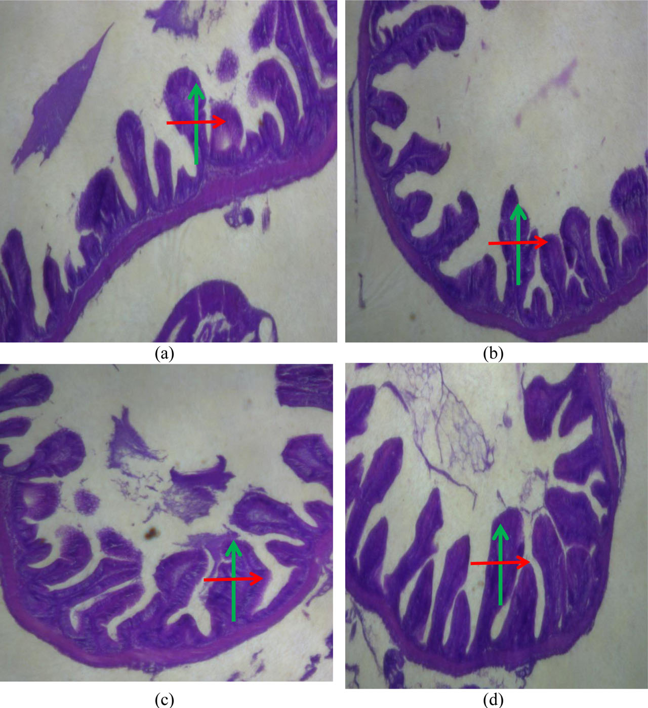
Figure 1.
Gut morphology of Nile tilapia, Oreochromis niloticus, fed different levels of dietary HMLE for 8 weeks1. (a) Gut morphology of fish fed the control (0) diet, (b) 2 g/kg, (c) 4 g/kg, and (d) 6 g/kg of HMLE-based diet. Arrow (green colour) = villus length, arrow (red colour) = villus width (HE ×40, 100) (n=3).
1HMLE = T. procumbens, M. scaber, M. pruriens, and C. papaya leaves extract
Post-parasitic infestation mortality of Nile tilapia fed HMLE-based diets
Figure 2 depicts the post-parasitic infestation fish mortality (P<0.05). There was significant variation (P<0.05) in the post-parasitic infestation in Nile tilapia fed with fortified diets. The maximum fish mortality (54.6%) was recorded in fish fed the control diet while the minimum (3.0%) was detected in fish treated with 6 HMLE g kg−1 (Figure 2).
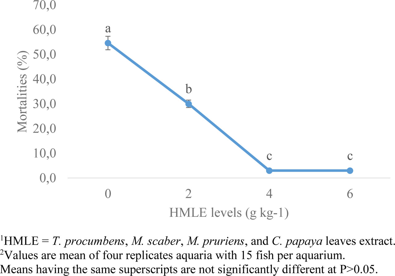
Figure 2.
Post-infestation mortality of Nile tilapia fed with diets containing various levels of HMLE1 for 8 weeks2 (n=3)
Discussion
The enhanced growth performance observed in Nile tilapia fed with different levels of HMLE-based diets underscores the potential of HMLE as a growth-promoting feed additive. Significant high FBW, BWG, DBWG, and SGR was witnessed in HMLE-fed fish, when compared to the control group, suggesting HMLE supplementation may improve nutrient assimilation, utilization, and metabolic efficiency. In similar studies, improved growth was reported in African catfish, Clarias gariepinus (Fawole et al., 2022), Japanese seabass, Lateolabrax japonicus (Wang et al., 2018), and European eel, Anguilla anguilla (Huang et al., 2020) fed diets supplemented with herbal medicine mixture. Also, diet containing 5 g kg−1 of oak acorn, coriander, and common mallow mixture extract fed to common carp (Raissy et al., 2022) and 5 g kg−1 Cnidium officinale, Massa medicata fermentata, Crataegi fructus, and Artemisia capillaries fed to olive flounder, Paralichthys olivaceus, resulted in great growth performance. The increase in FI and lower FCR in the HMLE-fed groups suggests improved palatability and feed efficiency, possibly due to the bioactive compounds in HMLE, which may enhance digestive enzyme activities and nutrient absorption. The increase in FI, FCR and consequently growth recorded in this study is not unconnected to the presence of aromatic compounds in HMLE especially p-cymene, quercetin, and chlorogenic acids which have been identified as an appetite stimulant, palatability and nutrients absorption enhancer (Balahbib et al., 2021). It is also suggested that biologically active compounds present in HMLE do not antagonize each other but rather work in synergy to improve the growth of Nile tilapia. The lack of significant differences in SR might indicate that HMLE is not toxic and has no ill effects on the overall health and wellbeing of the fish, reinforcing its potential as a safe dietary additive for Nile tilapia aquaculture. The study aligns with data reported by Ji et al. (2007) who demonstrated that dietary herbal mixture did not affect the survival of olive flounder.
Earlier study reported the use of blood to explain the pathophysiological condition of fish as well as nutritional status and health in response to feed supplements (Fawole et al., 2022). In the current study, haematological analysis revealed a noteworthy elevation in key haematological parameters, such as PVC, Hb, RBCs, and PLT in a dose-dependent manner in Nile tilapia nourished with HMLE-based diets while WBCs were not greatly affected. An increased PCV indicates a higher concentration of red blood cells, which enhances the fish’s ability to transport oxygen throughout its body (Oyelese et al., 1999). This is particularly beneficial for fish in environments especially when the oxygen levels are low and/or during activities that require more oxygen, thus helps in coping with temperature fluctuations. Further, high PCV can be associated with robust fish health, better growth performance, and an enhanced immune response, making fish more resistance to diseases. Similarly, Hb is an important oxygen carrier and therefore an increase in Hb level enhances the fish ability to transport oxygen adequately, metabolic efficiency, and consequently stress resistance. In the same vein, RBCs are responsible for transporting oxygen, so an increase in their count amplifies the fish’s ability to meet oxygen demands and recover quickly from stressors including infections. WBCs on the other hand, hold a critical role in combating pathogenic threats. So, an increase in WBCs indicates a robust immune system capable of fighting infections. No significant variations in the WBCs level presented in the current study showed that the fish are not exposed to pathogens. Platelets are involved in clot formation. An increase ensures efficient blood clotting and rapid wound healing. The increase in the PCV, Hb, RBCs and PLT counts suggests improved haematopoiesis and enhanced oxygen-carrying capacity, which could support better metabolic activity and overall physiological functions. Similar results were reported in common carp (Raissy et al., 2022) and African catfish (Fawole et al., 2022) when fed dietary-herbal mixtures. The rise in LYM and BAS with a concurrent decrease in HET and MON further indicates an improved immune status and a potential reduction in stress-related inflammation in HMLE-fed fish.
Protein metabolism enzymes like ALP, AST, and ALT, are commonly used to assess liver health issues because they are produced as a result of a malfunctioning liver (Abarike et al., 2020). ALP catalyses the hydrolysis of phosphate esters and participates in phosphorus metabolism. AST and ALT facilitate the transmission of an amino group from aspartate to α-ketoglutarate for the formation of glutamate and oxaloacetate. In this study, reduced activities of liver enzymes (alkaline phosphatase, aspartate aminotransferase, and alanine aminotransferase), especially in the group fed 6 g HMLE kg−1, point to potential hepatoprotective effects of HMLE, suggesting that it may reduce hepatic stress and promote liver health. The reduction in their levels suggests that the liver is not undergoing significant damage or stress and thus reflects normal physiological conditions. Fawole et al. (2022) reported that African catfish fed dietary polyherbal mixture had reduced liver enzymes activities and that it had no hepatotoxic effect on fish.
Antioxidant enzymes such as CAT, SOD, and MDA are involved in enzymatic mechanisms and their levels indicate the oxidative stress levels in fish because they possess a vital part in maintaining the normal redox homeostasis and neutralizing reactive oxygen species (ROS) in biological systems (Abdel-Tawwab and Wafeek, 2017; Livingstone, 2001). CAT and SOD are antioxidant enzymes that convert superoxide radicals (O2−), and hydrogen peroxide (H2O2) into water and oxygen and defend cells against the damage caused by ROS. The results of this study demonstrate that HMLE supplementation significantly enhances antioxidant defences, as indicated by increased levels of CAT, SOD, RBA, and LYZ. These enzymes are imperative for neutralizing reactive oxygen species, thereby protecting fish from oxidative stress and improving overall cellular health. The increased values of CAT and SOD may indicate enhanced ability to neutralize oxidative stress and suggest improved capacity to mitigate oxidative damage at the cellular level. MDA, on the other hand, is a byproduct of lipid peroxidation, which occurs when reactive oxygen species damage the polyunsaturated fatty acids in cell membranes.
In this study, MDA level was reduced. The reduction in malondialdehyde (MDA), a marker of lipid peroxidation, supports the antioxidant efficacy of HMLE. The lower MDA in fish fed dietary HMLE levels indicated that the fish’s antioxidant defences are promoted and are effectively neutralizing ROS, thereby preventing oxidative damage which could stimulate resilience against stress and diseases (Magnadóttir, 2006; Secombes and Wang, 2012). In this study, the elevated immune responses, as shown by increased RBA and LYZ activities, particularly at the 6 g HMLE kg−1 diet level, suggest that HMLE fortification enhances the fish’s immune defences against pathogens, likely by modulating innate immune pathways. Previous researches had illustrated that LYZ and RBA status in fishes showed significant increase when they were supplemented with plants (powder/extracts) based diet (Abdel-Latif et al., 2020; Abdel-Tawwab et al., 2020, 2018; Abdel-Tawwab and El-Araby, 2021; Adeniyi et al., 2024; Adeshina et al., 2020, 2019 b; Elumalai et al., 2020). The higher values of RBA and LYZ indicate an active and robust innate immune response and enhance the ability to eliminate pathogens quickly and effectively. The LYZ is an antimicrobial enzyme that destroys the cell wall of bacterial cell by degrading the β-1,4-glycosidic bond in peptidoglycan and therefore reflects a heightened antiparasitic defence mechanism.
The significant increases in histomorphometric parameters such as VH, VW, and AA in HMLE-fed fish reveal that HMLE promotes intestinal health and morphological integrity. Enhanced intestinal structures contribute to better nutrient absorption and overall growth performance. The improvements in intestinal histomorphometry suggest that HMLE may foster gut health, possibly through its bioactive components that support epithelial integrity and enhance nutrient transport efficiency. These results advocated that the increase in villi height and villi width leads to increase in absorption area, which increased absorption of nutrients. Several recent researches have shown that plants extract in diet greatly modified the morphometry of fish intestines (Abdel-Latif et al., 2020; Abdel-Tawwab et al., 2020, 2018; Adeshina et al., 2020, 2019 a).
The reduction in post-parasitic infestation mortality in HMLE-fed groups, with the least mortality rate detected in the group receiving 6 g HMLE kg kg−1 diet, underscores the protective effects of HMLE against parasitic threats. This outcome aligns with the observed enhancements in immune responses, suggesting that HMLE fortification may bolster the resilience of Nile tilapia against parasitic infections. The findings indicate that HMLE supplementation not only improves growth and health metrics but also contributes to reduced vulnerability to parasitic mortality, offering promising implications for disease management in aquaculture. In similar studies, grass carp fed 4 mg L−1 herbal mixture extract was protected against I. multifiliis infestation up to 71.1% (Fu et al., 2021; Liu et al., 2017). The variation in the post-infestation results was due to different combination of the herbs. When each plant material of HMLE was used separately, 6 g kg−1 of T. procumbens leaves extract reduced mortalities to 10.0% in African catfish exposed to D. vastator infestation (Adeshina et al., 2021). Also, 6 g kg−1 M. scaber leaves extract decreased the post-G. malalai infestation to 5.0% (Adeshina et al., 2021 b), 200 g kg−1 of M. pruriens extract resulted in 90% mortality reduction in gold fish against I. multifiliis (Ekanem et al., 2004), and 200 g kg−1 of C. papaya extracts induced protection up to 90% post I. multifiliis infestation in gold fish (Ekanem et al., 2004). As against single herbal extract where the dosage is high, herbal mixture has appeared to be a promising agent as lower dosage caused better protection against parasites. This further suggests the bioavailability of the active compounds present in HMLE and a good synergistic interaction, hence resulting in better protection and lower inclusion level.
Conclusion
This study demonstrated that a herbal mixture of leaf extracts (Tridax procumbens, Mitracarpus scaber, Mucuna pruriens, and Carica papaya) enhanced growth performance and the haemato-biochemical profile of Nile tilapia. Additionally, dietary HMLE exhibited anti-oxidative and immunostimulatory effects on Nile tilapia juveniles. Notably, incorporating HMLE at optimal level of 4.0 g per kg of diet enhanced the resistance to Dactylogyrus vastator infestation in Nile tilapia. However, future investigations should further explore long-term impacts of the herbal mixture of the leaves extracts and explore optimum limit.
Acknowledgments
The authors would like to extend their sincere appreciation to the Ongoing Research Funding Program, (ORF-2025-144), King Saud University, Riyadh, Saudi Arabia for their supports.
Notes
[36] Ethics approval
This project was submitted to the Animal Use Ethics Committee of the University of Ilorin, Ilorin, Nigeria, and was approved under UIL/2023/022.
[37] Data and model availability statement
None of the data was deposited in an official repository. Information can be made available from the authors upon request.
[38] CRediT authorship contribution statement
Bilal Ahamad Paray: Conceptualization, Methodology, Formal Analysis, Funding acquisition; Eijaz Ahmed Bhat: Original Draft Writing, Review & Editing; Sahih Sherzada: Project Administration; Olarinke V. Adeniyi: Original Draft Writing, Review & Editing; Ibrahim Adeshina: Investigation, Validation, Data curation, Software, Formal analysis, Original Draft Writing, Review & Editing; Supervision, Funding acquisition.