
Figure 1.
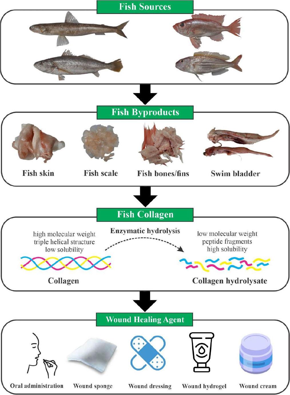
Figure 2.
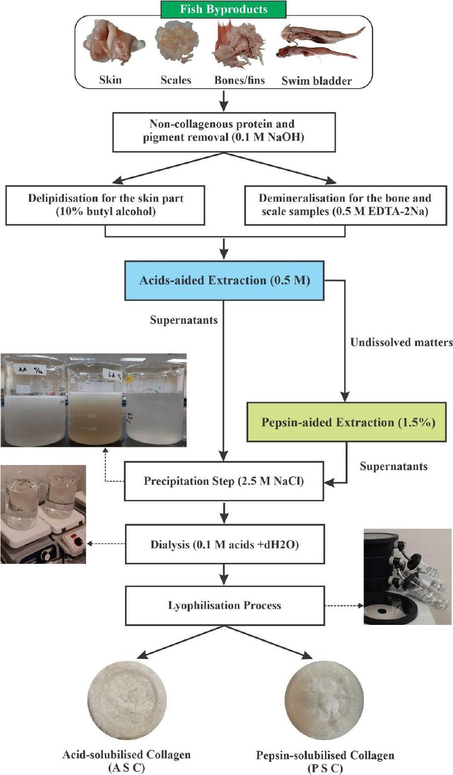
Figure 3.
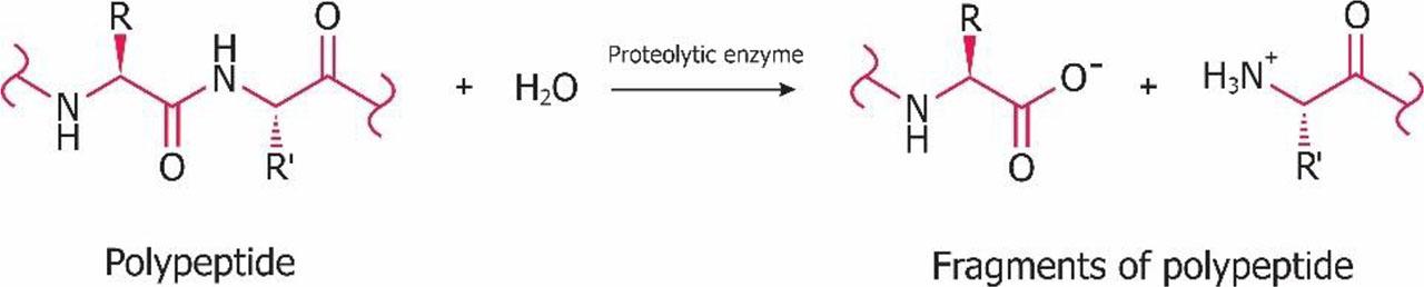
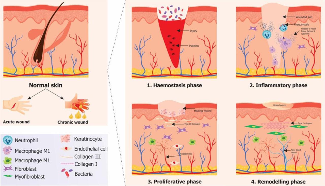
Figure 4.
Extraction, yield, type, solubility, and colour of collagens derived from tropical marine fish sources
| Source | Portion | Extraction | Time | Yield | SDS-PAGE data | Solubility | Colour attributes | Reference | |||||
|---|---|---|---|---|---|---|---|---|---|---|---|---|---|
| α1 (kDa) | α2 (kDa) | Type | NaCl | pH | *L | *a | *b | ||||||
| 1 | 2 | 3 | 4 | 5 | 6 | 7 | 8 | 9 | 10 | 11 | 12 | 13 | 14 |
| Threadfin bream | Scale + fin | ASC-Ca | 12 h | 22% | ND | ND | ND | ND | ND | 93.7 | –1.84 | 13.44 | (Normah and Afiqah, 2018) |
| (Nemipterus japonicus) | Scale + fin | ASC-C | 12 h | 8.30% | ND | ND | ND | ND | ND | 94.82 | 0.31 | 0.2 | |
| Spotted golden goatfish | Scale | ASC | 48 h | 0.46% | 117 | 108 | I | 0–20 g/L | 2–4 | ND | ND | ND | (Matmaroh et al., 2011) |
| (Parupeneus heptacanthus) | Scale | PSC | 48 h | 1.20% | 117 | 108 | I | 0–30 g/L | 2–4 | ND | ND | ND | |
| Bigeye snapper | Skin | ASC-A | 72 h | 5.79% | 118 | 106 | I | 0–3% | 1–5 | ND | ND | ND | (Oslan et al., 2022) |
| (Priacanthus tayenus) | Skin | ASC-L | 72 h | 3.19% | 118 | 106 | I | 0–3% | 1–5 | ND | ND | ND | |
| Skin | ASC-C | 72 h | 4.15% | 118 | 106 | I | 0–3% | 1–5 | ND | ND | ND | ||
| Skin | PSC | 48 h | 6.65% | 118 | 106 | I | 0–3% | 1–5 | ND | ND | ND | ||
| Barracuda | Skin | ASC-A | 72 h | 6.77% | 143.2 | 136.6 | I | 0–20 g/L | 1–5 | 78.54 | −0.05 | 0.64 | (Matarsim et al., 2023) |
| (Sphyraena sp.) | Skin | ASC-L | 72 h | 10.06% | 143.2 | 136.6 | I | 0–20 g/L | 1–3 | 56.88 | 0.6 | 5.83 | |
| Skin | ASC-C | 72 h | 8.35% | 143.2 | 136.6 | I | 0–20 g/L | 1–3 | 54.34 | 0.66 | 3.78 | ||
| Unicorn fish | Bone | ASC-A | 72 h | 0.40% | 138 | 118.3 | I | 0–10 g/L | 1–5 | 81.44 | −0.19 | 0.79 | (Fatiroi et al., 2023) |
| (Naso reticulatus) | Bone | ASC-L | 72 h | 1.08% | 138 | 118.3 | I | 0–20 g/L | 1–5 | 82.55 | 0.4 | 6.51 | |
| Bone | ASC-C | 72 h | 1.36% | 138 | 118.3 | I | 0–10 g/L | 1–3 | 79.35 | 0.04 | 3.26 | ||
| Sin croaker (Johniecop sina) | Bone + scale | ASC | 72 h | 2.74% | 120 | 100 | I | 0–2% | 1–2 | ND | ND | ND | (Normah and Afiqah, 2018) |
| Miiuy croaker | Swim bladder | ASC | 60 h | 1.33% | ND | ND | I | 0–2% | 1–4 | ND | ND | ND | (Li et al., 2018) |
| (Miichthys miiuy) | Swim bladder | PSC | 48 h | 8.37% | ND | ND | I | 0–2% | 1–4 | ND | ND | ND | |
| Chu’s croaker (Nibea coibor) | Swim bladder | ASC | 48 h | 7.33% | 130 | 110 | I | 0–3% | 1–5 | ND | ND | ND | (Xiao et al., 2023) |
| Blackspotted croaker | Swim bladder | ASC | 48 h | 7.15% | 130 | 110 | I | 0–2% | 1–5 | ND | ND | ND | (Xiao et al., 2023) |
| (Protonibea diacanthus) | |||||||||||||
| Barramundi | Skin | ASC | 48 h | 8.12% | ND | ND | I | 0–2% | 1–5 | 65.41 | 0.14 | 3.16 | (Bakar et al., 2013) |
| (Lates calcarifer) | Skin | PSC | 24 h | 43.63% | ND | ND | I | 0–2% | 1–5 | 61.33 | 2.59 | 5.35 | |
| Seabass | Scale | ASC | 48 h | 0.38% | 118 | 105 | I | ND | 1–4 | ND | ND | ND | (Chuaychan et al., 2015) |
| (Lates calcarifer) | Scale | PSC | 48 h | 1.06% | 118 | 105 | I | ND | 1–4 | ND | ND | ND | |
| Seabass | Skin | ASC | 48 h | 8.32% | 135.8 | 122.5 | I | ND | ND | 82.27 | 4.4 | 1.9 | (Razali et al., 2023) |
| (Lates calcarifer) | Skin | UAE | 48.3 h | 56.61% | 139.5 | 127 | I | ND | ND | 72.45 | 5.75 | 7.39 | |
| Emperor fish (Lethrinus lentjan) | Skin | ASC | 48 h | 7.70% | 176 | 150 | I | ND | ND | ND | ND | ND | (Firdayanti et al., 2023) |
| Parrotfish | Scale | ASC | 48 h | 1.17% | 118.1 | 107.7 | I | 0–20 g/L | 1–5 | 61.74 | 2.61 | 6.15 | (Jaziri et al., 2023) |
| (Scarus sordidus) | Scale | PSC | 48 h | 1.00% | 118.1 | 107.7 | I | 0–30 g/L | 1–5 | 74.81 | 1.09 | 6.14 | |
| Grouper (Epinephelus sp.) | Swim bladder | PSC | 48 h | 18.16% | 133 | 123 | I | ND | ND | ND | ND | ND | (Dong and Dai, 2022) |
| Golden pompano | Skin | ASC | 48 h | 21.81% | 105 | 120 | I | 0–30 g/L | 1–4 | ND | ND | ND | (Cao et al., 2019) |
| (Trachinotus ovatus) | Bone | PSC | 48 h | 1.25% | 105 | 120 | I | 0–30 g/L | 1–4 | ND | ND | ND | |
| Shortfin scad | Bone + skin | ASC | 24 h | 3.35% | ND | ND | I | ND | 1–10 | ND | ND | ND | (Sulaiman and Sarbon, 2020) |
| (Decapterus macrosoma) | Bone + skin | PSC | 30 h | 0.10% | ND | ND | I | ND | 1–3 | ND | ND | ND | |
| Mahi mahi | Skin | ASC | ND | 5.90% | 120 | 110 | I | ND | ND | ND | ND | ND | (Akita et al., 2019) |
| (Coryphaena hippurus) | Skin | PSC | ND | 4.00% | 120 | 110 | I | ND | ND | ND | ND | ND | |
| Fringescale sardinella | Scale | ASC | 24 h | 7.48% | ND | ND | I | ND | 1–5 | ND | ND | ND | (Hamdan and Sarbon, 2019) |
| (Sardinella fimbriata) | Scale | PSC | 30 h | 0.94% | ND | ND | I | ND | 1–6 | ND | ND | ND | |
| Horse mackerel | Scale | ASC | 96 h | 0.64% | ND | ND | I | 0–0.4 M | 1–5 | ND | ND | ND | (Minh et al., 2014) |
| (Trachurus japonicus) | |||||||||||||
| Grey mullet (Mugil cephalis) | Scale | ASC | 96 h | 0.43% | ND | ND | I | 0–0.4 M | 1–5 | ND | ND | ND | (Minh et al., 2014) |
| Flying fish | Scale | ASC | 96 h | 0.72% | ND | ND | I | 0–0.4 M | 1–5 | ND | ND | ND | (Minh et al., 2014) |
| (Cypselurus melanurus) | |||||||||||||
| Yellowback seabream | Scale | ASC | 96 h | 0.90% | ND | ND | I | 0–0.4 M | 1–5 | ND | ND | ND | (Minh et al., 2014) |
| (Dentex tumifrons) | |||||||||||||
| Needle fish | Skin | ASC-A | 72 h | 3.13% | 130 | 100 | I | 0–10 g/L | 1–3 | 69.77 | 71.89 | 57.14 | (Ramle et al., 2022) |
| (Tylosurus melanotus) | Skin | ASC-L | 72 h | 0.56% | 130 | 100 | I | 0–10 g/L | 1–3 | 0.15 | 0.92 | 0.73 | |
| Skin | ASC-C | 72 h | 1.03% | 130 | 100 | I | 0–10 g/L | 1–5 | 5.52 | 5.2 | 4.69 | ||
| Mackerel | Bone | PSC | 72 h | 1.75% | 123 | 116 | I | ND | ND | ND | ND | ND | (Asaduzzaman et al., 2020) |
| (Scomber japonicus) | Skin | PSC | 72 h | 8.10% | 123 | 116 | I | ND | ND | ND | ND | ND | |
| Sharpnose stingray | Skin | ASC | 24 h | 20.48% | ND | ND | I | ND | 1–6 | ND | ND | ND | (Ong et al., 2021) |
| (Dasyatis zugei) | Skin | PSC | 30 h | 34.84% | ND | ND | I | ND | 1–5 | ND | ND | ND | |
| Sharpnose stingray | Skin | ASC+UEA | 1 h | 42.34% | ND | 100 | I | ND | 1–6 | ND | ND | ND | (Shaik et al., 2021) |
| (Dasyatis zugei) | Skin | PSC+UEA | 1 h | 61.50% | ND | 95 | I | ND | 1–5 | ND | ND | ND | |
| Silvertip shark | Skeletal | PSC | 96 h | ND | ND | ND | II | 0–1% | 3–6 | ND | ND | ND | (Jeevithan et al., 2014) |
| (C. albimarginatus) | Head bone | PSC | 96 h | ND | ND | ND | II | 0–1% | 3–6 | ND | ND | ND | |
Optimisation methods for collagen derived from fish byproducts
| Source | Fish portion | Optimisation procedure | Yield | Characterization remarks | References |
|---|---|---|---|---|---|
| Milkfish | Scale | CCD with the optimal conditions: extraction time (X1) = 61.30 h; AcOH concentration (X2) = 0.66 M. | 0.73% | FTIR, DSC and analysis amino acid show that milkfish scale collagen in this study resembles that of commercial collagen | (Isnainita and Bambang, 2018) |
| Small-spotted catshark | Skin | CCD with the optimal conditions: temperature (X1) = 25°C; extraction time (X2) = 34.2 h; AcOH concentration (X3) = 1.0 M. | 61.24% | It contained two identical α1 chains (120 kDa) and one α2 chain (110 kDa) in the molecular form of [α1(I)]2 α2(I)], | (Blanco et al., 2019) |
| Yellowfin tuna | Dorsal skin | CCD with the optimal conditions: NaOH concentration (X1) = 0.92 N; NaOH treatment time (X2) = 24 h; pepsin concentration (X3) = 0.98% (w/v); hydrolysis time (X4) = 23.5 h. | 27.10% | The obtained type I PSC had a 20.5% imino acid and the optimal extraction process did not affect the helical structure of collagen. | (Woo et al., 2008) |
| Sole fish | Skin | Initiated with OVAT, and followed by BBD with the optimal conditions: AcOH concentration (X1) = 0.54 M; salt concentration (X2) = 1.90 M; solvent/solid ratio (X3) = 8.97 mL/g; extraction time (X4) = 32.3 h. | 19.27% | Extracted collagen was categorised as type I and IR analysis showed the existence of helical arrangements of collagen | (Arumugam et al., 2018) |
| Cuttlefish | Skin | BBD with the optimal conditions: pH value (X1) = 1.5; solid-liquid ratio (X2) = 20 mL/g; pepsin concentration (X3) = 15 U/mg | 8.79% | Considered as type I, and it confirmed the presence of collagen fibrils in the cuttlefish skin | (Hou et al., 2022) |
| Giant croaker | Skin | Initiated with OVAT, and followed by BBD with the optimal conditions: pepsin concentration (X1) = 1389 U/g; solid-liquid ratio (X2) = 1:57; extraction time (X4) = 8.67 h. | 84.85% | Characterized as type I collagen, its triple helical structure was maintained, and had a high solubility at pH 1.0–4.0 | (Yu et al., 2018) |
| Nile tilapia | Skin | Two-level 23 factorial experimental with the optimal conditions: AcOH = 0.35 M; temperature: 20°C; extraction time: 65 h. | 19.00% | The triple helical structure of ASC from tilapia skin collagen did not change after being confirmed by FTIR. | (Menezes et al., 2020) |
| Sea eel | Swim bladder | Initiated with OVAT and followed by BBD with the optimal conditions: pepsin concentration (X1) = 2067 U/g; solid-liquid ratio (X2) = 1:83; extraction time (X4) = 10 h. | 93.76% | Classified as type I and it displayed a fibrous structure under electron microscopy | (Li et al., 2023) |
| Grass carp | Swim bladder | CCD with the optimal conditions: liquid-solid ratio (X1) = 17.85; AcOH concentration (X2) = 0.54 M; extraction time (X3) = 34 h. | 8.21% | – | (Zhang et al., 2010) |
FTIR spectra peak area of collagen derived from tropical marine fish byproducts
| Source | Byproduct | Method | FTIR | References | ||||||
|---|---|---|---|---|---|---|---|---|---|---|
| Amide A | Amide B | Amide I | Amide II | Amide III | Δv | A/T | ||||
| 1 | 2 | 3 | 4 | 5 | 6 | 7 | 8 | 9 | 10 | 11 |
| Spotted golden goatfish | Scale | ASC | 3296 cm−1 | 3081 cm−1 | 1646 cm−1 | 1549 cm−1 | 1237 cm−1 | 97 | 0.9 | (Matmaroh et al., 2011) |
| (Parupeneus heptacanthus) | Scale | PSC | 3296 cm−1 | 3081 cm−1 | 1631 cm−1 | 1536 cm−1 | 1234 cm−1 | 95 | 0.9 | |
| Bigeye snapper | Skin | ASC-A | 3427 cm−1 | 2926 cm−1 | 1638 cm−1 | 1560 cm−1 | 1242 cm−1 | 78 | 0.9 | (Oslan et al., 2022) |
| (Priacanthus tayenus) | Skin | ASC-L | 3435 cm−1 | 2928 cm−1 | 1643 cm−1 | 1563 cm−1 | 1240 cm−1 | 80 | 0.9 | |
| Skin | ASC-C | 3439 cm−1 | 2927 cm−1 | 1642 cm−1 | 1564 cm−1 | 1240 cm−1 | 78 | 0.9 | ||
| Skin | PSC | 3401 cm−1 | 2933 cm−1 | 1649 cm−1 | 1555 cm−1 | 1240 cm−1 | 94 | 0.9 | ||
| Barracuda (Sphyraena sp.) | Skin | ASC-A | 3278 cm−1 | 2921 cm−1 | 1629 cm−1 | 1541 cm−1 | 1234 cm−1 | 87.6 | 0.9 | (Matarsim et al., 2023) |
| Skin | ASC-L | 3278 cm−1 | 2920 cm−1 | 1629 cm−1 | 1541 cm−1 | 1237 cm−1 | 87.6 | 0.9 | ||
| Skin | ASC-C | 3283 cm−1 | 2921 cm−1 | 1629 cm−1 | 1541 cm−1 | 1235 cm−1 | 87.6 | 0.9 | ||
| Bigeye tuna | Skin | ASC | 3301 cm−1 | 2927 cm−1 | 1639 cm−1 | 1546 cm−1 | 1240 cm−1 | 93 | 0.9 | (Ahmed et al., 2020) |
| (Thunnus obesus) | Skin | PSC | 3299 cm−1 | 2931 cm−1 | 1639 cm−1 | 1546 cm−1 | 1240 cm−1 | 93 | 0.9 | |
| Scale | PSC | 3298 cm−1 | 2926 cm−1 | 1639 cm−1 | 1546 cm−1 | 1239 cm−1 | 93 | 0.9 | ||
| Bones | PSC | 3297 cm−1 | 2926 cm−1 | 1639 cm−1 | 1545 cm−1 | 1239 cm−1 | 94 | 0.9 | ||
| Skipjack tuna | Skull | ASC | 3397 cm−1 | 2926 cm−1 | 1645 cm−1 | 1548 cm−1 | 1240 cm−1 | 97 | 0.9 | (Ding et al., 2019) |
| (Katsuwonus pelamis) | Skull | PSC | 3411 cm−1 | 2926 cm−1 | 1636 cm−1 | 1548 cm−1 | 1240 cm−1 | 88 | 0.9 | |
| Spine | ASC | 3397 cm−1 | 2926 cm−1 | 1645 cm−1 | 1548 cm−1 | 1240 cm−1 | 97 | 0.9 | ||
| Spine | PSC | 3411 cm−1 | 2926 cm−1 | 1638 cm−1 | 1548 cm−1 | 1240 cm−1 | 90 | 0.9 | ||
| Starry triggerfish | Skin | ASC | 3416 cm−1 | 2911 cm−1 | 1634 cm−1 | 1545 cm−1 | 1237 cm−1 | 88.7 | 0.9 | (Ahmad et al., 2016) |
| (Abalistes stellatus) | Skin | PSC | 3433 cm−1 | 2909 cm−1 | 1636 cm−1 | 1547 cm−1 | 1237 cm−1 | 89.2 | 0.9 | |
| Unicorn leatherjacket | Skin | PSC | 3294 cm−1 | 3086 cm−1 | 1635 cm−1 | 1546 cm−1 | 1236 cm−1 | 88.5 | 0.9 | (Ahmad and Benjakul, 2010) |
| (Aluterus monocerous) | Skin | PSC-A | 3293 cm−1 | 3080 cm−1 | 1632 cm−1 | 1547 cm−1 | 1235 cm−1 | 85.1 | 0.9 | |
| Skin | PSC-Y | 3294 cm−1 | 3080 cm−1 | 1640 cm−1 | 1545 cm−1 | 1235 cm−1 | 94.4 | 0.9 | ||
| Unicorn fish | Bone | ASC-A | 3308 cm−1 | 2920 cm−1 | 1639 cm−1 | 1543 cm−1 | 1238 cm−1 | 95 | 0.9 | (Fatiroi et al., 2023) |
| (Naso reticulatus) | Bone | ASC-L | 3278 cm−1 | 2924 cm−1 | 1638 cm−1 | 1545 cm−1 | 1238 cm−1 | 93.2 | 0.9 | |
| Bone | ASC-C | 3278 cm−1 | 2924 cm−1 | 1618 cm−1 | 1542 cm−1 | 1236 cm−1 | 75.4 | 0.9 | ||
| Mackerel | Bone | PSC | 3283 cm−1 | 2922 cm−1 | 1650 cm−1 | 1537 cm−1 | 1237 cm−1 | 113 | 0.9 | (Asaduzzaman et al., 2020) |
| (Scomber japonicus) | Skin | PSC | 3285 cm−1 | 2922 cm−1 | 1651 cm−1 | 1548 cm−1 | 1238 cm−1 | 103 | 0.9 | |
| Giant croaker | Swim bladder | ASC | 3419 cm−1 | 2926 cm−1 | 1656 cm−1 | 1555 cm−1 | 1240 cm−1 | 101 | 0.9 | (Chen et al., 2019) |
| (Nibea japonica) | Swim bladder | PSC | 3443 cm−1 | 2927 cm−1 | 1654 cm−1 | 1556 cm−1 | 1240 cm−1 | 98.2 | 0.9 | |
| Miiuy croaker | Swim bladder | ASC | 3325 cm−1 | 2938 cm−1 | 1653 cm−1 | 1543 cm−1 | 1241 cm−1 | 109.8 | 0.9 | (Li et al., 2018) |
| (Miichthys miiuy) | Swim bladder | PSC | 3362 cm−1 | 2932 cm−1 | 1655 cm−1 | 1548 cm−1 | 1243 cm−1 | 107 | 0.9 | |
| Seabass | Scale | ASC | 3285 cm−1 | 3075 cm−1 | 1657 cm−1 | 1553 cm−1 | 1456 cm−1 | 104 | 1.0 | (Chuaychan et al., 2015) |
| (Lates calcarifer) | Scale | PSC | 3311 cm−1 | 3086 cm−1 | 1650 cm−1 | 1548 cm−1 | 1459 cm−1 | 102 | 1.0 | |
| Seabass | Skin | ASC | 3292 cm−1 | 2922 cm−1 | 1634 cm−1 | 1548 cm−1 | 1238 cm−1 | 86 | 0.9 | (Razali et al., 2023) |
| (Lates calcarifer) | Skin | UAE | 3307 cm−1 | 2923 cm−1 | 1651 cm−1 | 1548 cm−1 | 1235 cm−1 | 103 | 0.9 | |
| Seabass (Lates calcarifer) | Skin | PSC | 3379 cm−1 | 2931 cm−1 | 1657 cm−1 | 1553 cm−1 | 1241 cm−1 | 104.5 | 0.9 | (Liao et al., 2018) |
| Emperor fish (Lethrinus lentjan) | Skin | ASC | 3367 cm−1 | 2916 cm−1 | 1635 cm−1 | 1555 cm−1 | 1385 cm−1 | 80 | 1.0 | (Firdayanti et al., 2023) |
| Parrotfish (Scarus sordidus) | Scale | ASC | 3287 cm−1 | 2428 cm−1 | 1636 cm−1 | 1541 cm−1 | 1235 cm−1 | 95 | 0.9 | (Jaziri et al., 2023) |
| Scale | PSC | 3298 cm−1 | 2927 cm−1 | 1636 cm−1 | 1541 cm−1 | 1235 cm−1 | 95 | 0.9 | ||
| Grouper (Epinephelus sp.) | Swim bladder | PSC | 3336 cm−1 | 2923 cm−1 | 1647 cm−1 | 1548 cm−1 | 1238 cm−1 | 98.8 | 0.9 | (Dong and Dai, 2022 a) |
| Golden pompano | Skin | ASC | 3421 cm−1 | 2932 cm−1 | 1653 cm−1 | 1542 cm−1 | 1240 cm−1 | 110.5 | 0.9 | (Cao et al., 2019 a) |
| (Trachinotus ovatus) | Bone | PSC | 3423 cm−1 | 2926 cm−1 | 1656 cm−1 | 1548 cm−1 | 1241 cm−1 | 107.3 | 0.9 | |
| Puffer fish | Skin | ASC | ND | ND | 1640 cm−1 | 1546 cm−1 | 1247 cm−1 | 94.2 | 0.9 | (Iswariya et al., 2018) |
| (Lagocephalus inermis) | Skin | PSC | ND | ND | 1640 cm−1 | 1546 cm−1 | 1247 cm−1 | 94.2 | 0.9 | |
| Shortfin scad | Bone + skin | ASC | 3456 cm−1 | ND | 1637 cm−1 | 1590 cm−1 | 1262 cm−1 | 46.9 | 0.9 | (Sulaiman and Sarbon, 2020) |
| (Decapterus macrosoma) | Bone + skin | PSC | 3449 cm−1 | 2934 cm−1 | 1636 cm−1 | 1560 cm−1 | 1264 cm−1 | 76.4 | 0.9 | |
| Mahi mahi | Skin | ASC | 3325 cm−1 | 3083 cm−1 | 1654 cm−1 | 1543 cm−1 | 1240 cm−1 | 111 | 0.9 | (Akita et al., 2019) |
| (Coryphaena hippurus) | Skin | PSC | 3326 cm−1 | 3078 cm−1 | 1656 cm−1 | 1534 cm−1 | 1235 cm−1 | 122 | 0.9 | |
| Fringescale sardinella | Scale | ASC | 3417 cm−1 | ND | ND | 1590 cm−1 | 1414 cm−1 | ND | 1.0 | (Hamdan and Sarbon, 2019) |
| (Sardinella fimbriata) | Scale | PSC | 3424 cm−1 | ND | ND | 1401 cm−1 | 1265 cm−1 | ND | 0.9 | |
| Needle fish | Skin | ASC-A | 3290 cm−1 | 2939 cm−1 | 1640 cm−1 | 1541 cm−1 | 1232 cm−1 | 98.8 | 0.8 | (Ramle et al., 2022) |
| (Tylosurus melanotus) | Skin | ASC-L | 3290 cm−1 | 2917 cm−1 | 1640 cm−1 | 1541 cm−1 | 1232 cm−1 | 98.8 | 0.8 | |
| Skin | ASC-C | 3290 cm−1 | 2920 cm−1 | 1640 cm−1 | 1541 cm−1 | 1236 cm−1 | 98.8 | 0.9 | ||
| Sailfish | Skin | ASC | 3423 cm−1 | 2928 cm−1 | 1654 cm−1 | 1560 cm−1 | 1240 cm−1 | 94 | 0.9 | (Tamilmozhi et al., 2013) |
| (Istiophorus platypterus) | Skin | PSC | 3337 cm−1 | 2924 cm−1 | 1646 cm−1 | 1549 cm−1 | 1240 cm−1 | 97 | 0.9 | |
| Cobia | Skin | ASC | 3384 cm−1 | 2927 cm−1 | ND | ND | ND | ND | ND | (Zeng et al., 2012) |
| (Rachycentron canadum) | Skin | PSC | 3333 cm−1 | 2924 cm−1 | ND | ND | ND | ND | ND | |
| Sharpnose stingray | Skin | ASC | 3424 cm−1 | ND | 1627 cm−1 | 1553 cm−1 | 1239 cm−1 | 73.9 | 0.9 | (Ong et al., 2021) |
| (Dasyatis zugei) | Skin | PSC | 3439 cm−1 | ND | 1627 cm−1 | 1553 cm−1 | 1239 cm−1 | 74.1 | 0.9 | |
| Sharpnose stingray | Skin | ASC+UEA | 3449 cm−1 | 2997 cm−1 | 1632 cm−1 | 1573 cm−1 | 1263 cm−1 | 59 | 0.9 | (Shaik et al., 2021) |
| (Dasyatis zugei) | Skin | PSC+UEA | 3449 cm−1 | 2998 cm−1 | 1637 cm−1 | 1579 cm−1 | 1262 cm−1 | 58 | 0.9 | |
| Silvertip shark | Skeletal | PSC | 3331 cm−1 | 2932 cm−1 | 1660 cm−1 | 1551 cm−1 | 1240 cm−1 | 109.1 | 0.9 | (Jeevithan et al., 2014) |
| (Carcharhinus albimarginatus) | Head bone | PSC | 3415 cm−1 | 2957 cm−1 | 1655 cm−1 | 1548 cm−1 | 1242 cm−1 | 107.4 | 0.9 | |
UV-vis, XRD, DSC and microstructural properties of collagen derived from tropical marine fish byproducts
| Source | Part | Method | UV-vis | XRD | DCS | Microstructural results | References | ||
|---|---|---|---|---|---|---|---|---|---|
| Peak A | Peak B | Tmax | ΔH | ||||||
| 1 | 2 | 3 | 4 | 5 | 6 | 7 | 8 | 9 | 10 |
| Spotted golden goatfish | Scale | ASC | ND | ND | ND | 41.58°C | ND | ND | (Matmaroh et al., 2011) |
| (P. heptacanthus) | Scale | PSC | ND | ND | ND | 41.01°C | ND | ND | |
| Bigeye snapper | Skin | PSC | ND | ND | ND | 31.3°C | ND | ND | (Benjakul et al., 2010) |
| Bigeye snapper | Skin | PSC | ND | ND | ND | 31.15°C | ND | ND | (Benjakul et al., 2010) |
| Bigeye snapper | Skin | ASC-A | ND | ND | ND | 31.4°C | ND | ND | (Oslan et al., 2022) |
| (Priacanthus tayenus) | Skin | ASC-L | ND | ND | ND | 31.7°C | ND | ND | |
| Skin | ASC-C | ND | ND | ND | 31.5°C | ND | ND | ||
| Skin | PSC | ND | ND | ND | 33.2°C | ND | ND | ||
| Brownstripe red snapper | Skin | ASC | ND | ND | ND | 31.52°C | ND | ND | (Jongjareonrak et al., 2005 a) |
| (Lutjanus vitta) | Skin | PSC | ND | ND | ND | 31.02°C | ND | ND | |
| Barracuda | Skin | ASC-A | 230.5 nm | 7.48° | 20.02° | 41.29°C | 0.13 J/g | A multi-layered form with the irregular sheet-like film connected by random-coiled filaments | (Matarsim et al., 2023) |
| (Sphyraena sp.) | Skin | ASC-L | 230.5 nm | 7.26° | 19.16° | 40.69°C | 0.08 J/g | ||
| Skin | ASC-C | 230.5 nm | 7.64° | 19.12° | 40.16°C | 0.05 J/g | |||
| Bigeye tuna | Skin | ASC | ND | ND | ND | 32.07°C | - | ND | (Ahmed et al., 2019 a) |
| (Thunnus obesus) | Skin | PSC | ND | ND | ND | 33.73°C | - | ND | |
| Scale | PSC | ND | ND | ND | 31.63°C | - | ND | ||
| Bones | PSC | ND | ND | ND | 32.26°C | - | ND | ||
| Skipjack tuna | Skull | ASC | 220 nm | ND | ND | ND | - | ND | (Ding et al., 2019) |
| (Katsuwonus pelamis) | Skull | PSC | 220 nm | ND | ND | ND | - | ND | |
| Spine | ASC | 220 nm | ND | ND | ND | - | ND | ||
| Spine | PSC | 220 nm | ND | ND | ND | - | ND | ||
| Starry triggerfish | Skin | ASC | ND | ND | ND | 35.9°C | 2.4 J/g | ND | (Ahmad et al., 2016) |
| (Abalistes stellatus) | Skin | PSC | ND | ND | ND | 33.6°C | 2.1 J/g | ND | |
| Puffer fish | Skin | ASC | 230 nm | ND | ND | ND | ND | Both ASC and PSC showed fibrous and porous structure under SEM | (Iswariya et al., 2018) |
| (Lagocephalus inermis) | Skin | PSC | 230 nm | ND | ND | ND | ND | ||
| Unicorn leatherjacket | Skin | PSC | ND | ND | ND | 31.98°C | 0.60 J/g | ND | (Ahmad and Benjakul, 2010) |
| (Aluterus monocerous) | Skin | PSC-A | ND | ND | ND | 31.73°C | 0.75 J/g | ND | |
| Skin | PSC-Y | ND | ND | ND | 31.68°C | 0.76 J/g | ND | ||
| Unicorn fish (Naso reticulatus) | Bone | ASC-A | 231.2 nm | 7.22° | 21.33° | 33.51°C | 3.9 mJ/g | The PSCs had an irregular and dense flake structure with coiled filaments | (Fatiroi et al., 2023) |
| Bone | ASC-L | 229.8 nm | 7.24° | 21.74° | 33.39°C | 7.7 mJ/g | |||
| Bone | ASC-C | 230.8 nm | 6.66° | 20.11° | 33.34°C | 5.7 mJ/g | |||
| Miiuy croaker (Miichthys miiuy) | Swim bladder | ASC | 226 nm | ND | ND | ND | ND | ND | (Li et al., 2018) |
| Swim bladder | PSC | 226 nm | ND | ND | ND | ND | ND | ||
| Chu’s croaker (Nibea coibor) | Swim bladder | ASC | 201–220 nm | ND | ND | 79.74°C | ND | The ASC was uniform and porous | (Xiao et al., 2023) |
| Giant croaker (Nibea japonica) | Skin | PSC | 230 nm | ND | ND | ND | ND | ND | (Yu et al., 2018) |
| Giant croaker (Nibea japonica) | Swim bladder | ASC | ND | ND | ND | ND | ND | All samples showed multi-layered, fibrous, and porous structure | (Chen et al., 2019) |
| Swim bladder | PSC | ND | ND | ND | ND | ND | |||
| Sin croaker (J. sina) | Bone | ASC | ND | ND | ND | 31.31°C | 0.05 J/g | ND | (Normah and Afiqah, 2018) |
| Blackspotted croaker | Swim bladder | ASC | 201–220 nm | ND | ND | 85.93°C | ND | The extracted collagen appeared to be an irregular dense sheet-like film | (Xiao et al., 2023) |
| Seabass | Skin | PSC | 230.3 nm | 7.05° | 20.30° | 109.6°C | ND | The collagen presented like soft white sponge with l irregular dense sheet-like film | (Liao et al., 2018) |
| Seabass | Scale | ASC | ND | ND | ND | 38.17°C | 0.72 J/g | ND | (Chuaychan et al., 2015) |
| (Lates calcarifer) | Scale | PSC | ND | ND | ND | 39.32°C | 0.91 J/g | ND | |
| Giant grouper | Skin | ASC | 215–230 nm | ND | ND | 31.71°C | ND | ND | (Hsieh et al., 2016) |
| (E. lanceolatus) | Skin | PSC | 215–230 nm | ND | ND | 31.33°C | ND | ND | |
| Grouper (Epinephelus sp.) | Swim bladder | PSC | 234 nm | ND | ND | 33.84°C | ND | ND | (Dong and Dai, 2022 b) |
| Parrotfish | Scale | ASC | 230 nm | 7.65° | 19.71° | 37.78°C | 0.35 J/g | ND | (Jaziri et al., 2023) |
| (Scarus sordidus) | Scale | PSC | 232 nm | 7.59° | 19.37° | 36.22°C | 0.02 J/g | ND | |
| Golden pompano | Skin | ASC | ND | 7.97° | 20.8° | 37.04°C | ND | ND | (Cao et al., 2019 a) |
| (Trachinotus ovatus) | Bone | PSC | ND | 7.58° | 20.6° | 38.23°C | ND | ND | |
| Cobia (Rachycentron canadum) | Skin | ASC | ND | ND | ND | ND | ND | ND | (Zeng et al., 2012) |
| Skin | PSC | ND | ND | ND | ND | ND | ND | ||
| Sailfish (Istiophorus platypterus) | Skin | ASC | ND | ND | ND | ND | ND | The samples look like fine globular filaments | (Tamilmozhi et al., 2013) |
| Skin | PSC | ND | ND | ND | ND | ND | |||
| Narrow-barred Spanish mackerel | Skin | ASC | ND | 7.6° | 19.4° | ND | ND | The extracted collagen had irregular dense sheet-like structure | (Naderi Gharahgheshlagh et al., 2023) |
| Horse mackerel (T. japonicus) | Scale | ASC | ND | ND | ND | 28.1°C | 0.59 J/g | ND | (Minh et al., 2014) |
| Grey mullet (Mugil cephalis) | Scale | ASC | ND | ND | ND | 27.1°C | 0.28 J/g | ND | |
| Flying fish (Cypselurus melanurus) | Scale | ASC | ND | ND | ND | 29.2°C | 0.59 J/g | ND | |
| Yellowback seabream (D. tumifrons) | Scale | ASC | ND | ND | ND | 28.2°C | 0.56 J/g | ND | |
| Needle fish | Skin | ASC-A | 231.5 nm | ND | ND | 39°C | ND | ND | (Ramle et al., 2022) |
| (Tylosurus melanotus) | Skin | ASC-L | 231.5 nm | ND | ND | 38.6°C | ND | ND | |
| Skin | ASC-C | 231.5 nm | ND | ND | 38.15°C | ND | ND | ||
| Sharpnose stingray | Skin | ASC | ND | ND | ND | 31.94°C | ND | ND | (Ong et al., 2021) |
| (Dasyatis zugei) | Skin | PSC | ND | ND | ND | 31.76°C | ND | ND | |
| Sharpnose stingray | Skin | ASC+UEA | ND | ND | ND | 45.57°C | ND | ND | (Shaik et al., 2021) |
| (Dasyatis zugei) | Skin | PSC+UEA | ND | ND | ND | 45.55°C | ND | ND | |
| Silvertip shark | Skeletal | PSC | 237.7 nm | ND | ND | 58.07°C | ND | The collagens depicted a porous, fibrillary and multi-layered structure | (Jeevithan et al., 2014) |
| (Carcharhinus albimarginatus) | Head bone | PSC | 238 nm | ND | ND | 54.64°C | ND | ||
| Brownbanded bamboo shark (C. punctatum) | Cartilages | ASC | ND | ND | ND | 36.73°C | 1.55 J/g | ND | (Kittiphattanabawon et al., 2010) |
| Cartilages | PSC | ND | ND | ND | 35.98°C | 0.85 J/g | ND | ||
| Blacktip shark | Cartilages | ASC | ND | ND | ND | 36.28°C | 0.70 J/g | ND | (Kittiphattanabawon et al., 2010) |
| (C. limbatus) | Cartilages | PSC | ND | ND | ND | 34.56°C | 0.95 J/g | ND | |
Latest reports on wound healing treatment derived from fish collagens
| Source | Animal model used | Type and size of wound | Wound treatment | Findings | References |
|---|---|---|---|---|---|
| 1 | 2 | 3 | 4 | 5 | 6 |
| Collagen peptide from jellyfish (Rhopilema esculentum) | Male mice (26 g) | Excision wound (d = 8.5 mm2) | Collagen peptide administered intragastrically every morning for 6 days (0.3–0.9 g/kg b.w.) | Smaller wound closure on mice after 6 days post-injury was obtained from the collagen from jellyfish; showed remarkable sign of re-epithelialisation, tissue regeneration and increased collagen deposition; Significantly increased levels of β-FGF and TGF-β expression. | (Felician et al., 2019) |
| Sturgeon fish (Acipenser baerii×Huso huso) skin collagen peptide-based nanoemulsion | Male mice (8-week-old) | Excision wound (d = 0.85 cm2) | Tube feeding initiated at day 2 to day 14 posttraumatic (100 and 300 mg/kg/day) | Most effective in declining fasting blood glucose (46.75%) obtained from the treated group with low-MW and high-dose nanoemulsion; The low-MW and high-dose nanoemulsion also enhanced wound healing area (95.53%) compared to other treated groups. | (Hou and Chen, 2023) |
| Collagen sponge from the swim bladders of giant croaker (N. japonica) | Male ICR mice (6–8 weeks old, 22–24 g) | Excision wound model (d = 8 mm2) | Collagen sponge dressing | Quicker wound closure in the wounds treated with collagen sponges compared to the control group; significantly decreased the levels of IL-1β, IL-6 and TNF-α | (Chen et al., 2019) |
| Type II collagen from cartilage of Acipenser baerii | Male C57BL/6J mice (8–10 weeks old) | Excision wound model (d = 8 mm2) | Wound dressing, the dressings were changed every 2 days | Accelerated wound healing, associated with reducing inflammation, increasing granulation, tissue formation and collagen deposition; Upregulated the production of growth factors. | (Lai et al., 2020) |
| Tilapia collagen peptide mixture TY001 | Male C57BL/6 mice (3 months old, 25 g) | Excision wound model (d = 8 mm2) | Administrated via drinking water (15–45 g/L) | Enhanced wound healing rate after 5 days post-wounding; Increased collagen deposition and Hyp level; Improved IGF-1, FGF2, mRNA expression of growth factors, serum cytokine, NO, SOD and CAT. | (Xiong et al., 2020) |
| Swim bladder collagen from sea eel (Muraenesox cinereus) | Male ICR mice (22–24 g) | Excision wound model (1 cm2) | Collagen sponge dressing | Rapid wound healing was exhibited in the treated group; Enhanced activities of SOD, CAT, T-AOC and GSH-Px; Decreased the levels of MDA, IL-1β, IL-6, and TNF-α; Prevented scar formation in the later stage. | (Li et al., 2023) |
| Hydrolysed collagen from the skin of chum salmon (Oncorhynchus keta) | Male Sprague-Dawley rats (230–250 g) | Excision wound model (d = 2 cm2) and incision wound model (two 4-cm long, parallel) | Administered orally started 1 day before wounding (2 g/kg/day) | Twelve percent (p<0.01) increase in the % coverage of wound found in the treated group as compared to the control group (excision wound type); Increased fibroblast infiltration, vascularisation, epithelialisation, and collagen deposition in the wounds treated with collagen peptides (incision type); Strongest upregulation in the VEGF and FGF-2 genes expression. | (Zhang et al., 2011) |
| Pinctada martensii active peptides | Mice | Full cortical wound (d = 0.8 cm2) | Gavaged with samples daily (0.5–2.0 g/kg b.w.) | The active peptides with low dose (0.5 g/kg b.w.) exhibited a shortened epithelialisation time by inhibiting inflammatory response; Enhanced the proliferation of FGF, CD31 and EGF; Increased collagen synthesis via the TGF-β/Smad signalling pathway; Inhibited scar formation and improved healing quality. | (Yang et al., 2019) |
| Collagen peptides from the skin of chum salmon (Oncorhynchus keta) | Sprague-Dawley rats | Incision wound (2.0 cm2) | Administered intragastrically every morning after the surgery day (0–1.15 g/kg b.w.) | Higher values of the skin tensile strength, uterine bursting pressure, and Hyp in the collagen treated group; Increased formation of capillary, fibroblast, and collagen fibre; Enhanced TGF-β1, bFGF, and CD31 expression. | (Wang et al., 2015) |
| Collagen peptides with low MW from Alaska pollock (Theragra chalcogramma) | Male Sprague-Dawley rats (160–170 g) | Excision wound model (d = 1 cm2) and incision wound model (2 cm long) | Gavaged with samples daily (0.5–2.0 g/kg b.w.) | Faster wound healing rate was recorded in the treated group with collagen peptides on day 4–12 post-surgery; Presented a near-normal epidermis structure; Increased the rates of EGF, bFGF, TGF-β1 and TβRII; Decreased the levels of Smad7 in TGF/Smad signalling pathway. | (Yang et al., 2018) |
| Collagen peptides from Salmo salar and tilapia skin | Male Sprague-Dawley rats (150–170 g) | Incision wound model (4.0 cm2) and excision wound (1.0 cm2) | Daily oral administration with collagen peptides (2 g/kg b.w.) | Accelerated significantly wound healing, with a complete healed on day 12 post-injury; Decreased TNF-α, IL-6 and IL-8; Showed upregulated BD14, NOD2, IL-10, VEGF, and β-FGF; Altered cutaneous microbiome colonization through upregulated NOD2 and BD14. | (Mei et al., 2020) |
| Biomimetic tilapia skin nanofibres | Male Sprague−Dawley rats (6–8 weeks old; 200–250 g) | Full-thickness skin/excision wound (d = 1.8 cm2) | The wound area was covered with tilapia collagen nanofibres as a wound dressing for 14 days | Complete healing of the wound observed in the treated collagen nanofibers after 14 days; The lowest level of inflammatory response in the treated group; Enhanced the growth of new epidermis throughout the wounded skin. | (Zhou et al., 2017) |
| Starry puffer (Arothron stellatus) skin collagen coated nanofibrous scaffold | Wistar albino male rats (180–220 g) | Open excision-type wound (2 × 2 cm2) | Dressing the wound after wounding, and changed periodically at an interval of 4, 8 and 12 days | The collagen coated nanofibrous scaffold with addition of Coccinia grandis extract showed with rapid wound closure after 16 days of injury; Increased levels of Hyp, hexosamine and uronic acid; Higher expression of VEGF, EGF, and TGF-β. | (Ramanathan et al., 2017 a) |
| Starry puffer (Arothron stellatus) skin collagen film incorporated with Coccinia grandis | Wistar albino male rats (200–220 g) | Full thickness excision wound (2 × 2 cm2) | The wounds were dressed at day 0, and changed periodically at an interval of 4 days | A faster healing was detected in the treated group after 8th day up to the complete healing (at day 18); Increased collagen synthesis and re-epithelialisation; The highest expression levels of EGF, VEGF and TGF-β on day 4–8 in the treated group. | (Ramanathan et al., 2017 b) |
| Biocomposite of fish scale collagen and fibrin with Macrotyloma uniflorum | Wistar albino male rats (180–200 g) | Full thickness excision wound (2 × 2 cm2) | Wound dressing was replaced at an interval of 4 days | Complete healing was recorded at day 13 post-injury and considered faster compared to other samples; Enhanced expression levels of growth factors (i.e., VEGF, EGF, FGF, and TGF-β; Increase collagen synthesis and down regulated MMPs. | (Muthukumar et al., 2014 a) |
| Collagen sponge of mrigal fish (Cirrhinus cirrhosus) scale | Wistar rats (200 g) | Excision wound (2 × 2 cm (4 cm2)) | Wound dressing | Significant reduction in wound area was observed in the collagen-treated group (day 10 = 80.51%; day 15 = 98.97%); Especially at day 10, a new epithelium layer with well-grown hairs was noted in the treated group. | (Pal et al., 2016) |
| Nile tilapia (Oreochromis niloticus L.) skin collagen | Wistar male rats (110 g) | Full thickness skin wound excision (1.5 × 1.5 cm) | Wound covered with dressing, and changed every 2 days | Smaller wound areas on days 9, 12 and 15 post-wounding were noted in the Nile tilapia skin collagen-treated group; The adjacent skin of rats treated by the collagen gels became smoother compared to the control. Upregulation in the bFGF, VEGF and α-SME genes expression. | (Elbialy et al., 2020) |
| Collagen-chitosan from Scomberomorus guttatus and shrimp skin | Male Sprague-Dawley rats (300–350 g) | A second degree of burn injury (2 × 4 cm2) | Hydrogel dressing | A significant reduction in wound size in the group treated with collagen-chitosan (3:1) compared to silver sulfadiazine treated group on days 15 and 25 | (Fatemi et al., 2021) |
| Hydrolysed collagen from Nile tilapia (Oreochromis niloticus) skin | New Zealand White rabbits | Deep partial-thickness scald model (d = 4 cm2) | Hydrolysed collagen treated once daily for 4 weeks | Wound treated with collagen was almost healed (95.9%) after 21 days, while the control group was 72.1% healing rate. | (Hu et al., 2017) |
| Tilapia scale collagen nanoparticles | Rabbits (5–6 months old; 1.5–2 kg) | Full thickness opens wound excision (d = 2.5 cm2) | Dressing gel of collagen nanoparticles | Effective sealing was noted after postoperative days 7 to 14; Better and quicker re-epithelisation, compared to the control and positive groups; Remarkably decreased exudation, inflammation, and microbial contamination | (Shalaby et al., 2023) |
| Collagen sponge from small-spotted catshark (Scyliorhinus canicula) incorporated with plant extract | BALB/c mice | Excisional wound model (d = 6 mm2) | Collagen sponge dressing | Increased significantly healing rate in the wounds treated with spongy collagen scaffolds plus plant extracts after 15 days post-injury. | (Lahmar et al., 2022) |
In vitro studies on fish collagens and other components towards skin wound repair
| Source | Sample concentration | Cell lines used | In vitro assay | Results | References |
|---|---|---|---|---|---|
| 1 | 2 | 3 | 4 | 5 | 6 |
| Type I collagen from the skin of bigeye tuna (Thunnus obesus) | 0–100 μg/mL | NIH-3T3 fibroblasts | MTT test: 1×10⁵cells/well | Enhanced the growth of NIH-3T3 fibroblasts, albeit no significant cytotoxic effect (p>0.05); | (Lin et al., 2019) |
| Collagen peptide (CP) from the swim bladders of giant croaker (Nibea japonica) | 0–100 μg/mL | HUVECs | MMT test 1×10⁵cells/well | CP sample significantly minimized the oxidative stress damage caused by H2O2 in HUVECs | (Zheng et al., 2020) |
| Barramundi (Lates calcarifer) scale collagen incorporated with mupirocin and Macrotyloma uniflorum | ND | NIH 3T3 cells and HaCaTs | MTT test: 8×10³to 7×10⁶ cells/well | A great biocompatibility agent against the fibroblasts and keratinocyte cell lines | (Muthukumar et al., 2014 c) |
| Scaffold from scale of mrigal fish (Cirrhinus cirrhosus) | ND | Primary fibroblasts and keratinocytes from human | MTT test: 5×10⁴cells/scaffold | A highly efficient cell growth and proliferation found in the treated collagen scaffold; Enhanced development of stratified epidermal layer in vitro. | (Pal et al., 2016) |
| Hydrolysed collagen from the skin of unicorn leatherjacket (A. monoceros) | 0.2 mg/mL | 3T3-L1 cells 0.05×10⁶cells/well | Scratch assay (0.05×10⁶cells/well) | At concentration of 0.2 mg/mL, a greater migration of cells was noted in the collagen peptide 5 (CP-5) compared to other treated samples. | (Kumar et al., 2019) |
| Hydrolysed collagen from the skin of Nile tilapia (Oreochromis niloticus) | 6.25–50.0 μg/mL | HaCaT cells | Scratch test: 5×10⁵cells/well | A higher scratch closure presented in the hydrolysed collagen (50.0 μg/mL) then other treatments. | (Hu et al., 2017) |
| Nanofibres of collagen from Nile tilapia (Oreochromis niloticus) skin | ND | HaCaTs | MTT test (2×10⁴ cells/well) | The fabricated type I collagen nanofibres enhanced the adhesion, proliferation, and migration of HaCaTs. | (Zhou et al., 2017) |
| Narrow-barred Spanish mackerel (S. commerson) skin collagen-based films | ND | 3T3 cells | MTT assay: 5×10³ cells/well | Collagen-based films could increase the growth of 3T3-cell and proliferation, as compared to control; Enhanced cell attachment due to its hydrophilicity | (Naderi Gharahgheshlagh et al., 2023) |
| Hydrolysed collagen from the skin of pirapitinga (Piaractus brachypomus) | 0–3 mg/ml | L292 mouse fibroblast cells | MTT test: 1×10⁴ | At the highest concentration, increased % cell viability, and became non-toxic; A remarkable wound closure ability of more than 80% at 12 h and 100% within 24 h. | (Manjushree et al., 2023) |
| Type I ASC from flounder fish (Paralichthys sp.) skin | 25–100% | HFF-1 and L929 cell lines | MTT test (3×10³cells/well) | A minimum (70%) of cell viability was presented in the ASC groups for both HFF-1 and L929 cells | (Sousa et al., 2023) |
| Hydrolysed collagen from salmon skin | 0–1000 μg/mL | HaCaT cell lines | MTT assay: 1×10⁴cells/well | Enhanced proliferation and migration of keratinocytes; Promoted the second phase of wound healing process, and enhanced the KSC properties | (Woonnoi et al., 2021) |
| Collagen from jellyfish (Rhopilema esculentum) | 0–50 mg/mL | HUVECs | Scratch test: 1×10⁵cells | Significant effects on the scratch closure on cells treated with collagen (6.25 mg/mL for 48 h), as compared to the vehicle treated cells. | (Felician et al., 2019) |
| Arothron stellatus skin collagen scaffold loaded with extract of Coccinia grandis and drug ciprofloxacin | ND | NIH 3T3 fibroblast and HaCaT cell line | MTT test: 5×10⁴cells/mL | A good biocompatibility of the scaffolds was noted owing to the presence of grooved pattern of the collagen matrix with cellular growth and adhesion | (Ramanathan et al., 2017 b) |
| Collagen from the bones of silver carp (Hypophthalmichthys molitrix) | 0.03–1.5 mg/ml | HaCaTs | MTT and Scratch tests: 6×10⁴cells/ml | Collagen could increase cell viability, proliferation and migration on MTT test; A high efficiency in supporting wound healing, especially stimulation of keratinocytes metabolism. | (Iosageanu et al., 2021) |
| Swim bladder collagen of gurijuba (H. parkeri) incorporated with chitosan | 7.5–15,000 μg/mL | NIH-3T3 cell line | MTT test: 5×10⁵cells/well | A high biocompatibility with NIH-3T3 murine fibroblast cells was observed in the collagen product | (Ferreira et al., 2022) |
| Fish collagen scaffolds (SA*:FCOL:HA) | ND | HDFs and KCs | MTT test (1×10⁵cells/well) | No inhibition in the proliferation of HDFs and KCs after being treated with collagen scaffolds and considered as biocompatible scaffolds. | (Afzali and Boateng, 2022) |
| Collagen-coated silk fibroin nanofibers | ND | NIH-3T3 cells | MTT assay: 5×10⁴cells/mL | Increased biocompatibility was noted in the collagen coated silk fibroin nanofibre, and fibroblast viability over the uncoated scaffolds. | (Selvaraj et al., 2023) |
| Polysaccharides from jellyfish (Rhizostoma pulmo) | 0.125 mg/mL per well | BALB/3T3 clone A31 | MTT test: 1×10⁴cells/well; | An effective scratch repair in 2 days (80%) was noted in the treated groups, and supporting in the cell migration and proliferation | (Migone et al., 2022) |
| Fibrous star poly(ε-caprolactone) melt-electrospun scaffolds | ND | BALB/3T3 clone A31 fibroblast cells | WST-1 cell proliferation reagent: 2×10⁴/cm2 | Increased production of collagen shown in the 3T3 fibroblasts, and supported adhesion, proliferation, and spatial organization of cells | (Gazzarri et al., 2013) |
| Hyaluronic acid/chitosan/bacterial cellulose-based membrane | ND | BALB-3T3 clone A31 cells | MTT method: 104cells/well | Better biocompatibility was observed in the film membranes, and the cell viability was around 94% at 5 days, suggesting a safe material for wound dressing development | (Dechojarassri et al., 2023) |
| Aloe vera and copaiba oleoresin-loaded chitosan films | ND | Balb/c 3 T3 clone A31 fibroblast cells | MTS test: indirect (1.2×10⁴ cells/well) and direct cytotoxicity (5×10⁴cells/well) | Films obtained with either 0.5% chitosan or 0.5% copaiba oleoresin induced cell proliferation which anticipate their potential for closure of wound and for the healing process | (Genesi et al., 2023) |
Amino acid compositions of collagen derived from tropical marine fish byproducts
| Source | Portion | Method | Amino acid (residues/1000) | References | |||||||||||||||||||
|---|---|---|---|---|---|---|---|---|---|---|---|---|---|---|---|---|---|---|---|---|---|---|---|
| Ala | Arg | Asp | Cys | Glu | Gly | His | Hyl | Hyp | Ile | Leu | Lys | Met | Phe | Pro | Ser | Thr | Tyr | Val | Imino acid | ||||
| 1 | 2 | 3 | 4 | 5 | 6 | 7 | 8 | 9 | 10 | 11 | 12 | 13 | 14 | 15 | 16 | 17 | 18 | 19 | 20 | 21 | 22 | 23 | 24 |
| Spotted golden goatfish | Scale | ASC | 134 | 53 | 43 | 2 | 71 | 336 | 6 | 5 | 78 | 8 | 19 | 27 | 14 | 14 | 108 | 36 | 23 | 3 | 19 | 186 | (Matmaroh et al., 2011) |
| (P. heptacanthus) | Scale | PSC | 134 | 52 | 42 | 2 | 69 | 340 | 6 | 6 | 81 | 7 | 19 | 26 | 14 | 13 | 108 | 35 | 24 | 2 | 19 | 189 | |
| Bigeye tuna | Skin | ASC | 99 | 82 | 40 | 1 | 99 | 227 | 7 | 9 | 82 | 11 | 25 | 33 | 21 | 20 | 150 | 36 | 31 | 4.3 | 22 | 232 | (Ahmed et al., 2019) |
| (Thunnus obesus) | Skin | PSC | 97 | 80 | 42 | 1 | 99 | 222 | 7 | 9 | 82 | 12 | 27 | 33 | 20 | 20 | 148 | 38 | 33 | 5.5 | 23 | 230 | |
| Scale | PSC | 92 | 75 | 44 | 2 | 102 | 222 | 8 | 9 | 87 | 14 | 29 | 32 | 19 | 20 | 142 | 39 | 33 | 6.2 | 25 | 229 | ||
| Bones | PSC | 91 | 79 | 44 | 2 | 101 | 216 | 9 | 10 | 87 | 14 | 27 | 39 | 19 | 20 | 140 | 38 | 33 | 6.4 | 24 | 227 | ||
| Skipjack tuna | Skull | ASC | 127 | 41 | 46 | 0 | 67 | 337 | 4 | 4 | 73 | 16 | 27 | 30 | 14 | 15 | 102 | 31 | 26 | 3.9 | 26 | 176 | (Ding et al., 2019) |
| (Katsuwonus pelamis) | Skull | PSC | 111 | 50 | 43 | 0 | 77 | 330 | 3 | 5 | 69 | 24 | 30 | 27 | 4 | 20 | 100 | 36 | 27 | 1.9 | 32 | 170 | |
| Spine | ASC | 126 | 48 | 46 | 0 | 66 | 339 | 5 | 4 | 73 | 12 | 26 | 29 | 14 | 14 | 104 | 33 | 25 | 2.9 | 26 | 178 | ||
| Spine | PSC | 110 | 51 | 44 | 0 | 78 | 332 | 2 | 5 | 70 | 21 | 28 | 31 | 0 | 23 | 98 | 39 | 29 | 0 | 28 | 168 | ||
| Unicorn leatherjacket | Skin | PSC | 141 | 53 | 47 | 0 | 72 | 325 | 6 | 4 | 83 | 7 | 16 | 27 | 12 | 12 | 109 | 36 | 27 | 2 | 21 | 192 | (Ahmad and Benjakul, 2010) |
| (Aluterus monocerous) | Skin | PSC-A | 140 | 53 | 45 | 0 | 74 | 320 | 6 | 4 | 81 | 8 | 18 | 28 | 12 | 12 | 106 | 38 | 27 | 2 | 22 | 187 | |
| Skin | PSC-Y | 134 | 53 | 55 | 0 | 86 | 290 | 8 | 4 | 72 | 12 | 27 | 33 | 13 | 15 | 97 | 39 | 30 | 5 | 26 | 169 | ||
| Starry triggerfish | Skin | ASC | 144 | 50 | 46 | 0 | 73 | 322 | 6 | 5 | 84 | 10 | 16 | 26 | 13 | 14 | 109 | 35 | 25 | 4 | 18 | 193 | (Ahmad et al., 2016 a) |
| (Abalistes stellatus) | Skin | PSC | 140 | 52 | 50 | 0 | 75 | 319 | 7 | 4 | 79 | 8 | 19 | 30 | 12 | 12 | 107 | 37 | 26 | 3 | 20 | 186 | |
| Puffer fish | Skin | ASC | 139 | 53 | 47 | 0 | 72 | 329 | 6 | 0 | 73 | 10 | 20 | 24 | 13 | 9 | 117 | 36 | 29 | 4 | 19 | 190 | (Iswariya et al., 2018) |
| (Lagocephalus inermis) | Skin | PSC | 132 | 51 | 46 | 0 | 70 | 334 | 8 | 0 | 79 | 12 | 22 | 20 | 11 | 11 | 119 | 31 | 27 | 4 | 23 | 198 | |
| Brownstripe red snapper | Skin | ASC | 143 | 65 | 50 | 0 | 81 | 252 | 7 | 9 | 81 | 7 | 24 | 33 | 15 | 15 | 131 | 37 | 29 | 4 | 18 | 212 | (Jongjareonrak et al., 2005 b) |
| (Lutjanus vitta) | Skin | PSC | 142 | 68 | 49 | 0 | 79 | 235 | 6 | 15 | 86 | 8 | 24 | 34 | 16 | 16 | 135 | 39 | 30 | 2 | 17 | 221 | |
| Seabass | Skin | ASC | 70 | 57 | 43 | 0 | 69 | 126 | 4 | 0 | 47 | 10 | 19 | 28 | 15 | 13 | 80 | 17 | 17 | 3.8 | 18 | 128 | (Bakar et al., 2013) |
| (Lates calcarifer) | Skin | PSC | 77 | 60 | 48 | 0 | 74 | 129 | 7 | 0 | 56 | 9 | 18 | 30 | 14 | 13 | 83 | 19 | 19 | 2.5 | 18 | 140 | |
| Seabass | Scale | ASC | 133 | 52 | 44 | 0 | 71 | 327 | 7 | 6 | 85 | 11 | 21 | 27 | 15 | 14 | 108 | 28 | 24 | 5 | 22 | 193 | (Chuaychan et al., 2015) |
| (Lates calcarifer) | Scale | PSC | 133 | 51 | 42 | 0 | 69 | 337 | 7 | 6 | 89 | 9 | 19 | 26 | 14 | 12 | 106 | 33 | 24 | 3 | 20 | 195 | |
| Mahi mahi | Skin | ASC | 120 | 54 | 45 | 0 | 80 | 331 | 5 | 3 | 69 | 8 | 20 | 26 | 14 | 30 | 110 | 39 | 24 | 2 | 19 | 179 | (Akita et al., 2019) |
| (Coryphaena hippurus) | Skin | PSC | 122 | 55 | 45 | 0 | 80 | 338 | 5 | 3 | 72 | 9 | 20 | 27 | 13 | 13 | 113 | 40 | 25 | 2 | 19 | 185 | |
| Pompano | Skin | ASC | 137 | 53 | 45 | 1 | 71 | 332 | 5 | 0 | 73 | 8 | 21 | 27 | 9 | 12 | 124 | 31 | 25 | 2.1 | 20 | 198 | (Cao et al., 2019) |
| (Trachinotus ovatus) | Bone | PSC | 130 | 54 | 43 | 1 | 70 | 343 | 5 | 0 | 77 | 9 | 22 | 26 | 9 | 12 | 120 | 31 | 24 | 2.3 | 21 | 197 | |
| Sailfish | Skin | ASC | 113 | 58 | 44 | 2 | 76 | 318 | 3 | 0 | 94 | 14 | 25 | 26 | 11 | 13 | 118 | 37 | 23 | 4 | 20 | 212 | (Tamilmozhi et al., 2013) |
| (I. platypterus) | Skin | PSC | 111 | 58 | 40 | 3 | 72 | 325 | 3 | 0 | 99 | 12 | 23 | 25 | 11 | 12 | 122 | 35 | 23 | 5 | 21 | 221 | |
| Cobia | Skin | ASC | 134 | 52 | 46 | 0 | 70 | 329 | 5 | 0 | 94 | 11 | 21 | 25 | 14 | 14 | 109 | 24 | 23 | 4 | 25 | 203 | (Zeng et al., 2012) |
| (R. canadum) | Skin | PSC | 135 | 51 | 45 | 0 | 69 | 342 | 5 | 0 | 79 | 12 | 21 | 26 | 14 | 14 | 112 | 25 | 23 | 2 | 25 | 191 | |
| Giant grouper | Skin | ASC | 131 | 54 | 48 | 0 | 71 | 337 | 6 | 0 | 70 | 9 | 20 | 26 | 12 | 14 | 119 | 34 | 24 | 3 | 21 | 189 | (Hsieh et al., 2016) |
| (E. lanceolatus) | Skin | PSC | 123 | 50 | 55 | 0 | 71 | 319 | 6 | 0 | 67 | 15 | 26 | 25 | 10 | 16 | 114 | 43 | 30 | 6 | 24 | 181 | |
| Miiuy croaker | Swim bladder | ASC | 96 | 41 | 44 | 0 | 90 | 321 | 9 | 0 | 89 | 11 | 34 | 32 | 6 | 26 | 107 | 28 | 21 | 7 | 23 | 196 | (Zhao et al., 2018) |
| (Miichthys miiuy) | PSC | 95 | 55 | 39 | 0 | 84 | 334 | 8 | 0 | 87 | 13 | 26 | 24 | 5 | 23 | 111 | 27 | 21 | 2 | 32 | 199 | ||
| Giant croaker | Swim bladder | ASC | 98 | 46 | 43 | 0 | 46 | 322 | 9 | 0 | 73 | 8 | 16 | 23 | 9 | 9 | 107 | 25 | 16 | 2 | 13 | 181 | (Chen et al., 2019) |
| (Nibea japonica) | PSC | 100 | 47 | 43 | 0 | 45 | 328 | 8 | 0 | 74 | 8 | 15 | 24 | 8.4 | 9 | 112 | 25 | 16 | 1 | 13 | 187 | ||
| Brownbanded bamboo | Cartilages | ASC | 104 | 51 | 42 | 1 | 78 | 317 | 7 | 7 | 94 | 18 | 24 | 28 | 13 | 14 | 109 | 41 | 23 | 3 | 25 | 203 | (Kittiphattanabawon et al., 2010) |
| shark (C. punctatum) | Cartilages | PSC | 104 | 51 | 43 | 1 | 77 | 317 | 7 | 7 | 94 | 19 | 25 | 27 | 12 | 13 | 110 | 41 | 24 | 3 | 25 | 204 | |
| Blacktip shark | Cartilages | ASC | 119 | 54 | 42 | 1 | 77 | 317 | 8 | 7 | 91 | 19 | 25 | 27 | 14 | 14 | 105 | 30 | 21 | 3 | 26 | 196 | (Kittiphattanabawon et al., 2010) |
| (C. limbatus) | Cartilages | PSC | 118 | 54 | 43 | 1 | 77 | 316 | 8 | 8 | 91 | 20 | 26 | 26 | 14 | 13 | 106 | 31 | 22 | 3 | 26 | 197 | |
| Silvertip shark | Skeletal | PSC | 133 | 49 | 45 | 4 | 76 | 320 | 9 | 0 | 49 | 21 | 29 | 29 | 13 | 14 | 106 | 38 | 25 | 7 | 25 | 156 | (Jeevithan et al., 2014) |
| (C. albimarginatus) | Head bone | PSC | 135 | 40 | 43 | 7 | 71 | 322 | 5 | 0 | 51 | 19 | 40 | 22 | 13 | 16 | 124 | 37 | 23 | 7 | 16 | 175 | |