
1. Introduction
Staphylococcus aureus (S. aureus) is a prominent opportunistic, multidrug-resistant (MDR) pathogen that causes nosocomial and community-related illnesses with considerable morbidity and mortality in both humans and animals (Felden et al. 2011; O'Neill 2014; Cheung et al. 2021). Because it carries a diverse set of virulence factors, this aggressive Gram-positive bacterium can circumvent all host defense barriers (Bien et al. 2011). S. aureus infections range from simple skin and soft tissue infections to potentially fatal invasive diseases such as osteomyelitis, endocarditis, pneumonia, and septicemia (Tong et al. 2015; Turner et al. 2019). Owing to biofilm formation, S. aureus is becoming a common contaminant of indwelling medical devices, increasing the economic burden by billions of dollars annually in the United States of America (USA) (Al-Mebairik et al. 2016). The mortality rate attributable to MRSA (Methicillin-resistant Staphylococcus aureus) infections in the USA is higher than that of HIV/AIDS (Bien et al. 2011). By 2050, annual mortality caused by MDR microbes (superbugs) is projected to reach 10 million, matching that of cancer, with an economic burden of about $100 trillion (O'Neill 2014; Ahmad-Mansour et al. 2021; Cassini et al. 2019; de Kraker et al. 2016). Fortunately, there have been considerable advances in our understanding of the molecular mechanisms underlying virulence factors in pathogenic bacteria, including S. aureus (Cheung et al. 2021; Ahmad-Mansour et al. 2021; Allen et al. 2014). S. aureus is penicillin-resistant due to production of β-lactamases. Moreover, resistance to methicillin (an altered penicillin-binding protein, PBP2a) has become a major public health concern, giving rise to the notorious MRSA strains, which are resistant to almost all β-lactam antibiotics. While MRSA infections have reached epidemic levels of up to 50% in some parts of the USA, they have been reported to be highly prevalent, up to 30%, in Saudi Arabia (Deurenberg and Stobberingh 2008). Additionally, S. aureus is part of the ESKAPE (Enterococcus faecium, Staphylococcus aureus, Klebsiella pneumoniae, Acinetobacter baumannii, Pseudomonas aeruginosa, and Enterobacter) group of pathogens that are commonly resistant to multiple antimicrobial agents (O'Neill 2014). Bacillus thuringiensis (Bt) is an entomopathogenic, Gram-positive, spore-forming bacterium that produces Cry and/or Cyt (δ-Endotoxins) parasporal crystal proteins (PCPs) during sporulation. For more than seven decades, Bt has been widely recognized as the most successful eco-friendly bioinsecticide available to humanity due to its selective toxicity against a wide range of crop pests and human disease vectors, including orders such as Lepidoptera, Coleoptera, and Diptera, as well as other invertebrates such as nematodes, mites, snails, and ticks (Bravo et al. 2007). Both the Cyt protein families (Cyt1 to Cyt3) and the Cry protein families (Cry 1 to Cry 74) (Crickmore et al. 2021) belong to a class of pore-forming toxins (PFTs), yet only the Cry proteins are target host-specific insect proteins, presumably due to the requirement for specific host receptors; hence, adverse effects on target vertebrate or invertebrate species are almost null or negligible. Such outstanding specificity prompted several investigators to elucidate the three-domain Cry protein structure, the required receptors, and the mode of action. In the target host insect, larvae ingest the crystalline Cry Bt-PCP, which is solubilized under the alkaline pH of the midgut. The released protoxins are activated by host-specific pro-teases in the lumen, which sequentially split the protoxin into the C-terminus and N-terminus halves. Proteolytic activity continues along the N-terminus half until it reaches a peptide region resistant to the acting protease; the resulting activated toxin (AT) is competent to bind to and insert into receptors in midgut microvilli; finally, the inserted AT oligomerizes, creating pores (cation-selective channels) that lead to cell membrane lysis and death (Palma et al. 2014, Das et al. 2021). The C-terminal half of the PCP Cry protoxin is not involved in specific killing activity, yet it is required for Cry protoxin crystallization during sporulation, thereby protecting PCPs from intrinsic Bt-producer proteases as a crystallization strategy (Li et al. 2013). By contrast, Cyt toxins are hydrophobic, show no homology to Cry proteins, do not require a specific receptor but bind directly to membrane lipids, and, even at low concentrations, they synergize with or bypass resistance to mosquito-larvicidal Cry proteins by acting as a membrane-bound receptor for Cry. These Cyt proteins are cytocidal to cells of both invertebrates and vertebrates, including mammalian red blood cells (RBCs) (Thomas and Ellar 1983). This explains why the non-insecticidal Bt-PCP-Cry protein exhibiting preferential cytocidal activity for human cancer cells (parasporins; P1 to P6 families) is devoid of such general (PFT)-hemolytic Cyt proteins (Aboul-Soud et al. 2019, Ohba et al. 2009). Additionally, the activated form of Cyt1Aa (23–24 kDa) from B. thuringensis subsp. israelensis (Bti) inhibited the growth of Escherichia coli (bactericidal) and S. aureus (bacteriostatic) with minimal inhibitory concentrations (MICs) of 1.25 and 5 μg/ml, respectively (Promdonkoy and Ellar 2003). Within B. thuringiensis serovar israelensis de Barjac (Bti), the genes that encode Cry and Cyt proteins are typically found on a large plasmid (128 kb) known as pBtoxis (Schnepf et al. 1998). In other Bt strains, however, the number of plasmids coding for Cry proteins is highly variable, ranging from 1 to 13 plasmids (Bravo et al. 2007), and these plasmids may harbor more than 700 genes coding Cry proteins (Palma et al. 2014). The number of genes in studied Bt strains has reached up to 7227 (Bravo et al. 2007), underscoring their enormous genetic diversity. Therefore, discovering more native Bt strains, which typically vary from one country to another and even from one region to another within the same country (Li et al. 2013), will likely yield novel Cry toxins and proteins with novel biological activity. In previous reports from our lab (Aboul-Soud et al. 2019; Ahmed et al. 2017; Ahmed et al. 2021), native Bt strains isolated from Saudi Arabian habitats exhibited significantly broader and more potent larvicidal activity than the reference Bti H14 against Anopheles gambiae, Culex pipiens and Aedes caspius. Moreover, we demonstrated preferential cytotoxic effects of activated PCPs (ATs) derived from native non-insecticidal B. thuringiensis strains against cervical cancer cells (Aboul-Soud et al. 2019). This study built on this work to examine the potential effects of trypsin-activated PCPs (ATs) isolated from 14 documented local non-mosquito-larvicidal B. thuringiensis isolates as novel natural inhibitors of expression of the virulence-associated hla, spa, and RNAIII genes and of biofilm development in S. aureus. It is anticipated that the antivirulence activities of PCPs may contribute, either alone or as an adjunct, to strategies for disarming S. aureus infections as well as other MDR bacterial pathogens. To the best of our knowledge, this is the first report on the use of activated PCPs (ATs) from native non-mosquito-larvicidal B. thuringiensis isolates to silence S. aureus gene(s) associated with virulence factors and inhibit biofilm formation.
2. Materials and methods
2.1. Isolation, culturing, and identification of native B. thuringiensis isolates
The 14 local Bt isolates were obtained from environmental samples collected across 14 regions in Saudi Arabia, as previously described by research group (El-Kersh et al. 2016; Aboul-Soudet al. 2019; Ahmedet al. 2021; El-kersh et al. 2014). Of the original 14 isolates, five (M11, M78, M91, M154t1, A11) were retained for detailed study due to long-term storage constraints. The remaining isolates were unavailable for further analysis. The samples were processed for Bt isolation, and colonies resembling the reference Bti H14 - white, large, with irregular margins, glossy or scalloped, or nonglossy (Figure 1A and 1B) were purified and further processed for identification using morphological and biochemical characterization (API 20E and API CH50 systems, BioMerieux, Marcyle Etoile, France) and by examining parasporal crystal shapes under both phase-contrast and scanning electron microscopes. The biochemical profiles of recovered Bt isolates, including lecithinase, motility, and hemolytic activities, as well as molecular typing via 16S rDNA gene sequencing for nineteen insecticidal Bt isolates and four non-insecticidal Bt isolates, namely: M78, GenBank: KF026328.1; M154t1, GenBank: KC960017.1; M242, GenBank: KC527056.1; M300 (KF971833.1); and B. cereus (ATCC1177, GenBank: KC84954.1), were compared with the Bti H14, (GenBank: KJ722438.1) reference strain and have also been previously published (Ahmed et al. 2017). In this study, 14 documented local non-insecticidal Bt isolates were included and maintained as stock cultures at −20°C in sterile liquid nutrient broth (NB; Oxoid, UK) containing 20% glycerol, as well as freeze-dried (lyophilized) cultures at 4 °C. None of these 14 selected native Bt isolates exhibited larvicidal activity against Aedes caspius, even at remarkably high concentrations (El-Kersh et al. 2016; Ahmed et al. 2021).
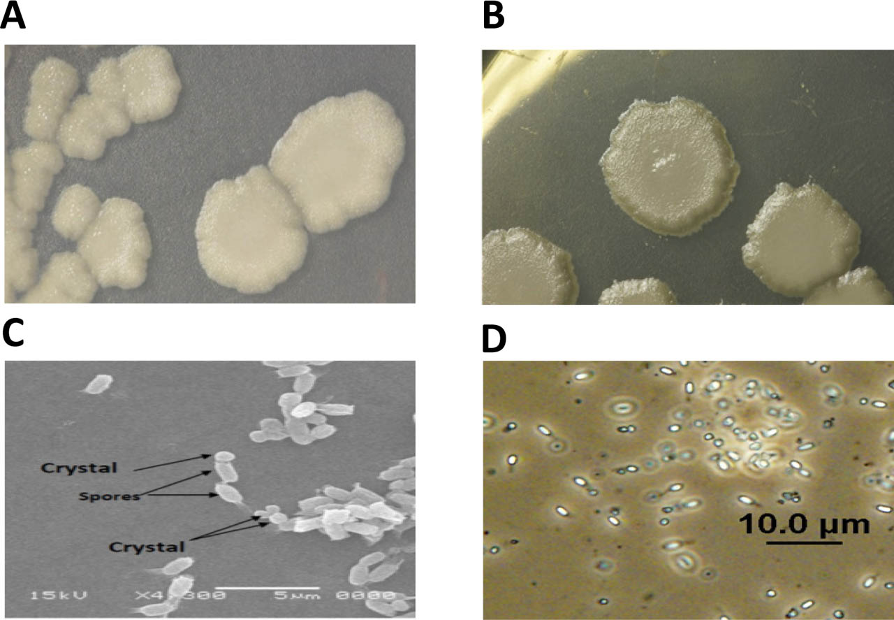
Figure 1:
Microscopic examination of the Bti H14 reference strain and Bt isolate M91. Photomicrographs showing colony morphology of Bti-H14 (A), whereas (B) shows colony morphology of native Bt M91. Colonies were seeded onto Luria Broth (LB) agar medium supplemented with MnCl2, exhibiting large, white, nearly circular colonies with fine, irregular margins and a glistening or non-glistening appearance. Bti H14 scanning electron microscopy (SEM) (C) and phase-contrast (D) at magnifications of X4, 300, and X1000, respectively.
2.2. Preparation of Bt-parasporal crystal proteins (PCP)
From revived lyophilized cultures or glycerol-revived stalk subcultures, a pure colony of each of 14 native non-insecticidal Bt isolates was streaked onto fresh Luria Broth (LB) agar supplemented with manganese (II) chloride tetrahydrate (MnCl2) (Loba Chemie) and incubated at 37 °C for 72 h (Palma et al. 2014; El-Kershet al. 2014). After incubation, Bt colonies of each isolate were suspended in sterile distilled water (SDW) as a wet mount and examined under a phase-contrast microscope (100X oil-immersion objective) to confirm the presence of spores and crystals (Figures 2A to 2H). Next, Bt growth was scraped from each plate, resuspended in 5 mL of SDW, vortexed, and 5 mL of 1 M NaCl was added, then centrifuged at 7000 rpm for 15 min. The pellet was resuspended in SDW with 1 M NaCl, then washed twice in SDW by centrifugation. A 6% sterile lactose solution was added to the washed pellet to reach approximately 2 McFarland tubes (Priest et al. 2004). The suspension was stirred for 30 min on a magnetic stirrer, and four volumes of acetone (Win Lab) were slowly added. Stirring continued for an additional 45 min. The mixture was allowed to stand at room temperature (RT) for 10 min, then filtered through Whatman No. 1 filter paper. The residues on the filter paper were left to aseptically dry overnight in an incubator at 37 °C. The white crystal-spore mixture pellets obtained after drying were suspended in SDW and washed twice with SDW by centrifugation. The washed pellets were then dissolved in an alkaline buffer (Na2CO3, 0.256 g) and dithiothreitol (DTT) (Loba Chemie PVT. LTD., India), 0.08 g, in 50 ml SDW with a few drops of 1 N NaOH (pH 12) for 3 h in a shaking incubator (Shel Lab) at 37 °C, followed by centrifugation at 7000 rpm for 20 min. The pH of the resultant supernatant (containing protoxin) was adjusted to 7.0–8.0 with 1N HCl, followed by membrane sterilization (Merck, Millipore, 0.45 μm), thereby removing spores and/or sporangium fragments. The protein content was then assayed using the Lowry method (El-Kersh et al. 2012) with a calibration curve constructed with bovine serum albumin (BSA) (Sigma-Aldrich; A9647). The protein concentrations for each of the 14 non-insecticidal Bt isolate PCPs were also adjusted to 1 mg/ml with SDW to minimize the influence of possible solubilized spore-protein impurities, given that the maximum protein concentration used in all experiments of this study did not exceed 50 to 100 μg, with corresponding volumes of 50 to 100 μl. The obtained solutions were then aliquoted into small volumes (0.5 ml) in sterile Eppendorf tubes and stored at −20 °C. Some aliquots were subjected to trypsin activation (AT), while others remained unactivated as protoxin samples (PT). For trypsin activation, 1 μg of trypsin (Type I; Sigma) was mixed with 20 μg of protoxin, and the mixture was digested with trypsin for 3 h at 37 °C. Trypsin was then inactivated by adding 0.5 mg of trypsin inhibitor (Type II; Sigma) per mg of trypsin. The trypsin-proteolysis fragments (trypsin-activated toxins, AT) were sterilized by bacterial membrane filtration and stored at −20 °C until testing. The effects of non-trypsin-activated protoxins (PT) on S. aureus virulence factor expression were also examined (Aboul-Soud et al. 2019; Ammouneh et al. 2011; Nethravathi et al. 2010; Naimov et al. 2008). To minimize contamination and remove spores and cellular debris, all AT and PT protein preparations were filtered through a 0.22 μm membrane filter before use in subsequent assays. This step effectively removes intact spores, sporangial fragments, and insoluble debris, ensuring that the tested solutions contain primarily soluble proteins.
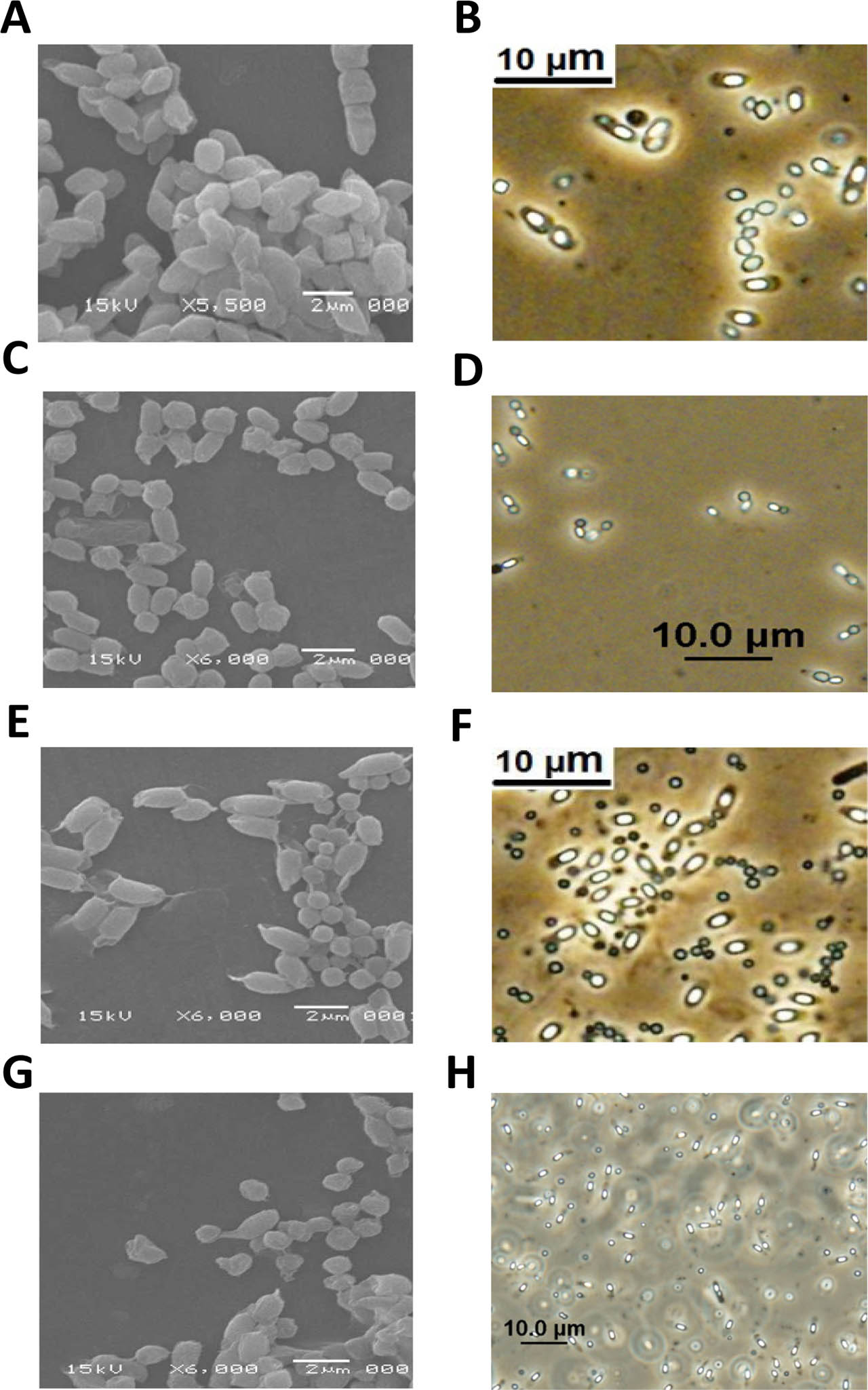
Figure 2.
Scanning electron microscopy and phase-contrast micrographs of native, non-insecticidal Bt isolates illustrating spores and the diversity of crystal shapes. (A and B) Bt-78 (bipyramidal-oval-square, non-spore-attached). (C and D) Bt-M91 (hexagonal, spore-attached). (E and F) Bt-M154t1 (spherical, non-spore-attached). (G and H) Bt-M11 (bipyramidal-square, spore-attached). In all cases, spores appear non-swollen, bright, and cylindrical, resembling those of the reference Bti H14 in Figure 1. 16S rRNA sequences, with reference accession numbers in NCBI GenBank® (https://www.ncbi.nlm.nih.gov/nuccore), when applicable, showed 99% similarity to our native Bt isolates (Li et al. 2013).
2.3. Hemolytic activity
To assess the inherent hemolytic activity of native Bt strains, fresh overnight cultures were streaked onto sheep blood agar plates. Plates were incubated aerobically at 30°C for 48 hours. Hemolytic zones and their types were recorded. S. aureus (MSSA, methicillin-sensitive Staphylococcus aureus) ATCC 29213 was used as the positive control.
2.4. SDS-PAGE
Sodium dodecyl sulfate-polyacrylamide gel electrophoresis (SDS-PAGE) was performed according to standard procedures. Protein bands of activated (AT) and unactivated PCP (PT) proteins were resolved on a 12% SDS-PAGE gel and subsequently visualized by silver staining (Aboul-Soud et al., 2019).
2.5. S. aureus hla and spa expression reporter assay and stock cultures of S. aureus strains
The reporter assay was conducted using S. aureus PC203 (spa::lacZ) and S. aureus PC322 (hla::lacZ) strains, as previously described and kindly provided by Nielsen and co-workers (Porcar and Juárez-Pérez 2003). These 2 fusion strains were derived from the S. aureus wild-type strain (8325-4), which had been cured of known prophages and was successfully used to screen compounds that influence virulence gene expression in S. aureus (Porcar and Juárez-Pérez 2003). The two S. aureus strains were grown on Tryptic Soy Agar (TSA; Mast Laboratories, Merseyside, UK) containing erythromycin (5 μg/ml) and 5-bromo-4-chloro-3-indolyl-β-D-galactopyranoside (X-Gal) (150 μg/ml). The PC203 (spa::lacZ) strain appears light blue, while the PC322 (hla::lacZ) strain displays blue colonies. For the hla and spa expression assay, each of the two S. aureus strains was grown over-night on TSA, and a pure colony of each strain was suspended in sterile saline to a 0.5 McFarland standard. An aliquot of 125 μl of the suspension was placed in a Petri dish and mixed with 25 mL of LB agar at 50 °C, supplemented with erythromycin (5 μg/mL) and X-gal (150 μg/mL). After solidification, wells were punched with a sterile drill (5 mm). Bt AT and PT isolates at concentrations of 10, 50, and 100 μg/mL were added to the wells. As controls, equal volumes of saline, alkaline buffer (Na2CO3 and DTT), and trypsin (at the specified concentration) were used. The Bti H14 reference strain contains Cyt1Aa, Cyt1Ab, and Cyt2Aa proteins (Ahmed et al. 2017), and the trypsin-activated form exhibits strong antibacterial effects (Promdonkoy and Ellar 2003), precluding its use as a control in this study. Plates were incubated at 37 °C, and results (bleaching zones in mm) were recorded after 24 h. The appearance of a white zone surrounding the well indicates downregulation of virulence gene expression (Porcar and Juárez-Pérez 2003). The two S. aureus strains were maintained as stock cultures at 4 °C in TSA supplemented with 5 μg/mL erythromycin and stored, as well as at −20 °C in tryptone soya broth (TSB; SPML, Saudi Arabia) containing 15% glycerol, and sub-cultured on blood agar plates at 37 °C before testing. Additionally, MSSA strain (ATCC 29213) and MRSA strain (ATCC 43300) were obtained from King Fahad Medical City (KFMC) and maintained as stock cultures at −20 °C in tryptone soya broth (TSB) containing 15% glycerol (SPML, Saudi Arabia) and sub-cultured on blood agar plates at 37 °C before testing.
2.6. Quantitative β-galactosidase activity
A quantitative β-galactosidase assay was conducted in liquid culture with the two S. aureus reporter strains PC203 (spa::lacZ) and PC322 (hla::lacZ) to evaluate the impact of Bt-derived PCPs on virulence gene expression and to compare the findings with those of the zone-inhibition assay. We diluted the overnight cultures 1:100 in fresh TSB containing 5 μg/ml erythromycin and allowed them to grow to the early log phase (OD600 = 0.3). Aliquots (1 mL) were treated with Bt-derived AT proteins (10, 25, and 50 μg/ml) or an equivalent volume of alkaline buffer (control) and incubated at 37°C with shaking for 3 h. We collected the cells, rinsed them, and then lysed them using both lysozyme (1 mg/ml) and lysostaphin (10 μg/ml) in Z-buffer (60 mM Na2HPO4, 40 mM NaH2PO4, 10 mM KCl, 1 mM MgSO4, pH 7.0). We used o-nitrophenyl-β-D-galactopyranoside (ONPG, 4 mg/mL) as the substrate to assess β-galactosidase activity. Reactions were terminated by adding 1 M Na2CO3, and absorbance was measured at 420 nm and 550 nm. β-galactosidase activity was expressed as Miller units (MU). Three replicates were performed, and the experiment was repeated twice.
2.7. Biofilm assay
The biofilm assay was performed using a 96-well flat-bottom microtiter plate. S. aureus PC203 (spa::lacZ), PC322 (hla::lacZ), and methicillin-resistant S. aureus (MRSA) ATCC 43300 were cultured overnight in TSB (SPML, Saudi Arabia) at 37°C and then diluted 1:200 in sterile TSB. To assess the effect of diluted (50 μg/mL) and undiluted (UD, 100 μg/mL) Bt AT and PT from the five selected (M11, M78, M91, M154t1, and A11) Bt isolates (based on data on bleaching zones, differences in geographical origin, bio-typing, and crystal shape) on biofilm formation, wells of 96-well microtiter plates were filled with two hundred μl aliquots of the diluted cultures with AT, PT, or saline in triplicate. Two parallel plates were prepared: one for growth assessment and the other for the biofilm assay. Both plates were incubated overnight at 37 °C. For planktonic biomass growth assessment, absorbance at 600 nm (OD600) was measured. To assess biofilm formation, planktonic cells were aspirated, and the wells were washed three times with sterile saline. The plate was inverted on filter paper and allowed to dry for 1 h at RT. Biofilms attached to the plate were stained with 200 μL of crystal violet (0.1% in SDW) for 15 min at RT. Excess stain was removed by washing three times with sterile saline, and the remaining crystal violet bound to biofilms was dissolved in 230 μL of 95% ethanol. Finally, absorbance at 550 nm (A550) was measured as an indicator of biofilm formation (Head et al. 2004; Kaur et al. 2009; Upadhyaya et al. 2010). To distinguish between growth inhibition and specific antibiofilm activity, the Normalized Biofilm Index (NBI) was calculated using the following formula:
Calculated NBI values were analyzed using oneway ANOVA with Tukey's post hoc test in GraphPad Prism v10.4.2. Data are presented as mean ± SEM.
2.8. Minimum Inhibitory Concentration (MIC)
According to Clinical and Laboratory Standards Institute (CLSI) recommendations, broth microdilution assays were used to determine MIC values. We tested two-fold serial dilutions of PCPs (100, 50, and 25 μg/mL) against MSSA (ATCC 29213), using Bti H14 AT as the positive control (Promdonkoy and Ellar 2003) at concentrations of 10, 5, 2.5, and 1.25 μg/mL.
2.9. PCR screening for Cyt1 gene
Bacterial plasmid DNA was extracted using an alkaline lysis protocol as previously described. Plasmid DNA was obtained from five native B. thuringiensis isolates (i.e., M11, M78, M154t, M91, and A11), and the reference strain H14 served as a positive control for gene amplification. Universal (UN) primers [Forward: 5′-CCT CAA TCA ACA GCA AGG GTT ATT-3′; Reverse: 5′-TGC AAA CAG GAC ATT GTA TGT GTA ATT-3′] were used to amplify the Cyt1 genes (Cyt1Aa, Cyt1Ab, and Cyt1Ba) by PCR (Aboul-Soud et al. 2019). The PCR reaction mixture (25 μL) comprised the following final concentrations: 1× PCR buffer, 1.5 mM MgCl2, 0.2 mM dNTPs, 0.2 μM of each primer, 1 U of Taq DNA polymerase, and 50 ng of template DNA. Amplification conditions were as follows: an initial denaturation at 94°C for 5 minutes, 35 cycles of 94°C for 40 seconds, 52°C for 60 seconds, and 72°C for 2 minutes, followed by a final extension at 72°C for 5 minutes. A 1.5% agarose gel stained with ethidium bromide was prepared to visualize the PCR results. Bti H14 was used as a positive control for Cyt1 amplification.
2.10. MTT cytotoxicity assay
The cervical cancer cell line HeLa was used to assess the toxicity of AT toxins. Cells were routinely grown in standard DMEM supplemented with glucose, pyruvate, and 10% fetal bovine serum (FBS). Cells were grown in T75 tissue culture flasks to confluence, then trypsinized and plated in 96-well plates at 1x10⁴ cells per well in DMEM supplemented with 5% FBS for 24 hours. Next, cells were treated with two-fold serial dilutions of AT toxins at concentrations of 200, 100, 50, 25, 12.5, and 6.25 μg/mL overnight. The next day, the medium was replaced with 150 μL of MTT solution (5 mg/mL), and the plates were washed with isopropanol to solubilize the formazan crystals. Plates were read at 570 nm, and cell viability (%) was calculated after subtracting the background level measured at 630 nm. DMSO-treated cells at the highest AT concentration were set to 100% viability (basal growth control). The final DMSO concentration was less than 2%, which is safe for the cells and does not cause toxicity.
2.11. RT-qPCR
Overnight cultures of MSSA (ATCC 29213) were diluted 1:200 in fresh, sterile Todd-Hewitt broth with 2% yeast extract (THY) containing 0.5% glucose. OnemL aliquots of the diluted cultures were mixed with 100 μg/mL of PCPs Bt AT toxins or DMSO and then cultured at 37 °C in triplicate. Samples were collected by centrifugation at OD600 nm ~0.5 (corresponding to the ML growth phase) and after an overnight culture (S phase). Next, RNA-protect Cell Reagent (Qiagen, Hilden, Germany) was added, and lysostaphin (Sigma) was used to lyse the cells. RNA was extracted using Trizol® (Schnelldorf) according to the manufacturer's protocol, followed by a TURBO DNA-free kit (Sigma) to remove residual DNA contamination in the RNA samples (Al-Mebairik et al. 2016). The purified RNA was reverse transcribed to cDNA, and RT-qPCR was performed using the Light Cycler 2.0 System and the 2(−ΔΔCt) method (Livak and Schmittgen 2001). Pre-designed, validated primers were purchased from TIB Molbiol (Berlin, Germany), and their sequences are shown in Table 1.
Table 1.
Oligonucleotide primers in the RT-qPCR assay (Al-Mebairik et al. 2016).
| Gene | Sequence (5′–3′) |
|---|---|
| spa | F: GCGCAACACGATGAAGCTCAACAA |
| R: ACGTTAGCACTTTGGCTTGGATCA | |
| hla | F: CTGAAGGCCAGGCTAAACCACTTT |
| R: GAACGAAAGGTACCATTGCTGGTCA | |
| 16S rRNA | F: CTGGTAGTCCACGCCGTAAAC |
| R: CAGGCGGAGTGCTTAATGC | |
| RNAIII | F: GCACTGAGTCCAAGGAAACTAACTCT |
| R: AGCCATCCCAACTTAATAACCATGT |
The expression levels of the tested genes were normalized using 16S rRNA expression as an internal standard (Al-Mebairik et al. 2016). A positive reaction was indicated by the accumulation of fluorescent SYBR Green I signal. In this experiment, four Bt isolates: M11, M78 (GenBank: KF026328.1), M91, and M154t1 (GenBank: KC960017.1) were used to study the effect of their AT on the expression of S. aureus hla, spa, and RNAIII genes.
2.12. Statistical Analysis
Data were analyzed using SPSS PC+ version 21.0. Normality was assessed using the Shapiro–Wilks test, and the assumption of homogeneity of variance was evaluated using Levene's test. One-way analysis of variance (ANOVA) was used to analyze biofilm formation in control versus experimental treatments with PCPs. Tukey's non-parametric post hoc test was used for multiple comparisons. Student's t-test was used for RT-qPCR experiments to profile expression of virulence genes. A P value of less than 0.05 was considered statistically significant.
3. Results
3.1. Morphological descriptions of Bt colonies and crystal shapes
In this work, the colony development of the 14 non-insecticidal Bt isolates on LB agar/MnCl2 showed strong resemblance to that of the reference Bti H14, appearing as raised, large, white, almost circular colonies with fine, irregular margins and a glistening or non-glistening scalloped appearance (Figures 1A and 1B). Moreover, strains and crystals were examined morphologically and compared to the reference Bti H14 strain using both scanning electron and phase-contrast microscopy (Figures 1C and 1D).
Wet-mount phase-contrast microscopy enabled differentiation of Bt colonies from those of B. cereus, which are closely related, except for the presence of parasporal crystals within the sporangia of only Bt isolates, which display shapes and sizes unique to each native Bt strain (Figures 2A to 2H).
Like the Bti H14 reference strain, native Bt isolates exhibited many shared morphological features, including cell chain arrangements and cylindrical, non-swollen spores, but showed substantial variation in the shapes of the parasporal crystals formed. None of the 14 recruited Bt isolates exhibited mosquito-larvicidal activity against Aedes aegypti, even at remarkably high concentrations (data not shown) (El-Kersh et al. 2016; Ahmed et al. 2021). As presented in Table 2, most of the 14 native Bt isolates exhibited crystals attached to spores, with shapes ranging from small to large spherical, cubic, square, or bipyramidal, as clearly visualized by scanning electron microscopy of the examined Bt isolates (i.e., Bti H14, Bt M11, Bt 78, Bt 91, and Bt 154t1) (Figure 2A to 2H). By contrast, the characteristics of Bti H14 parasporal crystals, i.e., amorphous irregular, bisected spherical, conical-budding, or merged triangular, not attached to spores (Figure 1E and 1F), were not observed among the investigated 14 non-insecticidal native Bt isolates.
Table 2:
Sampling type of the 14 non-insecticidal B. thuringiensis isolates (lab code), their crystal shapes, and the qualitative and quantitative quenching effects of different AT protein concentrations on the expression of S. aureus spa and hla strains.
| Bt. Isolate (Code) sample | Crystal-Shape | Protein Conc. ug/ml | Spa | hla | ||||
|---|---|---|---|---|---|---|---|---|
| Effect | Bleaching zone (mm) | β-galactosidase Activity (MU) | Effect | Bleaching zone (mm) | β-galactosidase Activity (MU) | |||
| (M11) soil | Oval-spherical Nonattached to spores | 100 | + | 20±0.5 | 90±2.3 | + | 20±1.2 | 92±5.5 |
| 50 | + | 16±1 | 163±7.2 | + | 19±0.7 | 108±4.3 | ||
| 10 | − | 0 | 452±0 | − | 0 | 338±0 | ||
| (M13m) soil | Small spherical, not attached | 100 | + | 18±1 | 126±7.1 | + | 19±1.2 | 108±7.4 |
| 50 | + | 15.3±1.2 | 176.13.9 | + | 15±0.5 | 155±5.2 | ||
| 10 | − | 0 | 452±0 | − | 0 | 388±0 | ||
| (M67t) Soil | Small spherical | 100 | - | 0 | 452±0 | - | 0 | 388±0 |
| 50 | + | 25±1.2 | 0±0 | + | 20±.2 | 92±1.9 | ||
| 10 | − | 0 | 452±0 | − | 0 | 388±0 | ||
| (M 78) soil | Irregular, bi-pyramidal/cubic like crystals | 100 | + | 19.6±.3 | 100±1.4 | + | 21±1.2 | 62±3.6 |
| 50 | + | 16±0.5 | 163±5.1 | + | 14.3±1.2 | 167±14.1 | ||
| 10 | − | 0 | 452±0 | − | 0 | 388±0 | ||
| (M91) soil | Nearly cubic, attached to spore | 100 | + | 20±1.5 | 90±6.8 | + | 19.3±1.3 | 89±6.1 |
| 50 | − | 16.3±0.8 | 158±7.8 | − | 15.6±0.7 | 147±6.6 | ||
| 10 | − | 0 | 452±0 | − | 0 | 388±0 | ||
| (M154t1) Leaves/soil | Small, spherical non-attached to spore | 100 | + | 21.6±0.8 | 63±2.3 | + | 18.6±1.4 | 101±7.6 |
| 50 | + | 17±1.5 | 145±12.8 | + | 16.3±1.8 | 136±15.1 | ||
| 10 | − | 0 | 452±0 | − | 0 | 388±0 | ||
| (M157) Dried leaves | Bipyramidal, attached to spore | 100 | + | 19±1 | 109±5.8 | + | 19.6±0.8 | 85±3.5 |
| 50 | + | 15.3±0.8 | 176±9.2 | + | 17±1.2 | 124±8.7 | ||
| 10 | − | 0 | 452±0 | − | 0 | 388±0 | ||
| (M160) sheep manure | Blunt end bipyramidal attached to spore | 100 | + | 20.6±0.8 | 81±3.2 | + | 21±1 | 62±30 |
| 50 | + | 19±0.5 | 109±2.9 | + | 20±1.2 | 92±5.5 | ||
| 10 | − | 0 | 452±0 | − | 0 | 388±0 | ||
| (M224) soil | Big spherical and hexagonal with inclined spore | 100 | + | 18.3±1.2 | 122±8.0 | + | 20.6±1.5 | 70±5.1 |
| 50 | + | 15±1.2 | 181±14.5 | + | 16.6±1.2 | 132±9,6 | ||
| 10 | − | 0 | 452±0 | − | 0 | 388 | ||
| (M242) soil | bipyramidal, with blunt edges | 100 | + | 21.3±0.8 | 68±2.6 | + | 22.3±0.8 | 43±1.5 |
| 50 | − | 0 | 452±0 | − | 0 | 388±0 | ||
| 10 | − | 0 | 452±0 | − | 0 | 388±0 | ||
| (M268P) Sewage W | Hexagonal attached to spores in long chains) | 100 | − | 0 | 452±0 | − | 0 | 388±0 |
| 50 | − | 0 | 452±0 | − | 0 | 388±0 | ||
| 10 | − | 0 | 452±0 | − | 0 | 388±0 | ||
| (M296) soil | Large and small bipyramidal | 100 | − | 0 | 452±0 | − | 0 | 388±0 |
| 50 | + | 20.3±0.8 | 68±3.4 | + | 18.6±0.8 | 101±4.4 | ||
| 10 | − | 0 | 452±0 | − | 0 | 388±0 | ||
| (M300) Rain W | Dark and bright ovoid (attached to spore) | 100 | + | 24±0.5 | 18±0.4 | + | 23.3±0.8 | 27±0.9 |
| 50 | − | 0 | 452±0 | − | 0 | 388±0 | ||
| 10 | − | 0 | 452±0 | − | 0 | 388±0 | ||
| A11 soil | Spherical non-spore attached | 100 | + | 16.6±1.2 | 154±11.2 | + | 19±0.5 | 93±2.5 |
| 50 | + | 13±1.7 | 217±29.7 | + | 15.6±0.8 | 147±7.6 | ||
| 10 | − | 0 | 452±0 | − | 0 | 388±0 | ||
3.2. PCR amplification of Cyt1 gene
To assess potential Cyt toxin production, we conducted PCR amplification of the universal Cyt1UN genes to screen for their presence in the five strains under investigation, namely: M11, M78, M154t, M91, and A11. Results indicated that while four out of five strains were negative for Cyt1, isolate M91 exhibited a PCR product of approximately 400 bp, indicating the presence of Cyt1 genes. The product size for Cyt1 was confirmed by an identical amplicon of the same size as the Bti H14 positive control, which confirms the validity of the assay (Figure 3).
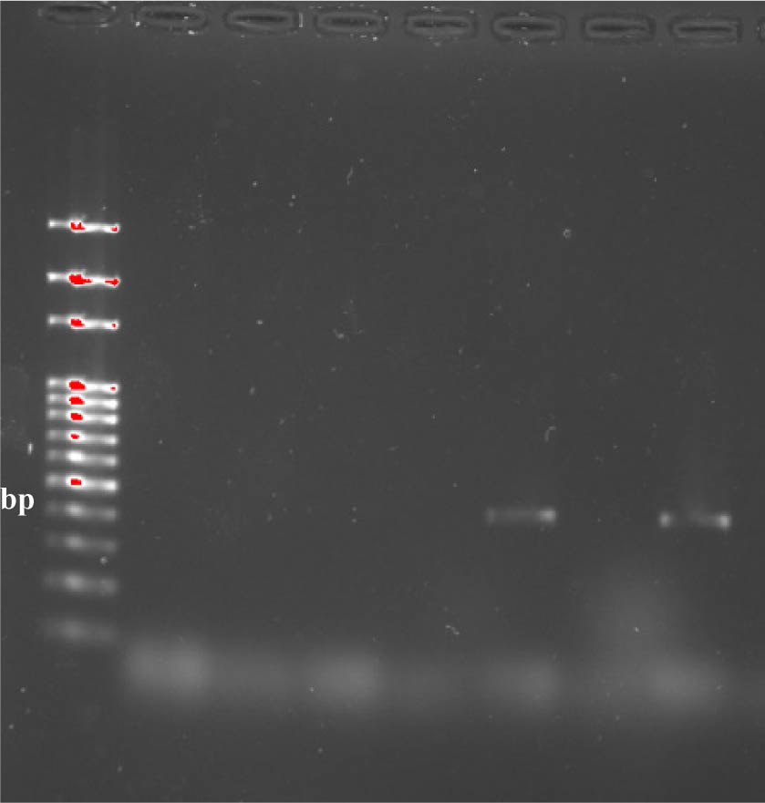
Figure 3.
PCR screening of Bt isolates for Cyt1 genes. Amplicons were resolved on 1.5% agarose gels containing ethidium bromide at 80 V for 40 min. Lane (L) contains the 1500 bp DNA standard marker for band size identification. Four Bt strains were negative for Cyt1 genes, except for isolate M91. The reference strain Bacillus thuringiensis subsp. israelensis (Bti H14) and deionized water were included as positive and negative controls, respectively.
3.3. SDS-PAGE of parasporal crystal proteins (PCPs)
SDS-PAGE analysis of activated and unactivated parasporal crystal proteins was conducted to highlight differences in solubilized PC proteins derived from the Bt isolates under investigation. As expected, a clear protein band profile was obtained, showing a distinct pattern for the unactivated PC proteins (PT) compared with the activated PC form (AT). Two strains, M11 and M78, were investigated by SDS-PAGE as representatives of all Bt strains under investigation (Figure 4). A protein band of about 28 kDa was consistently observed in the AT lanes of all five isolates exhibiting antivirulence activity (i.e., M11, M78, M91, M154t1, and A11; Figure 4). This suggests that it may be an active toxin fragment that should be purified and identified in the future.
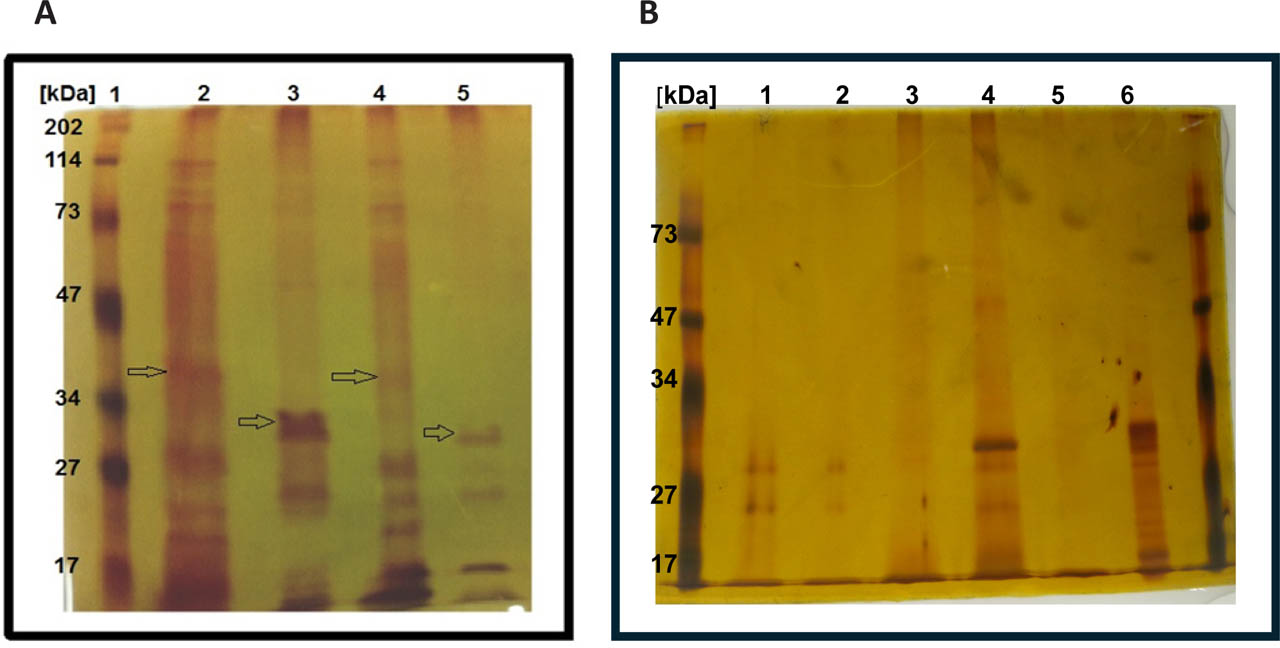
Figure 4.
Protein profiles of unactivated (PT) and activated (AT) parasporal crystal proteins (PCPs). Samples from strains M11, M78, M91, M154t, and A11 were run on a 12% SDS-PAGE gel and silver-stained to visualize protein bands. (A): M11 and M78. Lane 1, molecular marker; Lane 2: M11 strain, unactivated PC proteins loaded 100μl (100μg), bands at 111 kDa, 82 kDa, 58 kDa, 35 kDa, 27 kDa, 24 kDa, and 21 kDa; Lane 3, M11 strain, activated PC proteins (trypsinized) loaded 100μl (60μg/ml), bands at 28 kDa, 24 kDa, and 17 kDa; Lane 4: M78, unactivated PC proteins loaded 100μl (100μg), bands at 111 kDa, 82 kDa, 58 kDa, 35 kDa, 27 kDa, and 21 kDa; Lane 5, M78 strain, activated PC proteins (trypsinized) loaded 100μl (30μg), bands at 28 kDa, 24 kDa, and 17 kDa. (B): M91, M154t, and A11. Lane 1: M91 strain, unactivated PC protein, loaded 100μl (20μg), bands at 28 and 26 kDa; Lane 2: M91 activated PC proteins (trypsinized) loaded 100μl (20μg/ml), very faint bands at 28 and 26 kDa; Lane 3: M154t activated PC proteins (trypsinized) loaded 100μl (50μg/ml), faint bands at 28 and 26 kDa. Lane 4: M154t unactivated PC proteins loaded 100μl (50μg), bands at 30, 28, and 26 kDa; Lane 5: A11 activated PC proteins (trypsinized) loaded 100μl (50μg), faint bands at 28 and 26 kDa. Lane 6: A11 unactivated PC proteins loaded 100μl (50μg), bands at 32, 30, 28, 26, 24, 22, and 17 kDa.
3.4. Bt-derived AT proteins modulate the expression of virulence genes of S. aureus
To assess the effect of Bt-derived AT proteins on the modulation of virulence genes in S. aureus, we employed bleaching zone (X-gal) and β-galactosidase assays as qualitative and quantitative methods, respectively. We used two reporter S. aureus fusion strains (i.e., PC203, spa::lacZ and PC322, hla::lacZ) to screen for the dose-dependent transcriptional quenching effect of AT derived from the five non-insecticidal Bt isolates profiled by SDS-PAGE (Figure 3) at three concentrations (10, 50, and 100 μg/mL). Representative results of the quenching effect of AT derived from Bt isolate M91 at the three concentrations are shown (Figure 5A and 4B). In parallel, three controls, namely C1: saline, C2: alkaline buffer, and C3: trypsin, were used to validate the qualitative and quantitative quenching effects of the tested ATs from the five screened Bt isolates.
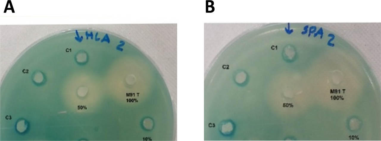
Figure 5.
Representative results showing the transcriptional effects of the Bt (M91) isolate AT (activated toxin, undiluted; M91 T (100%); and 50%-diluted AT) on S. aureus virulence genes (A) hla and (B) spa. To agar plates containing PC322 (hla::lacZ) (A) or PC203 (spa::lacZ) (B), 50 μL of Bt AT at the indicated concentrations [(100% (100 μg); 50% (50 μg); 10% (10 μg)] was added. Plates were incubated for 24 h at 37 °C. Controls: C1: saline; C2: alkaline buffer; C3: trypsin.
Data in Table 2 indicate that the AT toxins of nine Bt isolates (M11, M13m, M78, M91, M154t1, M157, M160, M224, and A11 at 100 and 50 μg/mL) produced dose-dependent reductions in transcription of the hla and spa genes in both strains. In contrast, AT toxins from Bt isolate M268P, at the three concentrations used (100, 50, and 10 μg/mL) (Table 2), showed no suppression of transcription of both hla and spa genes, reflecting the specificity of AT toxins. Likewise, none of the 14 Bt isolates tested exhibited changes in transcription of the two S. aureus genes (hla and spa) at the lowest concentration of 10 μg/mL, again reflecting a dose-dependent response. AT toxins from the M67t and M296 isolates, however, exhibited reduced mRNA transcription only at the second-highest dose of 50 μg/mL, but not at the lowest dose of 10 μg/mL, as expected (Table 2). These findings indicate that certain trypsin-activated Bt PCP (AT) toxins suppress transcription of both hla and spa genes; this suppression is dose-dependent and specific to the PCS protein type of each respective Bt isolate. This hypothesis remains to be further verified.
To confirm the qualitative findings from the plate-based reporter assay and mitigate potential issues related to diffusion and precipitation artifacts, we conducted quantitative β-galactosidase assays in liquid culture using sub-inhibitory concentrations (10, 50, and 100 μg/mL) of AT PCPs derived from all 14 native B. thuringiensis isolates. Table 2 shows β-galactosidase activity (in Miller units) for the five active antivirulence isolates: M11, M78, M91, M154t1, and A11. All five isolates dramatically decreased spa and hla expression in a dose-dependent manner. For instance, M78 AT at 100 μg/mL decreased hla and spa expression by 82.3% and 84.0%, respectively. M11 and M91 ATs also exhibited substantial repression at 100 μg/mL, lowering hla expression by 73.1% and 73.5%, respectively, and spa expression by 78.9% and 73.8%. For the other nine isolates, which showed different or no activity in the plate assay, the quantitative β-galactosidase results matched their qualitative profiles. Isolates M242 and M300, which had large bleaching zones in the plate test, also exhibited substantial suppression in liquid culture (M300: 93% reduction in hla; 96% in spa at 100 μg/mL). Isolates M268P and M296, which had no zones at 100 μg/mL, did not show a substantial drop in β-galactosidase activity.
3.5. Normalized biofilm index reveals genuine anti-biofilm activity of Bt-derived AT proteins
To test the effects of diluted (D=50 μg/mL) and undiluted (UD=100 μg/mL) Bt AT and PT on biofilm formation in three S. aureus strains, including a MRSA strain, wells of 96-well microtiter plates were filled with 200 μl of diluted cultures containing AT, PT, or saline, in triplicate. Two plates were prepared: one for growth assessment and the other for biofilm formation. S. aureus PC203 (spa::lacZ) and PC322 (hla::lacZ), as well as MRSA (ATCC 43300), were randomly assigned to the five selected Bt isolates (M11, M78, M91, M154t1, and A11) to highlight a wide spectrum of AT and PT effects on biofilm formation across different S. aureus strains. To account for possible growth-inhibitory effects, biofilm data were standardized to planktonic growth (OD600) and expressed as the Normalized Biofilm Index (NBI). Figure 6 shows that the NBI values for all PT-treated samples were highly similar to those of controls (NBI = 0.93–0.96, p > 0.05). This indicates that the protoxin itself had no specific antibiofilm activity. Conversely, UD AT treatments markedly reduced NBI in all S. aureus strains. For example, M91 UD AT lowered NBI from 1.05 ± 0.06 (control) to 0.46 ± 0.03 (p < 0.001). Similarly, M11 and M78 UD AT lowered NBI to 0.50 ± 0.03 and 0.53 ± 0.03, respectively (p < 0.001). Moreover, D AT treatments showed a moderate yet statistically significant reduction in NBI (e.g., M154t1 D AT: 0.74 ± 0.05, p < 0.01). These results corroborate that activated PCPs (AT) interfere with biofilm formation independently of bacterial growth suppression.
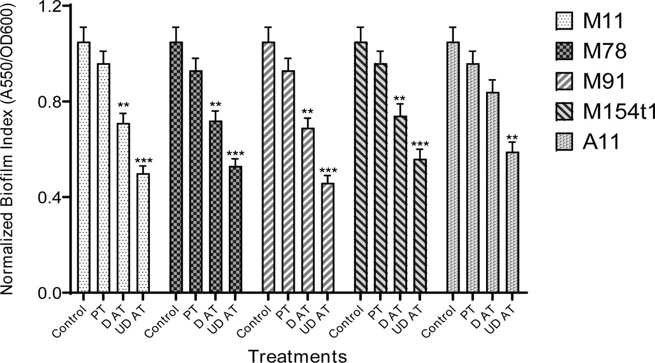
Figure 6.
The effect of Bt-derived PCPs either AT (diluted, D or undiluted, UD) or PT on S. aureus normalized biofilm index (NBI), compared to saline control. Biofilm formation was determined by an OD550 nm reading of crystal violet stain solubilized by ethanol with saline treatment as a control. NBI was calculated by dividing A550 over OD600. NBI for the isolate M11 was tested on PC322 (hla::lacZ). Data are presented as the mean ± standard error of means (SEMs). One-way ANOVA was conducted followed by Tukey's multiple comparison test to compare the mean of each treatment with the control. *p<0.05, **p<0.01, ***p<0.001. NBI for the isolate M78 was tested on PC322 (hla::lacZ). NBI for the isolate M91 was tested on PC203 (spa::lacZ) biofilm formation. NBI for the isolate M154t1 was tested on PC203 (spa::lacZ). NBI for the of Bt A11 isolate was tested on MRSA (ATCC 43300). Data points are the average of two independent growth experiments and three technical replicates.
3.6. The hemolytic activity of native Bt isolates
To assess potential Cyt toxin production, the five Bt strains selected for antiviral evaluation were streaked onto sheep blood agar. Notably, none of the tested strains exhibited α- or β-hemolysis after 48 hours of incubation (Table 3), indicating that either they lack the capacity to produce cytolytic toxins or the conditions used did not permit their generation.
3.7. MIC results
MIC tests showed that none of the Bt PCP formulations were bactericidal at levels up to 100 μg/mL. This demonstrates that the antivirulence and antibiofilm activities reported at ≤100 μg/mL are not attributable to growth inhibition.
3.8. MTT toxicity against HeLa cells
The cytotoxicity of AT PCPs on immortal mammalian cells was evaluated using HeLa cells. The colori-metric MTT viability assay was used to assess the toxic potential of AT PCPs derived from four Bt strains: M11, M78, M91, M154t1, and A11. AT PCPs were serially diluted twofold, with concentrations ranging from 200 to 6.25 μg/mL. The results were then compared with cell viability in the DMSO control (Figure 7). The cell viability graph indicated that AT PCPs exhibited no significant cytotoxicity at concentrations up to 100 μg/mL. However, at the highest concentration of 200 μM, they exhibited significant cytotoxicity toward HeLa cells, reducing cell viability by approximately 30% across all five strains. These results clearly support the safety profile of the concentrations used, up to 100 μg/mL, in mammalian cells.
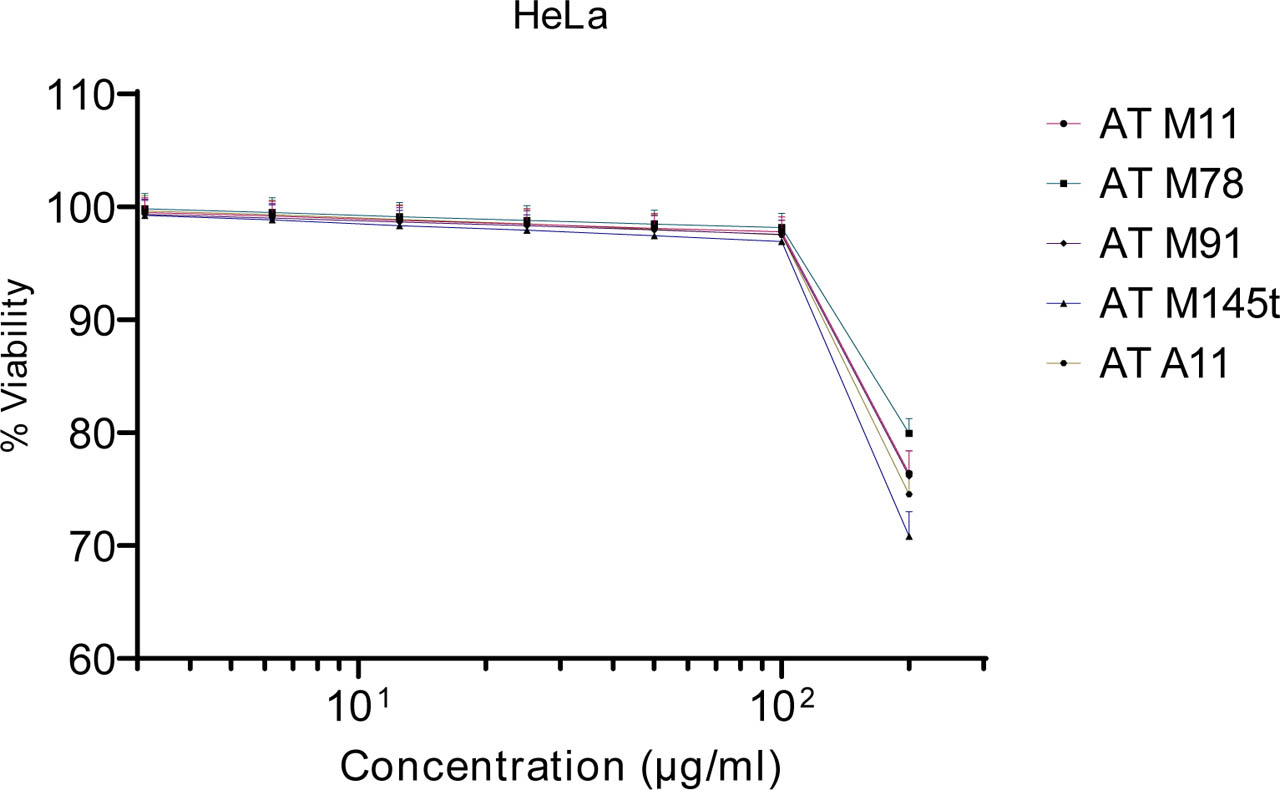
Figure 7.
MTT toxicity evaluation of AT PCPs derived from five Bt isolates against HeLa cervical cancer cells. Cell viability was normalized to the DMSO-treated control, whose final concentration didn't exceed 2%. Yellow MTT is enzymatically reduced by cellular dehydrogenases to form purple formazan crystals. Absorbance was measured at 570 nm, and background was subtracted by deducting the absorbance at 630 nm. Data represent means±SD of two experiments with 8 replicates for each concentration.
3.9. Bt AT inhibits the transcription of spa, hla and RNAIII in S. aureus
The virulence of S. aureus is governed by cell wall-associated proteins and secreted toxins, which are regulated and expressed in accordance with its growth environment and/or growth phase. Bt A11 was not included in qPCR due to sample limitations but showed strong antibiofilm activity in normalized assays (Figure 5). Therefore, the effect of a dose of 100 μg/ml of PCPs AT toxins derived from four Bt isolates (M11, M78, M91 and M154t1) on the transcriptional expression of hla, spa and RNAIII genes in S. aureus was investigated at mid-logarithmic phase and at stationary phase of growth by quantitative reverse transcription PCR (RT-qPCR). The expression levels of these genes were normalized using 16S rRNA for these Bt isolates as an internal standard. A positive reaction was detected by the accumulation of SYBR Green I fluorescence. For statistical analysis to obtain the mean ± standard error, the mean was calculated after the 2−ΔΔCt transformation was performed, and data were represented in the form of the log2 fold change (log2FC), relative to the DMSO control (Figure 8).
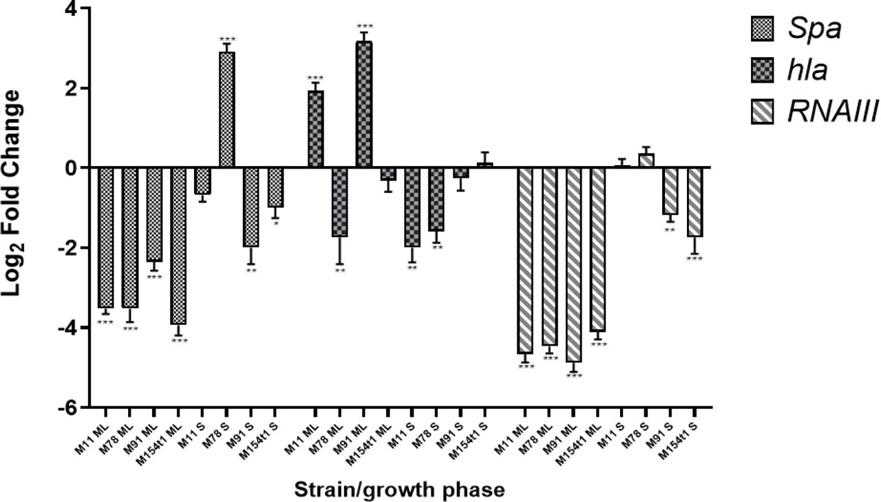
Figure 8.
Quantitative RT-PCR expression profiling of S. aureus MSSA (ATCC 29213) spa, hla, and RNAIII genes. S. aureus was treated with a single concentration of 100 μg/ml of PCPs AT toxins derived from M11, M78, M91, and M154t1Bt isolates during the mid-log phase (ML-) or stationary phase (S). DMSO served as the negative control (basal expression level). Data are presented as log2 fold change, with mean ± SEMs from two independent experiments and three technical replicates, calculated by 2-ΔΔCT. *p<0.05, **p<0.01, and ***p<0.001.
3.9.1. spa gene expression
During the mid-log phase, all four Bt isolates markedly reduced spa gene expression in S. aureus MSSA (ATCC 29213), which encodes surface and secreted proteins involved in aggregation. The greatest suppression was observed with M154t1 (log2FC = −3.94 ± 0.26, p < 0.001), followed by M11 and M78 (log2FC = −3.52, p < 0.01), and then M91 (log2FC = −2.35 ± 0.22, p = 0.005). In the stationary phase, however, regulatory effects differed: M78 markedly up-regulated spa (log2FC = +2.91 ± 0.20, p = 0.003), whereas M91 and M154t1 maintained significant down-regulation (log2FC = −2.00 and −1.00, respectively, p < 0.05). M11 showed a substantial, non-significant decrease (log2FC = −0.68 ± 0.17, p = 0.081). These phase-dependent changes suggest that Bt PCPs might interact with the spa regulatory network differently across growth phases.
3.9.2. hla gene expression
Modulation of hla expression was significantly strain- and phase-specific. At the midpoint of the log phase, M91 caused a large increase in hla (log2FC = +3.17 ± 0.22, p = 0.004), whereas M78 and M154t1 caused decreases (log2FC = −1.74 ± 0.67, p = 0.029 and −0.32 ± 0.28, p = 0.420, respectively). M11 also increased hla (log2FC = +1.93 ± 0.21, p = 0.002). During the stationary phase, M11 and M78 significantly down-regulated hla (log2FC = −2.00 ± 0.37, p = 0.015 and −1.59 ± 0.29, p = 0.010), whereas M91 and M154t1 showed no significant change. These findings suggest that specific Bt PCPs can either activate or inhibit hla, potentially circumventing canonical agr-mediated control.
3.9.3. RNAIII gene expression
Consistent with the spa gene expression pattern, all four isolates significantly inhibited RNAIII expression during the mid-log phase with log2FC values ranging from −4.11 (M154t1) to −4.88 (M91) (p < 0.001). At the stationary phase, M91 and M154t1 exhibited significant down-regulation (log2FC = −1.18 ± 0.17, p = 0.003 and −1.74 ± 0.41, p < 0.001, respectively), whereas M78 showed a modest, non-significant up-regulation (log2FC = +0.36 ± 0.16, p = 0.160). M11 showed no significant difference relative to the control (log2FC = −0.06 ± 0.16, p = 0.850). The consistent reduction in RNAIII across isolates during log-phase growth suggests that Bt PCPs may disrupt the agr quorum-sensing network.
4. Discussion
In recent years, antibiotic resistance has become a major threat to public health (Turner et al. 2019). The overuse of conventional antibiotics has led to the increased emergence of multidrug-resistant bacteria (O'Neill et al. 2014; Kim 2019; Ahmad-Mansour et al. 2021; Allen et al. 2014; Lin et al. 2015; Ivanova et al. 2013). Specifically, for S. aureus, which is responsible for worldwide outbreaks of nosocomial infections, the emergence of multidrug-resistant strains poses a threat to the global population; therefore, new strategies are required to disarm pathogenic organisms and expand our existing arsenal of drugs against infectious disease. As an alternative strategy, targeting virulence factors without killing the pathogens has gained increasing attention in recent literature (Cheung et al. 2021; Kurlenda et al. 2012; Kim 2019; Ahmad-Mansour et al. 2021; Gill et al. 2015; Khodaverdian et al. 2013; Nielsen et al. 2012; Wang et al. 2015). The main objective of this study was to screen and analyze the in vitro effects of natural δ-endotoxins (PCPs) isolated from Saudi Arabian native non-insecticidal Bt isolates, with (AT) and without prior trypsin activation (PT), on the production of selected S. aureus virulence factors. To the best of our knowledge, this is the first study to report the anti-virulence properties of alkaline-solubilized and trypsin-activated PCPs derived from native non-insecticidal Saudi Arabian Bt isolates against S. aureus strains. In the present study, 14 documented non-insecticidal Bt isolates were selected, and their PCP toxins were acetone/lactose co-precipitated (Priest et al. 2004), then alkaline-solubilized and trypsin-activated in vitro. This was followed by qualitative and quantitative profiling of hla (α-hemolysin) and spa (protein A) gene expression using a reporter fusion assay, as detailed in the methods section. In an X-gal-based bleaching zone inhibition assay, ATs from the 14 tested Bt strains showed significant, dose-dependent down-regulation of hla and spa mRNA expression (Table 2). In addition to the plate-based reporter assay, quantitative β-galactosidase measurements in liquid culture confirmed that Bt-derived AT proteins repress spa and hla transcription in a dose-dependent manner (Table 2). These results mitigate concerns about potential artifacts in the agar diffusion assay and reinforce the conclusion that Bt-derived PCPs modulate virulence gene expression independently of bacterial killing. Notably, the effect of Bt PCPs on S. aureus hla and spa mRNA expression is further complicated by the fact that both genes are regulated by a complex network that includes agr and additional elements such as sar, sea, and srr (LaSarre and Federle 2013). Because agr decreases spa expression and increases hla expression during the transition from late-exponential growth to stationary phase, the observed decrease in both spa and hla suggests that our activated Bt AT (PCPs) affect virulence gene expression independently of agr (Nielsen et al. 2014; Nielsen et al. 2012a; Yarwood et al. 2003). The present findings resembled those previously reported by Nielsen and co-workers (Porcar and Juárez-Pérez 2003), who found that glucose (a fast-utilizable sugar, a catabolite) markedly reduced the expression of both hla and spa genes. Recently, it has been demonstrated that the catabolite control protein A (CcpA) regulates S. aureus biofilm formation by directly repressing staphylokinase expression, and the multifaceted roles and multiple networks involving CcpA have been postulated (Kalia 2013). The authors suggested using staphylokinase for both vascular occlusion and S. aureus-associated biofilm infections. Additionally, agrA, which positively regulates the expression of hla, was significantly inhibited in S. aureus after treatment with a sub-MIC of allicin (a derivative of garlic). Consequently, the mode of action (MOA) by which allicin reduces α-toxin production may, in part, involve inhibition of the agr regulatory system (Tong et al. 2015). In a similar study, nigribactin, a novel siderophore from Vibrio nigripulchritudo, and Solonamide B, a cyclo-depsipeptide isolated from Photobacterium halotolerans, reduced hla expression while increasing spa expression, indicating that they interfere with activation of the agr quorum sensing (QS) system (Nielsen et al. 2014; Nielsen et al. 2012a).
Bacterial biofilms are complex microenvironments composed of single or mixed species. These species adhere to one another on biotic or abiotic surfaces, adopting a multicellular lifestyle encased in extracellular polymeric substances. Key characteristics of bio-film-forming bacteria include resistance to host defenses and tolerance of antimicrobials. It has been estimated that 65% to 80% of bacterial infections in humans are due to biofilm formation (Kalia 2013). The present results revealed that, unlike ATs, biofilm formation by the tested S. aureus strains was unaffected by the non-activated PTs of all five Bt isolates tested (M11, M78, M91, M154t1, and A11). By contrast, these PTs exhibited increased S. aureus growth. This enhancement of bacterial growth is presumed to result from the auto-proteolysis of PT proteins by S. aureus intrinsic proteases, which in turn further enhances S. aureus growth. These findings are consistent with those previously reported by Beena and co-workers (Koo et al. 2017) for activated parasporins derived from Bt. The authors found that none of the activated PCPs (PT) of the non-insecticidal Bt KAU59 was toxic to Jurkat cells, a human lymphocytic leukemic cancer cell line, and also increased cell viability. The authors considered this finding evidence for the effectiveness of proteolytic activation of Bt-KAU59 PCPs by proteinase K, which thereby resulted in cytotoxicity against Jurkat cells at the selected dose (Koo et al. 2017). The absence of NBI decrease in PT-treated samples, even when growth increased in some cases, underscores the essential role of trypsin activation in antibiofilm efficacy. Notably, when biofilm data were normalized to planktonic growth (NBI), the antibiofilm action of AT proteins remained substantial, indicating that biofilm breakdown was not merely a consequence of growth suppression. This aligns with earlier studies showing that natural products, such as honey, at low concentrations can target biofilm matrix components without killing bacteria (Paharik and Horswill 2016). Additionally, in the present study, M91 was PCR profiled and confirmed to contain the Cyt1 gene. However, reductions in biofilm formation by S. aureus spa (PC203) were observed that were independent of growth inhibition, suggesting that this isolate, despite having the Cyt1 gene, might not be active under our current experimental conditions. The absence of the Cyt1 gene in four of five active isolates (M11, M78, M154t1, and A11) suggests that Cyt toxins do not contribute to their antivirulence efficacy. Although M91 tested positive for Cyt1, it did not show hemolytic activity on sheep blood agar. This indicates that the gene may be nonfunctional or that the Cyt protein it encodes does not function under the tested conditions. These findings suggest that future studies on the antivirulence of Bt PCPs (ATs) should begin with fully pure toxins. The cyt toxins are well known as non-receptor-specific, general pore-forming cytolytic (hemolytic) toxins that act in vertebrate and invertebrate species as well as in Gram-positive and Gram-negative bacteria (Palma et al. 2014; Cahan 2008). In contrast, 50 μg/mL ATs from both Bt-M78 and Bt M154t1 reduced biofilm formation and increased the growth yield of S. aureus spa (PC203). In a similar study, Norwegian forest honey (Medihoney™), Manuka honey, and one Yemeni honey reduced the growth rate of planktonic cultures of MRSA, Staphylococcus epidermidis, Klebsiella pneumoniae, and Pseudomonas aeruginosa in a concentration-dependent manner and inhibited both biofilm formation and established biofilms (Paharik and Horswill 2016). As an explanation for P. aeruginosa biofilm inhibition, it has been proposed that fructose in honey samples blocks PA-IIL lectin-mediated binding (Conlon et al. 2013). In the current study, the AT of Bt strains M78, M91, and M154t1 showed reduced bio-film formation and increased growth yield. Such growth enhancement suggests that, although AT decreased biofilm formation, many complex substances (proteins, DNA, and polysaccharides) were retained to support better growth (García-Contreras et al. 2016). This suggestion is well-substantiated, as sub-inhibitory antibiotic concentrations that inhibit protein synthesis usually decrease biofilm formation, whereas those that inhibit (wane) cell wall synthesis augment biofilm formation (Hoiby et al. 2010). However, several of the transcriptional patterns observed here, such as the simultaneous downregulation of RNAIII and biofilm formation, or the overexpression of hla with repression of RNAIII, don't align with the typical S. aureus regulatory logic. Although we hypothesize that these effects may engage other pathways (e.g., SaeRS activation or SarA/SarS inhibition), these ideas have not been verified in the current investigation. The intricate relationship between virulence regulation and biofilm dynamics in S. aureus suggests that Bt PCPs may interact with multiple regulatory nodes, a hypothesis that warrants focused examination in subsequent research. Interestingly, AT of Bt isolate A11 demonstrated a reduction in biofilm formation while S. aureus growth was not affected. Therefore, it is concluded that AT in Bt isolate A11 is considered a realistic model for biofilm inhibition. Our native Bt isolates did not exhibit hemolytic activity on blood agar, indicating that our PCP preparations do not contain significant quantities of Cyt toxins. This corroborates the assertion that the antiviral and antibiofilm effects are mediated by Cry-like proteins rather than by nonspecific cytolysis. In a similar study, two chemical compounds, CCG-203592 and CCG-205363, were reported to consistently inhibit S. aureus biofilm formation without affecting bacterial growth (Al-Mebairik et al. 2016). The authors stated that these inhibitions result from interference by the chemical compounds (CCG-203592 and CCG-205363) with the expression of genes involved in bio-film formation, including icaADBC, dltABCD, spa, sdrD, hla, and RNAIII. Additionally, researchers found that the fungal derivative apicidin attenuates MRSA virulence by inhibiting QS and enhancing host defense (Jamal et al. 2018). The same authors also proposed the clinical application of apicidin to promote immune clearance of the pathogen and block disease progression. It was demonstrated that virulence gene expression in S. aureus is controlled and coordinated by specific, sensitive cascade devices, primarily at the transcriptional level, rather than by translational and/or post-translational controls (Qvortrup et al. 2019). In the current study, three genes (spa, hla, and RNAIII) were selected to evaluate their susceptibility to inhibition of gene expression by a single dose of 100 μg/mL ATs from four Bt isolates, namely M11, M78, M91, and M154t1, using RT-qPCR. We observed profound alterations in the expression of virulence genes under investigation. To rule out the possibility that the observed antivirulence effect is merely due to a bactericidal effect, we conducted an MIC assay. AT proteins from the selected four strains exhibited no antibacterial activity (MIC >100 μg/mL) against MRSA (ATCC 43300) at the concentrations used in this study (25, 50, and 100 μg/mL), confirming that the profound alterations in virulence gene expression are a direct result of anti-virulence targeting and not a secondary effect of growth inhibition or cellular toxicity. In S. aureus, the spa gene encodes protein A, which is known for its ability to bind to the FC region of human Igs and inhibit opsono-phagocytosis. In addition, SPA protein A promotes cell-to-cell interactions and biofilm formation (Rabin et al. 2015). This gene is expressed during the logarithmic phase of growth and is downregulated as cells progress into the post-exponential phase. Regulation of spa expression has been found to be complex, involving multiple factors, including the positive regulators SarA and SarS, as well as the agr QS systems (Cheung et al. 2008; Gao et al. 2004). The present results showed that, except for AT Bt-M78, all tested ATs from Bt M11, M91, and M154t1 isolates at a concentration of 100 μg/mL downregulated spa gene expression in both growth phases evaluated. By contrast, 100 μg/mL AT from the Bt-M78 isolate decreased spa expression during the ML growth phase and increased it during the S phase. Thus, all tested Bt isolates are efficient at inhibiting spa expression, and these findings confirm our present results from both the reporter fusion assay and the biofilm assay). Nevertheless, with the current data, it remains unknown whether agr, sar, or other regulators are involved. Therefore, we presume that the reduction in spa expression may, in part, depend on inhibition of sarA and sarS, which are induced by ATs in tested Bt PCPs. Among the virulence factors contributing to the success of S. aureus infection is the expression of α-hemolysin (hla gene). hla gene expression is promoted by the agr QS system. On the contrary, the agr QS system down-regulates hla expression during the logarithmic phase of growth and up-regulates gene expression during the post-exponential phase of growth (Vestergaard et al. 2016). This reciprocal regulation facilitates the progression of an infection from the preliminary stages, when staphylococcal surface proteins are required to promote host tissue colonization, to the later stages, when exotoxins are required to combat host immune defenses (Qvortrup et al. 2019). In the present study, ATs from the Bt M78 and Bt M154t1 isolates both showed downregulation of hla gene expression across all tested growth phases. ATs from the Bt-M11 and Bt-M91 isolates showed upregulation of hla expression during the ML phase and downregulation during the S phase. However, inhibition of both spa and hla genes suggests that activated crystal proteins (ATs) affect virulence gene expression independently of agr (Cheung et al. 2021). Notably, M91 increased hla expression while suppressing spa and RNAIII, suggesting activation of the SaeRS system independent of agr. Similarly, M78 up-regulated spa in stationary phase despite RNAIII suppression, possibly via agr-independent enhancement of SarA/SarS. These patterns highlight the ability of Bt PCPs, such as those found in M91 and M78, to selectively perturb distinct nodes within the complex S. aureus regulatory network by bypassing or perhaps partially uncoupling agr-mediated control. In this context, and in support of our hypothesis, it has been reported that hla can be triggered independently of agr through the SaeRS and SigB systems in S. aureus, particularly under stressful conditions or in response to unfavorable stimuli, such as antimicrobial peptides (Montgomery et al. 2010).
The significant inhibition of RNAIII (the agr effector) alongside the down-regulation of spa suggests that M91 may predominantly affect the SarA/SarS regulators, which positively regulate spa, while preserving hla induction through SaeRS. Furthermore, RNAIII is the intracellular effector of the agr QS system that controls many virulence factors, including exoproteins and cell wall-associated proteins (Cheung et al. 2021; Chevalier et al. 2010). Therefore, any reduction in RNAIII transcription provides an additional route to block S. aureus virulence. In the present study, RNAIII was downregulated by Bt M78 and Bt M154t1 across the two growth phases evaluated, whereas isolate M91 showed reduced gene expression during the ML phase and induced expression during the S phase. The AT of Bt M11 downregulated RNAIII expression during the ML phase and showed no effect during the S phase. According to Cheung and co-workers (Cheung et al. 2021), RNAIII-dependent gene regulation occurs mostly via inhibition of a DNA-binding repressor protein (Rot). The extracellular QS signal of the agr system is a post-translationally modified, short thiolactone-containing autoinducing peptide (AIP) that requires prior cleavage for activation and recognition as a modification type. The blockage of such AIP activation has been shown to prevent S. aureus-induced abscess formation in mice (Baraniak et al. 2011). Likewise, the majority of non-self AIPs, such as those from other staphylococcal species or other bacteria, are inhibitory (Cheung et al. 2021). This would explain the observed inhibitory effects on the transcription of S. aureus hla, spa, and RNAIII at a concentration of 100 ug/ml of ATs isolated from the Bt PCPs tested. In Gram-negative bacteria, however, the most common QS auto-inducer is acyl-homoserine lactone (AHL), which is generated from S-adenosyl methionine by signaling pathway proteins analogous to LuxI or LuxR of Vibrio fischeri. Therefore, whole culture extracts of Bacillus species, including B. thuringiensis (Dong et al. 2004; El Aichar et al. 2022), which contains the enzyme lactonase, were proposed as anti-virulence agents for Pseudomonas aeruginosa and the tobacco plant pathogen Erwinia carotovora. This is attributed to the fact that such enzymes specifically inactivate AHL molecules, thereby inhibiting biofilm formation in the respective organisms. Our in vitro findings indicate that AT-PCPs from local Bt isolates attenuate virulence in S. aureus, suggesting they may serve as natural candidates for further exploration as antivirulence agents. Moreover, Bt is well recognized for its eco-friendliness and safety. Of interest are the enormous anti-virulence factors of natural origin that are intensely present in garlic extracts, essential oils, forest honey (Medihoney) (Paharik and Horswill 2016), whole culture extracts of various Bacillus species (Gutiérrez et al. 2017), or whole insecticidal B. thuringiensis co-cultured with the E. carotovora plant pathogen. Indeed, such approaches involve the use of non-specific, multi-active ingredients, including those with antibacterial activity. By contrast, our approach specifically utilizes ATs of Bt PCPs, which are well-recognized, target-specific proteins, are feasible for commercial production, cloning, and simple purification, thereby ultimately permitting effective dose-response assessments (Bravo et al. 2007).
5. Limitations and future directions
It is important to recognize that this study has several limitations. First, not all original isolates were available for complete analysis, which may affect reproducibility. Second, trypsin is commonly used to activate PCPs in vitro, but it doesn't fully recapitulate the protease environment present during infection. Insect midgut proteases naturally activate Cry proteins. However, it remains unclear how trypsin activation relates to human infection. Subsequent research should examine whether activation by host proteases, such as neutrophil elastase, cathepsins, or serum pro-teases, yields analogous antivirulence effects, thereby augmenting the translational significance of these results. Moreover, although trypsin-activated PCPs exhibit strong antiviral efficacy, their stability, immunogenicity, and potential cytotoxicity in mammalian systems need to be thoroughly examined before they can be incorporated into any therapeutic programs. Nonetheless, our results provide a fundamental proof of concept that native Bt PCPs, upon proteolytic activation, can interfere with critical virulence pathways in S. aureus, thereby warranting further mechanistic and preclinical exploration. Third, this study uses semi-purified PCP preparations, which are protein mixtures rather than purified toxins. While this approach enabled screening of native Bt isolates for antibiofilm formation and antivirulence activity against S. aureus, it precludes identification of the specific protein(s) responsible for the observed bioactivity. Future studies should include PCP protein purification, LC-MS/MS identification, cry gene sequencing, and recombinant expression to confirm the active moiety. The persistent ~28 kDa band in the SDS-PAGE profiles of active isolates suggests a viable candidate protein for subsequent purification and mechanistic studies. The present investigation uses semi-purified PCP mixtures; however, the consistent band across geographically diverse isolates indicates a conserved bioactive component, potentially an activated Cry or parasporin-like fragment. Fourth, the regulatory mechanisms underlying the observed gene expression patterns remain unresolved and would require transcriptional profiling of key regulators (e.g., agrA, sarA, saeR). Future research should incorporate qPCR analysis of essential regulators (i.e., agrA, sarA, sarS, saeR, and sigB) to accurately delineate the regulatory targets of each PCP. Fifth, all experiments were conducted in vitro; in vivo validation is needed to assess therapeutic potential. Sixth, not all original isolates remained available for complete analysis, which may affect reproducibility. Future investigations should focus on protein purification, mechanism-of-action mapping, and evaluation in infection models.
6. Conclusions
Our findings are promising and highlight the need for further evaluation of native trypsin-activated B. thuringiensis parasporal crystal proteins (AT-PCPs) against other clinically relevant pathogens and for detailed molecular characterization of Cry proteins to identify strains with broad-spectrum antivirulence potential. Results provide proof of concept that ATPCPs can attenuate virulence and biofilm formation in S. aureus, supporting further investigation of their potential as adjuvants to conventional antimicrobials. In conclusion, the present study models the activated ATs of PCPs from the studied native non-insecticidal B. thuringiensis isolates (M11, M78, M91, M154t1, and A11) as novel natural biodegradable inhibitors of virulence-associated hla, spa, and RNAIII gene expression and biofilm formation in S. aureus. Although filtration (0.22 μm) removed spores and particulate matter, the PCP preparations used in this study remain semi-purified and likely contain a mixture of solubilized Cry and possibly Cyt proteins. However, the absence of hemolytic activity in our PCP preparations supports the notion that the observed antivirulence effects are not due to nonspecific cytolysis mediated by Cyt toxins. Future studies involving protein purification and LC-MS/MS identification are needed to delineate the exact bioactive component and fully correlate parasporal protein composition with bioactivity. Further study is required to identify the involved Cry-gene(s), determine their sequences, and clone, express, and purify the corresponding PCPs. Our RT-qPCR results indicate that Bt derived PCPs can influence virulence gene expression through mechanisms that may circumvent or reconfigure canonical agr systems, which may present new avenues for antivirulence therapies. However, it is important to emphasize that these findings are based on in vitro experiments with semi-purified protein mixtures, and the specific active component(s) and regulatory mechanisms remain unidentified. Future work should focus on (i) purifying and identifying the specific active component(s), (ii) elucidating the molecular mechanisms using regulator-focused assays, and (iii) evaluating efficacy and safety in relevant in vivo infection models. Despite these necessary next steps, our results provide a proof-of-concept foundation for exploring Bt parasporal proteins as a new class of antivirulence leads against antibiotic-resistant S. aureus and potentially other priority pathogens.
Acknowledgment
This Project was supported by the National Plan for Science, Technology and Innovation (MAARIFAH), King Abdulaziz City for Science and Technology (KACST), Kingdom of Saudi Arabia, Award Number (13-MED1360-02). We thank Prof. Dr. Anita Nielsen, Department of Veterinary Disease Biology, University of Copenhagen, Denmark, for kindly providing us with the two strains of S. aureus: PC322 (hla::lacZ) and PC203 (spa::lacZ).
Notes
[3] Data availability statement
The five key Bt isolates are archived in our laboratory and will be made available upon request.