
Fig 1.
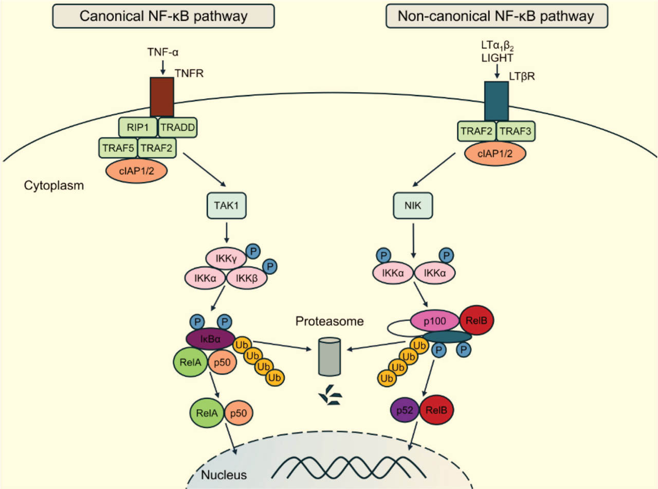
Activation of the canonical and noncanonical NF-κB pathways via TNFR (left) and LTβR (right). Pro-inflammatory cytokines such as TNF-α activate canonical NF-κB signaling through the recruitment of RIP1, TRADD, TRAF2/5, and cIAP1/2. TAK1 phosphorylates the IKK complex, composed of IKKα, IKKβ, and IKKγ. This results in phosphorylation, ubiquitination, and subsequent proteasomal degradation of IκBα. RelA:p50 dimers then translocate to the nucleus to initiate transcription. Noncanonical NF-κB signaling is mediated by LTβR and is activated by LTα1β2 or LIGHT. Receptor engagement induces the proteasomal degradation of TRAF3 and disrupts the TRAF2/3-cIAP1/2 complex, which normally targets NIK for degradation. NIK is thereby stabilized and activates IKKα, which phosphorylates p100, leading to its proteasomal processing into p52. RelB:p52 dimers translocate to the nucleus to drive transcription. cIAP1/2, cellular inhibitor of apoptosis protein 1/2; IKK, IκB kinase; IκBα, inhibitor of κBα; LIGHT, tumor necrosis factor ligand superfamily member 14; LTα1β2, lymphotoxin α1β2; LTβR, lymphotoxin β receptor; NIK, NF-κB-inducing kinase; P, phosphorylation; RIP1, receptor-interacting serine/threonine-protein kinase 1; TAK1, transforming growth factor-β-activated kinase 1; TNF-α, tumor necrosis factor α; TNFR, TNF receptor; TRADD, TNFR1-associated death domain protein; TRAF, tumor necrosis factor receptor-associated factor; Ub, ubiquitination.
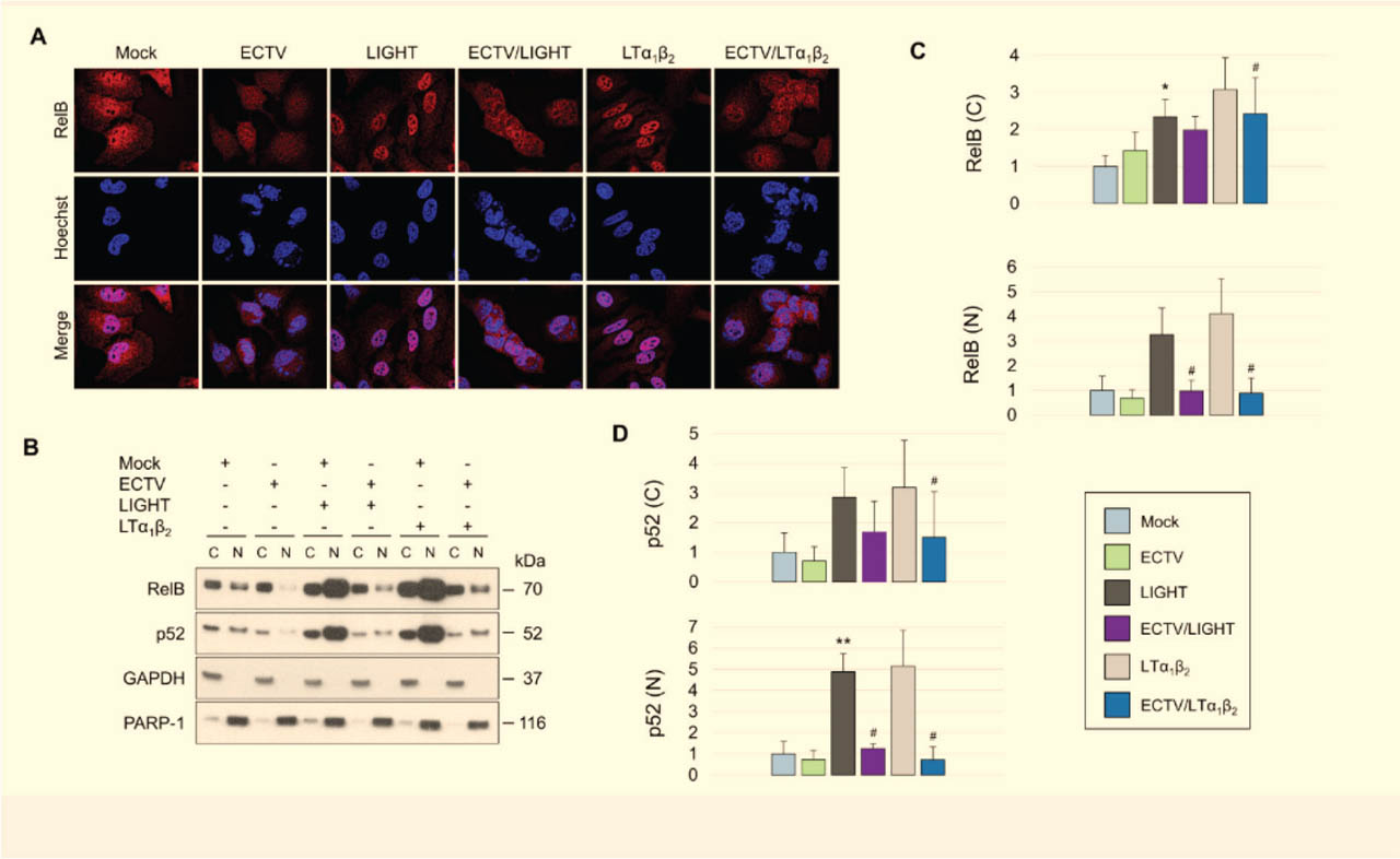
Fig 2.
ECTV inhibits LIGHT- and LTα1β2-induced nuclear accumulation of RelB and p52 in HeLa cells. (A) Representative immunofluorescence images of mock- and ECTV-infected HeLa cells that were left unstimulated or stimulated with LIGHT (TNFSF14) or LTα1β2. RelB was detected using a rabbit anti-RelB monoclonal antibody, followed by a rhodamine Red-X-conjugated goat anti-rabbit secondary antibody. Viral factories and nuclei were visualized with Hoechst. Images are representative of three independent biological replicates. (B) Western blot analysis of cytoplasmic and nuclear fractions from mock- and ECTV-infected HeLa cells that were unstimulated or stimulated with either LIGHT or LTα1β2. RelB and p52 were detected using rabbit anti-RelB and anti-p100/p52 primary antibodies, followed by HRP-conjugated goat anti-rabbit secondary antibodies. GAPDH and PARP-1 served as cytoplasmic and nuclear loading controls, respectively. (C,D) Densitometric quantification of cytoplasmic and nuclear RelB (C) and p52 (D) levels shown in (B). Data represent the mean of three independent biological experiments. Statistical significance was assessed using a paired t-test. *p ≤ 0.05 vs. mock; **p ≤ 0.01 vs. mock; #p ≤ 0.05 vs. LIGHT or LTα1β2. C, cytoplasmic; ECTV, ectromelia virus; GAPDH, glyceraldehyde 3-phosphate dehydrogenase; HRP, horseradish peroxidase; LTα1β2, lymphotoxin α1β2; N, nuclear; PARP-1, poly(ADP-ribose) polymerase 1; TNFSF14, tumor necrosis factor superfamily member 14.
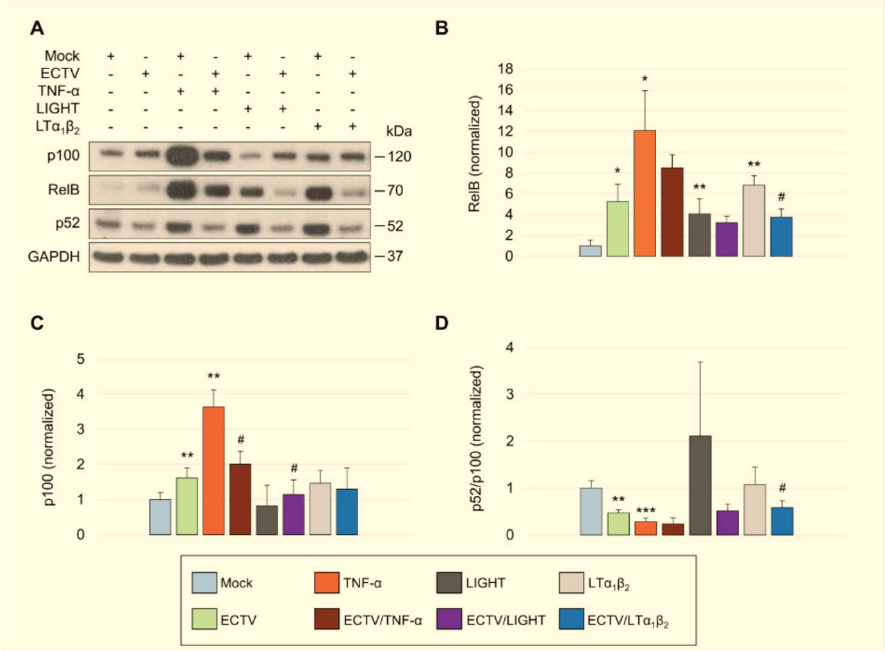
Fig 3.
ECTV differentially modulates RelB and p100 levels and the p52/p100 ratio in HeLa cells treated with TNF-α, LIGHT, or LTα1β2. (A) Western blot analysis of HeLa cells treated with TNF-α, LIGHT (TNFSF14), or LTα1β2 and either mock-infected or infected with ECTV. RelB, p100, and p52 were detected using rabbit anti-human primary antibodies followed by HRP-conjugated goat anti-rabbit secondary antibodies. GAPDH served as a loading control. (B–D) Densitometric quantification of RelB (B), p100 (C), and the p52/p100 ratio (D) from the blots shown in (A). Data represent the mean of two independent biological experiments. Statistical significance was assessed using a paired t-test. *p ≤ 0.05, **p ≤ 0.01, and ***p ≤ 0.001 vs. mock; #p ≤ 0.05 vs. TNF-α, LIGHT, or LTα1β2. ECTV, ectromelia virus; GAPDH, glyceraldehyde 3-phosphate dehydrogenase; HRP, horseradish peroxidase; LTα1β2, lymphotoxin α1β2; TNF-α, tumor necrosis factor α; TNFSF14, TNF superfamily member 14.
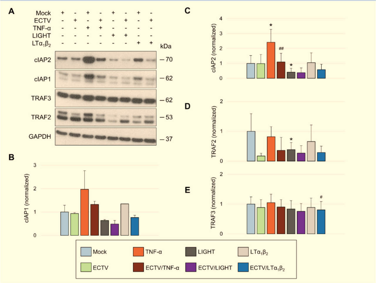
Fig 4.
ECTV reduces cIAP2 and TRAF3 levels in HeLa cells. (A) Western blot analysis of cIAP1, cIAP2, TRAF2, and TRAF3 in mock- and ECTV-infected HeLa cells that were left untreated or treated with TNF-α, LIGHT (TNFSF14), or LTα1β2. cIAP1/2 and TRAF2/3 were detected using rabbit anti-human primary antibodies followed by HRP-conjugated goat anti-rabbit secondary antibodies. GAPDH served as a loading control. (B–E) Densitometric quantification of cIAP1 (B), cIAP2 (C), TRAF2 (D), and TRAF3 (E) levels shown in (A). Data represent the mean of two independent biological experiments. Statistical significance was assessed using a paired t-test. *p ≤ 0.05 vs. mock; #p ≤ 0.05 vs. LTα1β2; ##p ≤ 0.01 vs. TNF-α. cIAP1/2, cellular inhibitor of apoptosis protein 1/2; ECTV, ectromelia virus; GAPDH, glyceraldehyde 3-phosphate dehydrogenase; HRP, horseradish peroxidase; LTα1β2, lymphotoxin α1β2; TNF-α, tumor necrosis factor α; TNFSF14, TNF superfamily member 14; TRAF2/3, TNF receptor-associated factor 2/3.
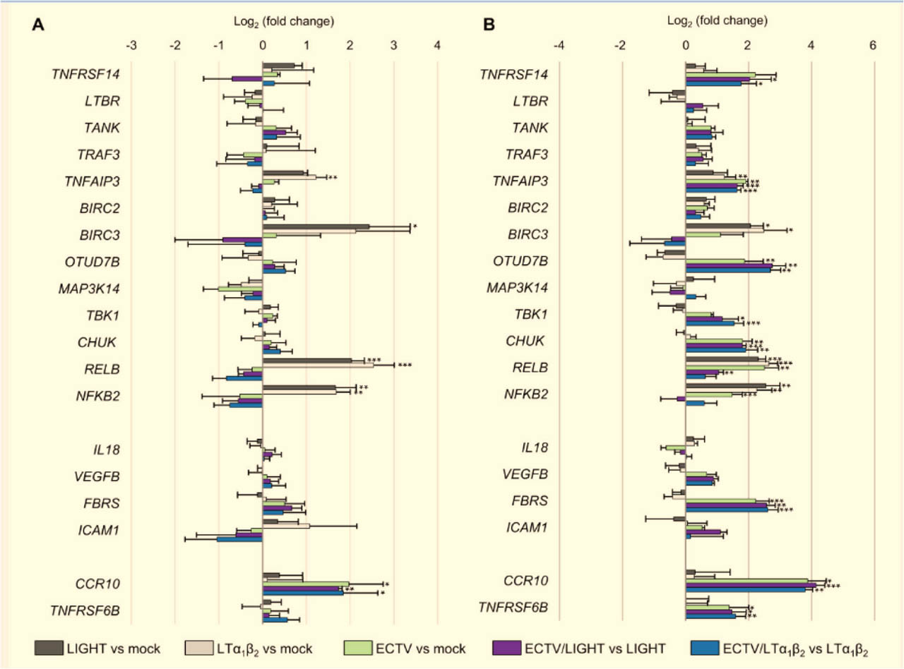
Fig 5.
ECTV alters the expression of genes encoding noncanonical NF-κB pathway components and selected RelB-regulated target genes in HeLa cells. RT-qPCR analysis of mock- and ECTV-infected HeLa cells that were left untreated or treated with LIGHT (TNFSF14) or LTα1β2 for 12 h (A) or 18 h (B). Data are presented as log2 fold changes in gene expression for the comparisons indicated in the figure. Results represent the mean of three independent biological experiments. Statistical significance was assessed using an unpaired Student's t-test (*p ≤ 0.05, **p ≤ 0.01, ***p ≤ 0.001). BIRC2/3, baculoviral IAP repeat-containing protein 2/3; CCR10, CC motif chemokine receptor 10; CHUK, conserved helix-loop-helix ubiquitous kinase; ECTV, ectromelia virus; FBRS, fibrosin; ICAM1, intercellular adhesion molecule 1; IL18, interleukin-18; LTα1β2, lymphotoxin α1β2; LTBR, lymphotoxin β receptor; MAP3K14, mitogen-activated protein kinase kinase kinase 14; NF-κB, nuclear factor κB; OTUD7B, OTU deubiquitinase 7B; RT-qPCR, reverse transcription quantitative PCR; TANK, TRAF family member-associated NF-κB activator; TBK1, TANK-binding kinase 1; TNFAIP3, TNF-α-induced protein 3; TNFRSF14, TNF receptor superfamily member 14; TNFRSF6B, TNF receptor superfamily member 6B; TNFSF14, TNF superfamily member 14; TRAF2/3, TNF receptor-associated factor 2/3; VEGFB, vascular endothelial growth factor B.