
1. Introduction
Nuclear factor κ-light-chain-enhancer of activated B cells (NF-κB) is a family of pleiotropic transcription factors comprising RelA (p65), RelB, c-Rel, NF-κB1 (p105/p50), and NF-κB2 (p100/p52) (Tas et al. 2023; Kaszycki and Kim 2025). Two major NF-κB signaling pathways have been described: the canonical and noncanonical pathways. The canonical NF-κB pathway is activated by pro-inflammatory cytokines, such as tumor necrosis factor (TNF)-α, microbe-associated molecular patterns, and cell-derived danger-associated molecular patterns (Kusiak and Brady 2022). Canonical NF-κB signaling involves activation of transforming growth factor β-activated kinase 1, which phosphorylates the IKKβ subunit of the inhibitor of κBα (IκBα) kinase complex (IKK). Activated IKK phosphorylates IκBα, an NF-κB inhibitor that sequesters RelA:p50 dimers in the cytoplasm of resting cells. Phosphorylation of IκBα at Ser32 and Ser36 triggers its ubiquitination and subsequent degradation by the 26S proteasome, allowing RelA:p50 dimers to translocate to the nucleus within minutes (Figure 1). Activated NF-κB dimers regulate expression of innate immunity genes across multiple cell types, coordinating diverse cell-mediated processes, including antiviral responses (Liu et al. 2017; Chawla et al. 2021; Kusiak and Brady 2022).
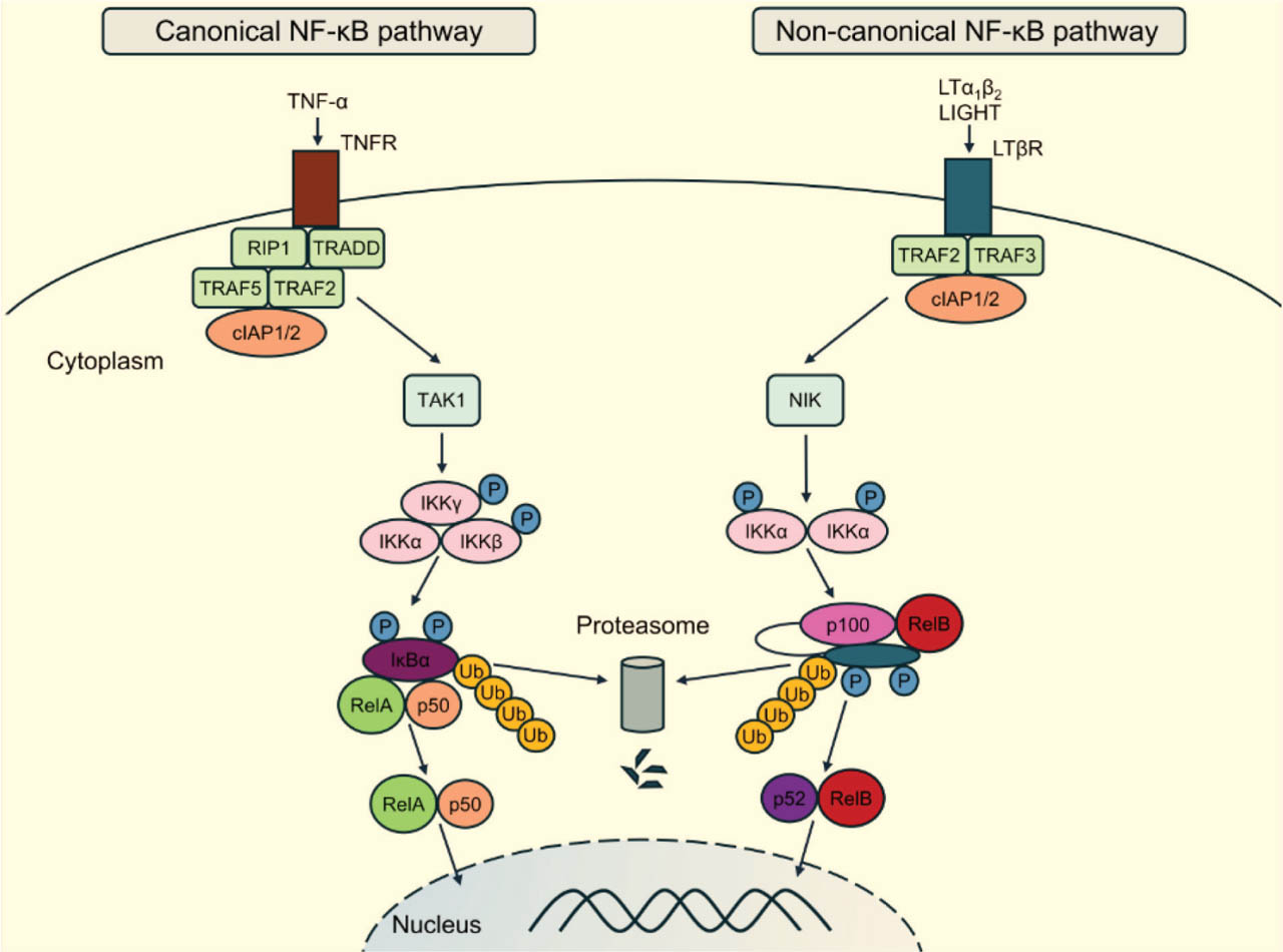
Fig 1.
Activation of the canonical and noncanonical NF-κB pathways via TNFR (left) and LTβR (right). Pro-inflammatory cytokines such as TNF-α activate canonical NF-κB signaling through the recruitment of RIP1, TRADD, TRAF2/5, and cIAP1/2. TAK1 phosphorylates the IKK complex, composed of IKKα, IKKβ, and IKKγ. This results in phosphorylation, ubiquitination, and subsequent proteasomal degradation of IκBα. RelA:p50 dimers then translocate to the nucleus to initiate transcription. Noncanonical NF-κB signaling is mediated by LTβR and is activated by LTα1β2 or LIGHT. Receptor engagement induces the proteasomal degradation of TRAF3 and disrupts the TRAF2/3-cIAP1/2 complex, which normally targets NIK for degradation. NIK is thereby stabilized and activates IKKα, which phosphorylates p100, leading to its proteasomal processing into p52. RelB:p52 dimers translocate to the nucleus to drive transcription. cIAP1/2, cellular inhibitor of apoptosis protein 1/2; IKK, IκB kinase; IκBα, inhibitor of κBα; LIGHT, tumor necrosis factor ligand superfamily member 14; LTα1β2, lymphotoxin α1β2; LTβR, lymphotoxin β receptor; NIK, NF-κB-inducing kinase; P, phosphorylation; RIP1, receptor-interacting serine/threonine-protein kinase 1; TAK1, transforming growth factor-β-activated kinase 1; TNF-α, tumor necrosis factor α; TNFR, TNF receptor; TRADD, TNFR1-associated death domain protein; TRAF, tumor necrosis factor receptor-associated factor; Ub, ubiquitination.
The noncanonical NF-κB signaling pathway is activated by certain TNF superfamily (TNFSF) ligands and by oncogenic viruses. TNFSF ligands, including TNF-α, LIGHT (TNFSF14), and lymphotoxin α1β2 (LTα1β2), are trimeric membrane-bound molecules expressed by lymphoid cells (Norris and Ware 2007; Šedý et al. 2014; Sun 2017; Suo et al. 2022). The lymphotoxin β receptor (LTβR), in turn, is present on macrophages, dendritic cells (DCs), epithelial cells, and stromal cells, enabling communication between lymphoid and nonlymphoid compartments of the host (Norris and Ware 2007; Koroleva et al. 2018).
LIGHT and LTα1β2 interact with LTβR, whose activation induces noncanonical NF-κB signaling through IKK and NF-κB-inducing kinase (NIK)-IKKα-mediated p100 processing (Saitoh et al. 2002; Piao et al. 2021). In resting cells, NIK is continuously degraded by an E3 ubiquitin ligase complex composed of cellular inhibitor of apoptosis proteins 1 (cIAP1) and cIAP2, TNF receptor-associated factor 2 (TRAF2), and TRAF3. During noncanonical signaling, TRAF3 is degraded by the cIAP1/2-TRAF2 complex, resulting in NIK stabilization. Stabilized NIK activates IKKα, which phosphorylates p100, an IκB-like inhibitor of RelB. Phosphorylated p100 is subsequently processed into p52, enabling RelB:p52 nuclear translocation and transcription of target genes (Yu et al. 2020) (Figure 1). Noncanonical NF-κB signaling is slower than canonical signaling and regulates lymphoid organogenesis, immune cell maturation, and antiviral innate immunity (Jin et al. 2014; Sun 2017; Chawla et al. 2021; Rodriguez et al. 2024; Kaszycki and Kim 2025).
Crosstalk between canonical and noncanonical pathways enhances immune responses. LIGHT can bind herpesvirus entry mediator (HVEM) on stromal and myeloid cells (Norris and Ware 2007; Šedý et al. 2014; Suo et al. 2022). LIGHT-HVEM signaling recruits TRAF2, leading to activation of serine kinases and NF-κB signaling. Simultaneous engagement of LTβR and HVEM, involving TRAF2 or TRAF5, amplifies NF-κB-mediated responses (Rooney et al. 2000). On the other hand, TRAF3 can inhibit HVEM-dependent activation of noncanonical NF-κB signaling (Hauer et al. 2005).
Noncanonical signaling is negatively regulated by proteins such as OTU deubiquitinase 7B (OTUD7B), which deubiquitinates TRAF3. In addition, IKKα and TRAF family member-associated NF-κB activator (TANK)-binding kinase 1 (TBK1) promote NIK degradation (Yu et al. 2020). In contrast, A20 acts as a negative regulator of canonical NF-κB signaling but promotes noncanonical signaling by stabilizing NIK by disrupting TRAF2-TRAF3 complexes upon LTβR stimulation (Yamaguchi et al. 2013).
In this study, we investigated how mousepox virus (ectromelia virus [ECTV]) modulates TNFSF-induced responses associated with the noncanonical NF-κB pathway in human HeLa epithelial cells. ECTV, a murine orthopoxvirus belonging to the Poxviridae family, is closely related to variola virus (VARV), the causative agent of smallpox (Shehata et al. 2025). Although smallpox was eradicated more than 40 years ago, VARV stocks are retained for biodefense purposes. Strains of vaccinia virus (VACV), a zoonotic orthopoxvirus closely related to VARV, were widely used as sources of vaccine antigens during the Smallpox Eradication Program launched by the World Health Organization (1966–1980) (Hsu et al. 2024). Zoonotic orthopoxviruses, including VACV, cowpox virus, and monkeypox virus, remain a global public health concern because of persistent animal reservoirs (Gieryńska et al. 2023; Hernaez and Alcamí 2024; Hsu et al. 2024; Gao et al. 2025). The recent global outbreak of monkeypox, with its human-to-human transmission and heightened risk for immunocompromised individuals, underscores the importance of orthopoxvirus research (Kangbai et al. 2025; Yahya et al. 2026).
Therefore, understanding orthopoxvirus pathogenesis is critical for developing antiviral countermeasures. VACV and ECTV are well-established models for studying host–pathogen interactions, immune evasion, and systemic infection (Brady and Bowie 2014; Garver et al. 2016; Huston et al. 2023; Hernaez and Alcamí 2024; Hsu et al. 2024). While poxvirus interference with canonical NF-κB signaling is well characterized (Brady and Bowie 2014; Lawler and Brady 2020; Reus et al. 2022), modulation of noncanonical signaling remains less understood. Our previous work showed that ECTV differentially modulates noncanonical NF-κB signaling in RAW 264.7 murine macrophages and JAWS II DCs (Struzik et al. 2020). Given the cell-type specificity of NF-κB signaling (Rodriguez et al. 2024) and the epitheliotropic nature of poxviruses (Diven 2001), we used HeLa cells as a nonimmune epithelial cell model. We show that ECTV suppresses LIGHT- and LTα1β2-induced nuclear accumulation of RelB and p52 while increasing RELB and NFKB2 gene expression and whole-cell RelB and NF-κB2/p100 protein levels. In contrast, ECTV infection alone did not significantly alter nuclear RelB accumulation. ECTV also alters the expression of genes and the abundance of proteins associated with noncanonical NF-κB signaling, apoptosis, and inflammation. Together, these findings suggest that ECTV modulates the expression and regulation of key molecular components of the noncanonical NF-κB pathway in epithelial cells in vitro.
2. Materials and Methods
2.1. Virus
This study used the Moscow strain of ectromelia virus (ECTV-MOS; ATCC VR-1374; ATCC, Manassas, VA, USA). The virus was grown and titrated in Vero cells (ATCC CCL-81; ATCC, Manassas, VA, USA). Following purification, viral stocks were stored at −70°C. Virus titer was quantified by plaque assay and reported as plaque-forming units per milliliter (PFU/mL).
2.2. Cell culture, infection, and treatment
HeLa cells (a human cervical carcinoma-derived epithelial cell line; ATCC CCL-2; ATCC, Manassas, VA, USA) were cultured as previously described (Boratyńska et al. 2010). Cells were seeded on 24-well plates with microscopic slides for immunofluorescence analysis (4 × 10⁴ cells/well), in 6-well plates for Western blotting or quantitative PCR with reverse transcription (RT-qPCR) (2 × 10⁵ cells/well), or in 25 cm2 flasks for nuclear and cytoplasmic fractionation (6 × 10⁵ cells/flask). Cells in all models were allowed to adhere for 24 h. Subsequently, cells were either left uninfected or infected with ECTV-MOS at a multiplicity of infection (MOI) of 5. Mock- and ECTV-infected cells were left untreated or stimulated with recombinant human (rh) LTα1β2 (100 ng/mL), LIGHT (100 ng/mL), or TNF-α (25 ng/mL) (all from Sigma-Aldrich, St. Louis, MO, USA). Cells were fixed or harvested after 12 h of stimulation and 12 h or 24 h of infection for immunofluorescence microscopy or Western blot analysis, respectively. For RT-qPCR, cells were stimulated with LIGHT or LTα1β2 and infected for 12 h or 18 h.
2.3. Fluorescence microscopy analysis
Staining for fluorescence microscopy analysis of RelB and DNA was performed as previously described (Struzik et al. 2020). Briefly, cells were fixed with 4% paraformaldehyde (Sigma-Aldrich) and incubated with anti-RelB rabbit monoclonal primary antibody (EPR7076, C-terminal; Abcam, Cambridge, MA, USA) followed by goat anti-rabbit IgG-Rhodamine Red-X secondary antibody (Jackson ImmunoResearch Laboratories, West Grove, PA, USA). Cellular and viral DNA was stained with Hoechst 33342 (Calbiochem, San Diego, CA, USA). Slides were mounted in Dako Mounting Medium (Agilent Technologies, Santa Clara, CA, USA) and imaged using an Olympus BX60 fluorescence microscope (Olympus, Tokyo, Japan) equipped with a PROMICAM 3-5CP camera (Promicra, Prague, Czech Republic). Image acquisition was performed with QuickPHOTO 2.3 (Promicra), and images were analyzed with CellSens Dimension 1.9 (Olympus) and ImageJ 1.48v (NIH, Bethesda, MD, USA).
2.4. Western blot
Total protein extracts, as well as nuclear and cytoplasmic fractions, were prepared and quantified as previously described (Struzik et al. 2020). Electrophoresis and transfer were performed using the Bolt™ system (Thermo Fisher Scientific, Waltham, MA, USA). Proteins were detected using the following rabbit primary antibodies: anti-RelB (EPR7076, C-terminal; Abcam), anti-NF-κB2 p100/p52, anti-c-IAP1, anti-cIAP2 (58C7), anti-TRAF2, anti-TRAF3, anti-poly(ADP-ribose) polymerase (PARP, 46D11), and anti-glyceraldehyde 3-phosphate dehydrogenase (GAPDH, D16H11) (all from Cell Signaling Technology, Danvers, MA, USA). Secondary goat anti-rabbit IgG-horseradish peroxidase (HRP) antibodies (Cell Signaling Technology) were used for chemiluminescence detection, as described previously (Struzik et al. 2020). Signal intensities of the analyzed bands were normalized to GAPDH or PARP and expressed relative to the corresponding control band (set to 1).
2.5. RNA isolation and RT-qPCR
RT-qPCR was performed in accordance with the Minimum Information for Publication of Quantitative Real-Time PCR Experiments (MIQE) guidelines (Bustin et al. 2009). Total RNA isolation, RNA concentration and purity assessment, reverse transcription to cDNA, and amplification were carried out as previously described (Struzik et al. 2020; Szulc-Dąbrowska et al. 2023). Genes associated with noncanonical NF-κB signaling were selected using the GeneCards and Harmonizome databases and analyzed with a RealTime ready Custom Panel (Roche Diagnostics GmbH, Mannheim, Germany). GAPDH, phosphoglycerate kinase 1, and acidic ribosomal phosphoprotein P0 were used as reference genes. Appropriate control genes were included to assess the presence of genomic DNA.
2.6. Statistical analysis
The results were analyzed using GraphPad Prism version 9 (GraphPad Software, La Jolla, CA, USA). Results from two (densitometric analysis of protein content in whole-cell lysates) or three independent experiments (densitometric analysis of cytoplasmic and nuclear fractions and RT-qPCR) are presented as the mean ± standard deviation. A paired t-test and an unpaired Student's t-test were used for statistical analysis. For all results, a p value ≤0.05 indicated statistical significance (*p ≤ 0.05, **p ≤ 0.01, ***p ≤ 0.001, #p ≤ 0.05, ##p ≤ 0.01, ###p ≤ 0.001).
3. Results
3.1. ECTV reduces LTα1β2- and LIGHT-induced nuclear accumulation of RelB and p52 in HeLa cells
We initially examined the subcellular distribution of RelB in HeLa cells. Immunofluorescence microscopy revealed RelB localization in both the cytoplasm and the nucleus of unstimulated cells. TNFSF ligands, including LIGHT and LTα1β2, induce activation of NIK, RelB, and p52 in HeLa cells (Mordmüller et al. 2003; Kim et al. 2005; Rauert et al. 2010; D'lgnazio et al. 2018). Accordingly, cells were treated with rhLTα1β2 or rhLIGHT for 12 h, at which time robust nuclear accumulation of RelB and p52 was observed. Since ECTV replicates in HeLa cells (Boratyńska et al. 2010; van Buuren et al. 2014), viral factories were detectable at 12 h post-infection (hpi). ECTV infection alone did not visibly alter the subcellular distribution of RelB. However, ECTV reduced the nuclear accumulation of RelB induced by LIGHT or LTα1β2 (Figure 2a).
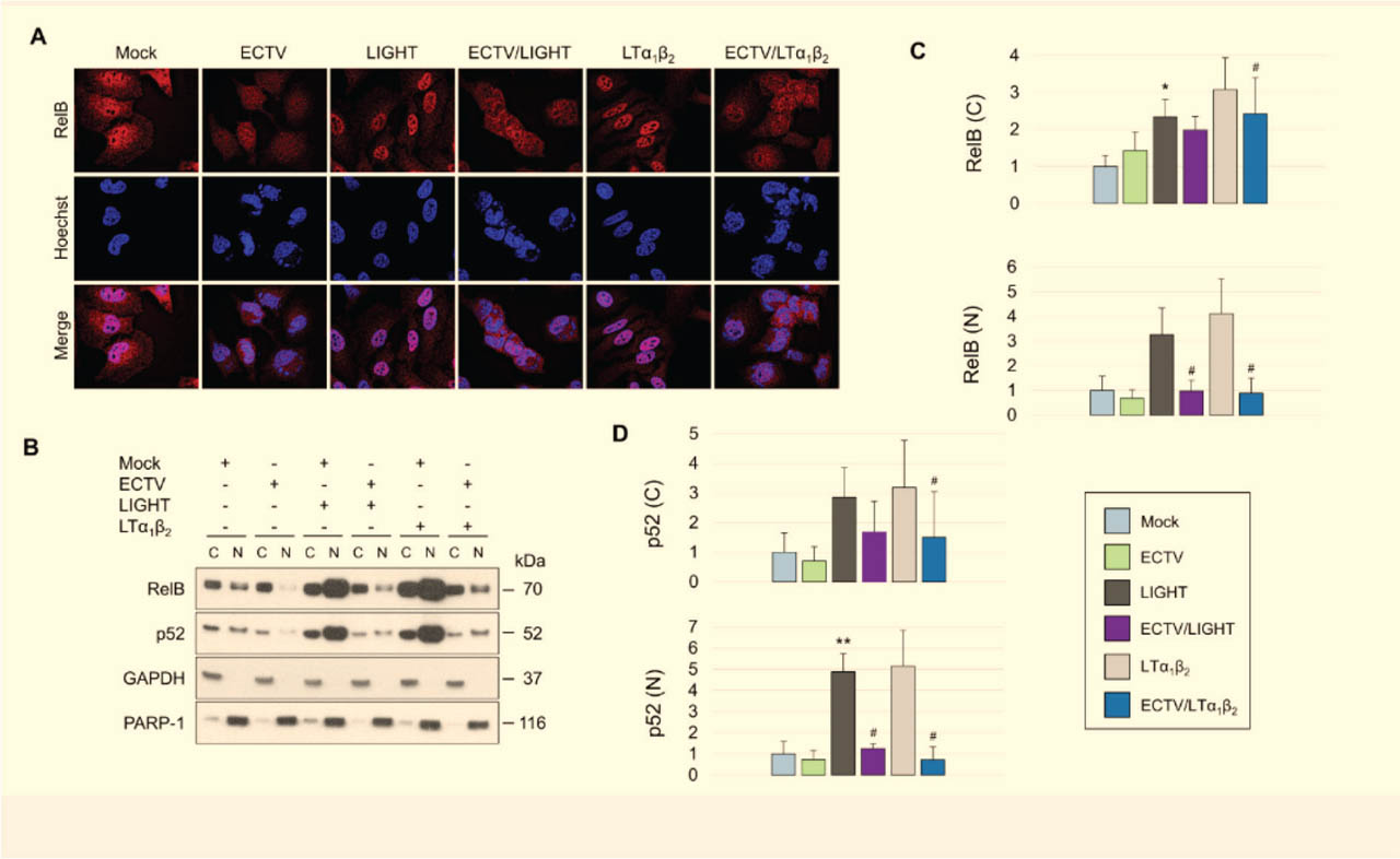
Fig 2.
ECTV inhibits LIGHT- and LTα1β2-induced nuclear accumulation of RelB and p52 in HeLa cells. (A) Representative immunofluorescence images of mock- and ECTV-infected HeLa cells that were left unstimulated or stimulated with LIGHT (TNFSF14) or LTα1β2. RelB was detected using a rabbit anti-RelB monoclonal antibody, followed by a rhodamine Red-X-conjugated goat anti-rabbit secondary antibody. Viral factories and nuclei were visualized with Hoechst. Images are representative of three independent biological replicates. (B) Western blot analysis of cytoplasmic and nuclear fractions from mock- and ECTV-infected HeLa cells that were unstimulated or stimulated with either LIGHT or LTα1β2. RelB and p52 were detected using rabbit anti-RelB and anti-p100/p52 primary antibodies, followed by HRP-conjugated goat anti-rabbit secondary antibodies. GAPDH and PARP-1 served as cytoplasmic and nuclear loading controls, respectively. (C,D) Densitometric quantification of cytoplasmic and nuclear RelB (C) and p52 (D) levels shown in (B). Data represent the mean of three independent biological experiments. Statistical significance was assessed using a paired t-test. *p ≤ 0.05 vs. mock; **p ≤ 0.01 vs. mock; #p ≤ 0.05 vs. LIGHT or LTα1β2. C, cytoplasmic; ECTV, ectromelia virus; GAPDH, glyceraldehyde 3-phosphate dehydrogenase; HRP, horseradish peroxidase; LTα1β2, lymphotoxin α1β2; N, nuclear; PARP-1, poly(ADP-ribose) polymerase 1; TNFSF14, tumor necrosis factor superfamily member 14.
To confirm the effect of ECTV on RelB subcellular distribution, we performed Western blot analyses of nuclear and cytoplasmic fractions. Levels of p52 were analyzed in parallel to assess the potential impact of ECTV on RelB:p52 dimer formation. Cells were stimulated with LIGHT or LTα1β2 to activate the noncanonical NF-κB pathway (Figure 2b).
Quantitative analyses revealed that ECTV significantly reduced cytoplasmic RelB and p52 levels in LTα1β2-treated cells (Figure 2c,d). In addition, nuclear RelB and p52 levels were significantly decreased in cells stimulated with either LIGHT or LTα1β2 (Figure 2c,d). In contrast, no significant changes in nuclear or cytoplasmic RelB levels were observed following ECTV infection alone, although a tendency toward decreased nuclear RelB and increased cytoplasmic RelB was noted (Figure 2c).
Collectively, these data demonstrate that ECTV suppresses LIGHT- and LTα1β2-induced nuclear accumulation of RelB and p52 in HeLa cells.
3.2. ECTV differentially modulates RelB and p100 levels and the p52/p100 ratio in HeLa cells treated with TNF-α, LIGHT, or LTα1β2
To investigate whether ECTV modulates the protein levels of RelB and p100/p52, key components of noncanonical NF-κB signaling, we performed Western blot analysis of whole-cell extracts from mock- and ECTV-infected HeLa cells. Cells were treated with TNF-α, a potent activator of canonical NF-κB signaling (Kusiak and Brady 2022), which induces transcription of the RELB and NFKB2 genes encoding RelB and p100, respectively (Madge and May 2010). Cells were also stimulated with either LIGHT or LTα1β2 for 12 h, since RelB regulation depends on RelB-containing signaling complexes (Bren et al. 2001).
We first assessed whole-cell RelB protein levels. Treatment with TNF-α, LIGHT, or LTα1β2 significantly increased RelB abundance. ECTV infection alone also significantly increased whole-cell RelB levels. In LTα1β2-stimulated cells, ECTV significantly reduced whole-cell RelB abundance compared with mock-infected stimulated controls (Figure 3a,b). Thus, although ECTV increased whole-cell RelB abundance, this increase was not accompanied by a significant increase in nuclear RelB accumulation under basal conditions (Figure 2), indicating that changes in total RelB levels did not directly translate into altered nuclear localization.
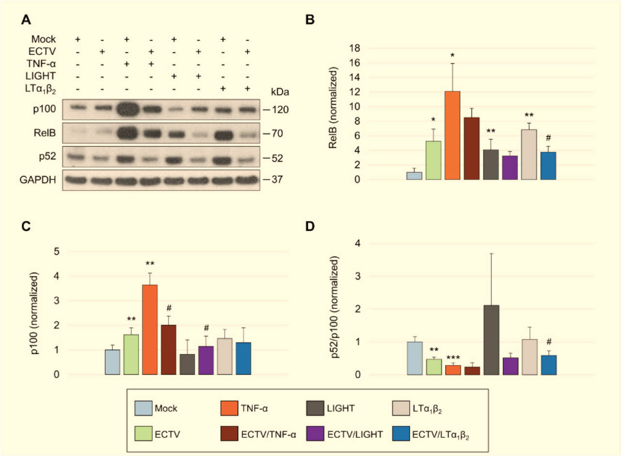
Fig 3.
ECTV differentially modulates RelB and p100 levels and the p52/p100 ratio in HeLa cells treated with TNF-α, LIGHT, or LTα1β2. (A) Western blot analysis of HeLa cells treated with TNF-α, LIGHT (TNFSF14), or LTα1β2 and either mock-infected or infected with ECTV. RelB, p100, and p52 were detected using rabbit anti-human primary antibodies followed by HRP-conjugated goat anti-rabbit secondary antibodies. GAPDH served as a loading control. (B–D) Densitometric quantification of RelB (B), p100 (C), and the p52/p100 ratio (D) from the blots shown in (A). Data represent the mean of two independent biological experiments. Statistical significance was assessed using a paired t-test. *p ≤ 0.05, **p ≤ 0.01, and ***p ≤ 0.001 vs. mock; #p ≤ 0.05 vs. TNF-α, LIGHT, or LTα1β2. ECTV, ectromelia virus; GAPDH, glyceraldehyde 3-phosphate dehydrogenase; HRP, horseradish peroxidase; LTα1β2, lymphotoxin α1β2; TNF-α, tumor necrosis factor α; TNFSF14, TNF superfamily member 14.
Whole-cell p100 protein levels were significantly elevated following TNF-α treatment (Figure 3c), whereas no significant changes were observed after stimulation with LIGHT or LTα1β2. ECTV infection also increased p100 levels. Notably, ECTV reduced the TNF-α-induced elevation in p100 protein levels. In contrast, p100 levels were modestly but significantly elevated in LIGHT-treated ECTV-infected cells compared with mock-infected LIGHT-treated controls (Figure 3c).
To evaluate the effect of ECTV on p100 processing, we next analyzed the p52/p100 ratio. Cell treatment with TNF-α resulted in a significant reduction in the p52/p100 ratio, whereas no significant changes were observed in LIGHT- or LTα1β2-treated cells. ECTV infection significantly decreased the p52/p100 ratio in untreated and LTα1β2-treated cells, indicating an altered balance between p100 and p52 (Figure 3d).
3.3. ECTV reduces cIAP2 and TRAF3 levels in HeLa cells
The cIAP1/2-TRAF2-TRAF3 complex regulates NIK stability and noncanonical NF-κB signaling (Yu et al. 2020). To determine whether ECTV alters the abundance of components of the NIK-regulatory complex, we evaluated the protein levels of cIAP1/2 and TRAF2/3 in mock- and ECTV-infected HeLa cells. Because canonical NF-κB target genes encode cIAP1, cIAP2, and TRAF2 (Wang et al. 1998; Bernard et al. 2001), cells were stimulated with TNF-α. In parallel, cells were treated with either LIGHT or LTα1β2 to determine whether stimulation of the noncanonical NF-κB pathway influences the abundance of NIK-regulatory complex components and whether these responses are altered by ECTV infection.
ECTV infection did not significantly affect cIAP1 protein levels in cells stimulated with TNF-α, LIGHT, or LTα1β2 (Figure 4a,b). In contrast, TNF-α stimulation significantly increased cIAP2 abundance, whereas ECTV infection markedly reduced cIAP2 levels in TNF-α-treated cells. LIGHT, but not LTα1β2, also decreased cIAP2 levels; however, ECTV infection did not further alter the reduced cIAP2 presence under these conditions (Figure 4c).
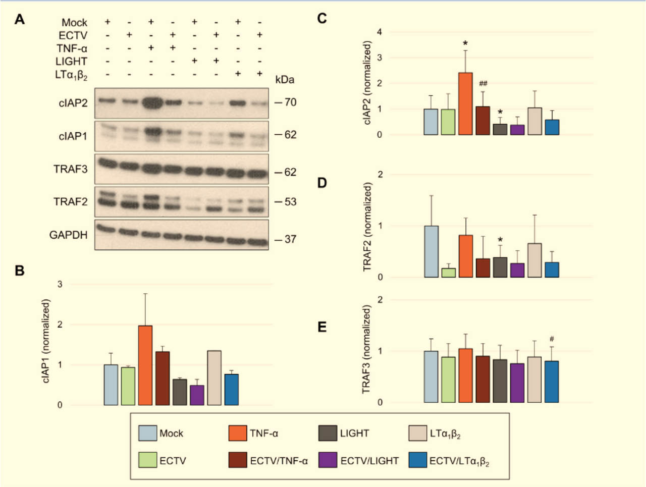
Fig 4.
ECTV reduces cIAP2 and TRAF3 levels in HeLa cells. (A) Western blot analysis of cIAP1, cIAP2, TRAF2, and TRAF3 in mock- and ECTV-infected HeLa cells that were left untreated or treated with TNF-α, LIGHT (TNFSF14), or LTα1β2. cIAP1/2 and TRAF2/3 were detected using rabbit anti-human primary antibodies followed by HRP-conjugated goat anti-rabbit secondary antibodies. GAPDH served as a loading control. (B–E) Densitometric quantification of cIAP1 (B), cIAP2 (C), TRAF2 (D), and TRAF3 (E) levels shown in (A). Data represent the mean of two independent biological experiments. Statistical significance was assessed using a paired t-test. *p ≤ 0.05 vs. mock; #p ≤ 0.05 vs. LTα1β2; ##p ≤ 0.01 vs. TNF-α. cIAP1/2, cellular inhibitor of apoptosis protein 1/2; ECTV, ectromelia virus; GAPDH, glyceraldehyde 3-phosphate dehydrogenase; HRP, horseradish peroxidase; LTα1β2, lymphotoxin α1β2; TNF-α, tumor necrosis factor α; TNFSF14, TNF superfamily member 14; TRAF2/3, TNF receptor-associated factor 2/3.
We next examined the effects of ECTV on the adaptor proteins TRAF2 and TRAF3. Stimulation with TNF-α or LTα1β2 did not significantly affect TRAF2 levels, whereas LIGHT treatment resulted in a reduction in TRAF2 protein levels. ECTV infection did not significantly alter TRAF2 levels under any of the conditions tested (Figure 4d). TRAF3 levels were comparable between control cells and cells stimulated with TNF-α, LIGHT, or LTα1β2. Notably, ECTV infection significantly reduced TRAF3 abundance only in LTα1β2-treated cells, without a significant effect observed in TNF-α- or LIGHT-stimulated cells (Figure 4e). These results indicate that ECTV differentially modulates pro-survival and adaptor protein levels involved in TNFSF signaling.
3.4. ECTV alters the expression of genes encoding noncanonical NF-κB pathway components and selected RelB-regulated target genes in HeLa cells
To assess whether ECTV alters the expression of genes associated with the noncanonical NF-κB pathway in HeLa cells, mock- and ECTV-infected cells were stimulated with either LIGHT or LTα1β2. RT-qPCR analysis was performed after 12 h and 18 h of stimulation and/or infection. Next, we assessed the expression of genes encoding key components and regulators of the noncanonical NF-κB pathway. These included genes encoding HVEM and LTβR (TNF receptor superfamily member 14 [TNFRSF14] and LTBR); the adaptor proteins TANK and TRAF3 (TANK and TRAF3); the regulatory enzyme A20 (TNF-α-induced protein 3 [TNFAIP3]); antiapoptotic cIAP1 and cIAP2 proteins (baculoviral IAP repeat-containing protein 2/3 [BIRC2 and BIRC3]); the deubiquitinase OTUD7B (OTUD7B); NIK, TBK1, and IKKα kinases involved in the noncanonical NF-κB pathway regulation (mitogen-activated protein kinase kinase kinase 14 [MAP3K14], TBK1, and conserved helix-loop-helix ubiquitous kinase [CHUK]); and transcriptional regulators (RELB and NFKB2). In addition, we analyzed selected RelB-regulated target genes, including interleukin-18 [IL18], vascular endothelial growth factor B [VEGFB], FBRS, intercellular adhesion molecule 1 [ICAM1], and the RelB-dependent receptor genes CC motif chemokine receptor 10 (CCR10) and TNFRSF6B.
At 12 h post-stimulation, LIGHT significantly increased the expression of BIRC3, RELB, and NFKB2, whereas LTα1β2 stimulation upregulated TNFAIP3, RELB, and NFKB2. At this time point, ECTV infection did not cause statistically significant changes in the expression of noncanonical NF-κB pathway components (Figure 5a).
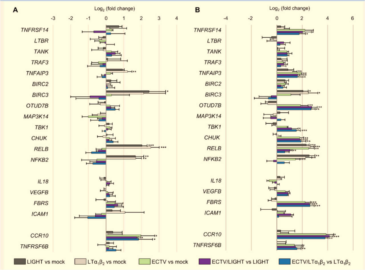
Fig 5.
ECTV alters the expression of genes encoding noncanonical NF-κB pathway components and selected RelB-regulated target genes in HeLa cells. RT-qPCR analysis of mock- and ECTV-infected HeLa cells that were left untreated or treated with LIGHT (TNFSF14) or LTα1β2 for 12 h (A) or 18 h (B). Data are presented as log2 fold changes in gene expression for the comparisons indicated in the figure. Results represent the mean of three independent biological experiments. Statistical significance was assessed using an unpaired Student's t-test (*p ≤ 0.05, **p ≤ 0.01, ***p ≤ 0.001). BIRC2/3, baculoviral IAP repeat-containing protein 2/3; CCR10, CC motif chemokine receptor 10; CHUK, conserved helix-loop-helix ubiquitous kinase; ECTV, ectromelia virus; FBRS, fibrosin; ICAM1, intercellular adhesion molecule 1; IL18, interleukin-18; LTα1β2, lymphotoxin α1β2; LTBR, lymphotoxin β receptor; MAP3K14, mitogen-activated protein kinase kinase kinase 14; NF-κB, nuclear factor κB; OTUD7B, OTU deubiquitinase 7B; RT-qPCR, reverse transcription quantitative PCR; TANK, TRAF family member-associated NF-κB activator; TBK1, TANK-binding kinase 1; TNFAIP3, TNF-α-induced protein 3; TNFRSF14, TNF receptor superfamily member 14; TNFRSF6B, TNF receptor superfamily member 6B; TNFSF14, TNF superfamily member 14; TRAF2/3, TNF receptor-associated factor 2/3; VEGFB, vascular endothelial growth factor B.
We next assessed gene expression at 18 h post-stimulation. LIGHT treatment significantly increased the expression of BIRC3, RELB, and NFKB2, whereas LTα1β2 stimulation upregulated TNFAIP3, BIRC3, RELB, and NFKB2. Compared with mock-infected cells, ECTV infection significantly increased the expression of TNFAIP3, OTUD7B, CHUK, RELB, and NFKB2. In LIGHT-treated cells, ECTV further upregulated TNFRSF14, TNFAIP3, OTUD7B, TBK1, CHUK, and RELB. Similarly, in LTα1β2-treated cells, ECTV increased the expression of TNFRSF14, TNFAIP3, OTUD7B, TBK1, and CHUK genes (Figure 5b).
We also examined the effect of ECTV on the expression of RelB-regulated target genes. No statistically significant changes were detected in IL18, VEGFB, or ICAM1 expression following LIGHT, LTα1β2, or ECTV treatment. In contrast, FBRS expression was significantly increased in ECTV-infected cells, irrespective of LIGHT or LTα1β2 stimulation at 18 hpi (Figure 5b).
We further analyzed the expression of RelB-dependent receptor genes CCR10 and TNFRSF6B at 12 hpi and 18 hpi. Although LIGHT or LTα1β2 stimulation alone did not affect CCR10 expression at either time point, ECTV challenge significantly increased CCR10 expression at both time points. Moreover, CCR10 expression was higher in ECTV-infected cells treated with LIGHT or LTα1β2 than in cells treated with either ligand alone (Figure 5a,b). At 18 hpi, ECTV also markedly increased TNFRSF6B expression in mock-, LIGHT-, and LTα1β2-treated cells (Figure 5b). Together, these findings demonstrate that ECTV broadly remodels the transcriptional landscape of genes associated with the noncanonical NF-κB pathway and selected RelB-responsive targets.
4. Discussion
Noncanonical NF-κB signaling orchestrates interactions between lymphoid and nonlymphoid cells through TNFSF ligands and plays a critical role in antiviral immunity (Norris and Ware 2007; Koroleva et al. 2018). Both oncogenic and non-oncogenic viruses exploit noncanonical NF-κB signaling to promote viral latency, oncogenic transformation, and replication (Sun 2017; Struzik and Szulc-Dąbrowska 2019).
Poxviruses encode viral TNF receptor homologs (vTNFRs) capable of binding TNFSF members, including TNF-α, LTα, and LTβ (Alejo et al. 2018). However, the extent to which poxviruses modulate TNFSF-induced noncanonical NF-κB signaling remains poorly defined. Because poxviruses exhibit pronounced epitheliotropism (Diven 2001), we employed a nonimmune epithelial HeLa cell model to investigate how ECTV modulates TNFSF-regulated noncanonical NF-κB signaling in vitro.
The inhibitory effect of ECTV on RelB nuclear translocation has previously been demonstrated in immune-derived RAW 264.7 macrophages treated with lipopolysaccharide (LPS), LPS plus interferon (IFN)-γ, or phorbol 12-myristate 13-acetate (PMA) plus ionomycin (Io), as well as in JAWS II DCs stimulated with PMA plus Io (Struzik et al. 2020). Similarly, ECTV inhibits p52 nuclear translocation in cells treated with LPS together with IFN-γ (Struzik et al. 2020). The present study extends these observations to epithelial cells stimulated with LIGHT and LTα1β2, suggesting that ECTV employs a broad strategy to suppress noncanonical NF-κB activation across multiple cell types and stimuli.
ECTV-mediated inhibition of cytokine-induced nuclear accumulation of RelB and p52 was accompanied by changes in whole-cell p100 abundance and a reduced p52/p100 ratio, suggesting that altered regulation of p100 processing may contribute to these effects. In this study, ECTV increased p100 protein levels in unstimulated and LIGHT-stimulated cells, but decreased p100 levels upon TNF-α treatment, and reduced the p52/p100 ratio in HeLa cells. Similar inhibition of p100 processing has been observed in ECTV-infected RAW 264.7 and JAWS II cells (Struzik et al. 2020). However, NFKB2 expression was elevated only in ECTV-infected cells. In contrast, ECTV suppresses Nfkb2 expression in RAW 264.7 cells stimulated with polyinosinic:polycytidylic acid (poly[I:C]) or IFN-γ together with LPS at 18 hpi (Struzik et al. 2020). These observations indicate that ECTV differentially modulates the balance between p100 and p52 in a cell type- and stimulus-dependent manner.
Notably, ECTV can also disrupt NF-κB signaling through proteasome inhibition mediated by the ankyrin/F-box protein EVM005, which blocks the Skp1-Cullin 1-F-box protein ubiquitin ligase complex containing β-transducin repeat-containing protein (β-TrCP), thereby preventing IκBα degradation and p65 nuclear translocation (van Buuren et al. 2014). Such a mechanism may also contribute to ECTV-mediated interference with p100 processing.
Regulation of p100 synthesis, and thus noncanonical NF-κB signaling, is linked to canonical NF-κB activation. TNF-α, a prototypical TNFSF cytokine, activates RelA:p50 dimers and increases p100 levels in HeLa cells (Mordmüller et al. 2003; D'lgnazio et al. 2018). TNF-α is a key mediator of early antiviral inflammation, promoting macrophage and DC activation and inducing antimicrobial responses in epithelial cells (Alvarez-de Miranda et al. 2021; Savenkova et al. 2024). Here, we demonstrate that ECTV inhibits TNF-α-induced p100 synthesis, indicating concurrent interference with both canonical and noncanonical NF-κB signaling. This effect may be mediated by the viral cytokine response modifier D (CrmD), a vTNFR homolog that inhibits murine and human TNF-α activity (Pontejo et al. 2015; Alvarez-de Miranda et al. 2021). CrmD is a critical determinant of mousepox lethality and confers protection when used as a vaccine antigen (Alejo et al. 2018).
Mechanistically, p100, the precursor of p52, functions as an inhibitor of RelB, whose expression is positively regulated by RelA:p50 and RelB:p50 dimers in HeLa cells (Bren et al. 2001). Although whole-cell RelB and p100 protein levels were increased, this was not accompanied by increased nuclear RelB in unstimulated ECTV-infected cells and, therefore, should not be interpreted as evidence of constitutive activation of the noncanonical NF-κB pathway. Rather, these findings indicate increased expression of pathway components without clear evidence of their nuclear activation. Although subcellular fractionation revealed a trend toward increased cytoplasmic RelB and decreased nuclear RelB, neither change reached statistical significance. Therefore, while the observed pattern is consistent with reduced nuclear translocation of RelB, the present data do not provide conclusive evidence of altered subcellular localization.
Consistently, ECTV inhibits RelB synthesis in unstimulated and poly(I:C)-stimulated RAW 264.7 and JAWS II cells, but not in cells treated with Pam3CSK4 or IFN-γ together with LPS (Struzik et al. 2020). Moreover, ECTV reduces RelB expression in granulocyte-macrophage colony-stimulating factor-derived bone marrow cells from BALB/c mice (Szulc-Dąbrowska et al. 2017). Given that RelB negatively regulates type I IFN (IFN-I) signaling in DCs (Saha et al. 2020) and coordinates pro-inflammatory responses in epithelial cells (Tully et al. 2012), dysregulation of RelB may represent a conserved viral strategy to impair antiviral immunity across diverse cell types.
Activation of noncanonical NF-κB signaling relies on NIK stabilization, regulated by cIAPs, TRAFs, and IKKα (Razani et al. 2010). Although ECTV modulates TRAF2 levels in immune cells (Struzik et al. 2020), our studies clearly show that ECTV selectively reduces TRAF3 levels in LTα1β2-stimulated HeLa cells. TRAF2 and TRAF3 regulate IFN-I responses in a cell-specific manner (Xie et al. 2019), and TRAF3 acts as a negative regulator of IFN responses during DNA virus infection (Parvatiyar et al. 2018). Because TRAF3 is regulated by the deubiquitinase OTUD7B, which negatively controls noncanonical NF-κB signaling and IFN-I responses (Hu et al. 2013; Xie et al. 2022), modulation of OTUD7B by ECTV may further refine antiviral signaling. Indeed, ECTV upregulated OTUD7B expression in HeLa cells but did not upregulate Otud7b expression in RAW 264.7 cells (Struzik et al. 2020).
The mRNA level of the gene encoding TBK1, which regulates both IFN production and NIK degradation (Yu et al. 2020), was also upregulated by ECTV in LIGHT- or LTα1β2-stimulated HeLa cells. TBK1 phosphorylation during ECTV infection has been shown to be cell type-dependent (Cheng et al. 2018), further supporting the notion that ECTV differentially shapes antiviral signaling in distinct tissues. Since IFN signaling exerts a protective role in skin poxvirus infection (Harbour et al. 2023), modulation of these pathways may be particularly relevant for viral immune evasion in epithelial cells.
ECTV also upregulated TNFAIP3, a RelB-regulated gene encoding the anti-inflammatory protein A20, an activator of noncanonical NF-κB signaling (Vereecke et al. 2011; Yamaguchi et al. 2013; Bai et al. 2022). Considering the increased susceptibility of A20-mutant mice to ECTV infection (Zammit et al. 2019), induction of A20 may represent a viral strategy to dampen inflammatory responses in epithelial cells.
Pro-survival genes, such as BIRC3, which encodes cIAP2, are additional viral targets. Because cIAP2 inhibits caspase-8-mediated apoptosis downstream of TNF-α signaling (Mahoney et al. 2008), ECTV-mediated reduction of TNF-α-induced cIAP2 levels may allow fine control of inflammation and cell survival. This is consistent with previous findings in RAW 264.7 cells (Struzik et al. 2020).
We also observed that ECTV increased expression of FBRS, encoding fibrosin (FBRS), a RelB:p52-regulated fibrogenic lymphokine produced by fibroblasts, CD4+ T cells, and other cell types (Wyler 1996; Prakash and Robbins 2001). FBRS has been implicated in chronic inflammation (Prakash et al. 1995). This finding suggests a potential role for FBRS in orthopoxvirus infection that warrants further investigation.
Finally, epithelial immunity relies on noncanonical NF-κB-regulated receptors such as CCR10, DcR3, and HVEM (Hsieh and Lin 2017; Hou et al. 2022; Willuveit et al. 2025). ECTV-induced upregulation of CCR10 and TNFRSF6B (DcR3) in HeLa cells suggests novel mechanisms by which orthopoxviruses may modulate epithelial immune responses. Because DcR3 neutralizes LIGHT activity and lacks a murine homolog, its induction may be particularly relevant to human orthopoxvirus infection (Hsieh and Lin 2017).
We also observed increased expression of TNFRSF14, encoding HVEM, in ECTV-infected cells treated with LIGHT or LTα1β2. LIGHT-HVEM signaling promotes the development of memory CD8+ T cells during VACV infection (Desai et al. 2018), highlighting the importance of TNFSF-NF-κB interactions in shaping antiviral immunity and vaccine responses.
A limitation of this study is the use of human epithelial cells to investigate a murine orthopoxvirus. Although this system enabled mechanistic analysis of virus-induced modulation of noncanonical NF-κB pathway components, future studies in murine epithelial cells and mousepox in vivo models will be important to validate these findings in the natural host context.
Collectively, these findings underscore the complexity of poxvirus-mediated modulation of NF-κB signaling and provide a framework for future studies aimed at developing antiviral and vaccine strategies.
5. Conclusions
In conclusion, this study identifies previously unrecognized strategies through which ECTV differentially regulates noncanonical NF-κB signaling in epithelial cells in vitro.
We show that ECTV suppresses LIGHT- and LTα1β2-induced nuclear accumulation of RelB and p52 while altering the expression of genes and proteins associated with the noncanonical NF-κB pathway. Our data do not provide evidence that the increased whole-cell expression of RelB and NF-κB2/p100 is accompanied by activation of the noncanonical NF-κB pathway. These findings demonstrate that ECTV remodels the molecular landscape of noncanonical NF-κB signaling and its associated inflammatory and apoptosis-related regulators in nonimmune cells. The precise molecular mechanisms and functional consequences of these alterations during orthopoxvirus infection remain to be elucidated.
Abbreviations
- CCR10,
CC motif chemokine receptor 10
- CHUK,
conserved helix-loop-helix ubiquitous kinase
- cIAP,
cellular inhibitor of apoptosis
- cIAP1/2,
cellular IAP protein 1/2
- CrmD,
cytokine response modifier D
- DC,
dendritic cell
- DcR3,
decoy receptor 3
- ECTV,
ectromelia virus
- ECTV-MOS,
ECTV strain Moscow
- FBRS,
fibrosin
- GAPDH,
glyceraldehyde 3-phosphate dehydrogenase
- hpi,
hours post-infection
- HRP,
horseradish peroxidase
- HVEM,
herpesvirus entry mediator
- ICAM1,
intercellular adhesion molecule 1
- IFN-I,
type I interferon
- IFN-γ,
interferon-γ
- IKK,
IκBα kinase complex
- IL,
interleukin
- Io,
ionomycin
- IκBα,
inhibitor of κBα
- LPS,
lipopolysaccharide
- LTα1β2,
lymphotoxin α1β2
- LTβR,
LTβ receptor
- MAP3K14,
mitogen-activated protein kinase kinase kinase 14
- MIQE,
minimum information for publication of quantitative real-time PCR experiments
- NF-κB,
nuclear factor κ-light-chain-enhancer of activated B cells
- NIK,
NF-κB-inducing kinase
- OTUD7B,
OTU deubiquitinase 7B
- PARP,
poly(ADP-ribose) polymerase
- PFU,
plaque-forming unit
- PMA,
phorbol 12-myristate 13-acetate
- poly I:C,
polyinosinic:polycytidylic acid
- rh,
recombinant human
- RT-qPCR,
quantitative PCR with reverse transcription
- SD,
standard deviation
- TAK1,
transforming growth factor β-activated kinase 1
- TANK,
TRAF family member-associated NF-κB activator
- TBK1,
TANK-binding kinase 1
- TNFAIP3,
TNF-α-induced protein 3
- TNFR,
TNF receptor
- TNFRSF,
TNFR superfamily
- TNFSF,
TNF superfamily
- TNF-α,
tumor necrosis factor α
- TRAF2/3,
TNFR-associated factor 2/3
- VACV,
vaccinia virus
- VARV,
variola virus
- VEGFB,
vascular endothelial growth factor B
- vTNFR,
viral TNFR
Acknowledgments
RT-qPCR experiments were performed with CePT infrastructure financed by the European Regional Development Fund allocated by the European Union (Innovative economy 2007– 13, Agreement POIG.02.02.00–14–024/08–00).
Notes
[1] Financial disclosure Funding
This work was supported by grant no. UMO-2015/19/D/NZ6/02873 from the National Science Centre, Poland.
[2] Contributed by Author Contributions
Conceptualization— JS; methodology and validation—JS, LS-D, and MK; formal analysis and investigation—JS and LS-D; resources—MK and MG; data curation, JS, LS-D, and MK; writing—original draft preparation, JS; writing— review and editing, LS-D, MK, and MG; supervision—JS; project administration—JS; funding acquisition—JS. All authors have read and agreed to the published version of the manuscript.