
1. Introduction
Sepsis is a fatal systemic inflammatory syndrome caused by a dysregulated host response to infection, often rapidly progressing to multiple organ dysfunction. Among the affected organs, the kidneys are particularly vulnerable due to their unique hemodynamic and hypermetabolic characteristics, leading to sepsis-associated acute kidney injury (SA-AKI), which is a detrimental complication determining patient prognosis (Mou et al. 2023; Zhang et al. 2023). SA-AKI not only significantly increases the risk of short-term mortality but is also closely associated with the subsequent development of chronic kidney disease and decreased long-term survival. However, current clinical management strategies primarily rely on supportive measures, such as renal replacement therapy, and effective interventions targeting its core pathogenesis remain limited, resulting in unsatisfactory therapeutic efficacy and a bottleneck in further improving patient prognosis (Jiang et al. 2020).
During the pathological process of sepsis, the inflammatory response is a core element driving disease progression and organ dysfunction (Li et al. 2024). It begins with the overactivation of the body’s innate immune system by pathogen-associated molecular patterns and damage-associated molecular patterns. This uncontrolled inflammatory cascade directly damages vascular and organ endothelial barriers, induces widespread apoptosis, and ultimately contributes to organ failure, with renal impairment being one of the manifestations (Liu et al. 2024).
Nimbolide, a limonoid compound isolated from Azadirachta indica, has attracted considerable attention for its anti-inflammatory and antioxidant effects. Recent research reveals that Nimbolide regulates the interaction between nuclear factor (NF)-κB and HDAC-3, thereby exerting multifaceted protective effects in a lipopolysaccharide-induced acute respiratory distress syndrome model, including inhibiting nitrogenous stress, alleviating oxidative damage, and reducing inflammatory responses (Pooladanda et al. 2019). Nimbolide has also been reported to alleviate the pathological process of intervertebral disc degeneration by inhibiting macrophage polarization toward the pro-inflammatory M1 phenotype and thereby downregulating related inflammatory signaling pathways (Teng et al. 2023). In addition, Nimbolide effectively blocked the pyroptosis pathway in a sepsis model by directly targeting and inhibiting the NLRP3 inflammasome, thereby alleviating acute lung injury (Wang et al. 2026).
Despite some preliminary research in this field, the role and mechanism of Nimbolide in SA-AKI remain unclear. In the present study, we reveal for the first time that Nimbolide treatment alleviates renal tissue damage, inhibits oxidative stress and inflammatory responses, reduces renal cell apoptosis, and suppresses activation of the nuclear factor kappa B (NF-κB)/STAT3 signaling pathway in renal tissue. These findings provide experimental evidence supporting Nimbolide as a potential therapeutic strategy for SA-AKI.
2. Methods
2.1. Animals
Male C57BL/6 mice (8–10 weeks old) were acclimatized for 7 days before the experiments. They were fasted for 6–12 h before surgery, with free access to water, to reduce intestinal contents and surgical complications. Anesthesia was induced with 3% isoflurane and maintained with 2% isoflurane inhalation anesthesia. After the mice were prepared and disinfected, the abdomen was opened to expose the cecum for cecal ligation and puncture (CLP). The cecum was gently exteriorized from the incision using a sterile cotton swab. A ligation was made distal to the cecum using 4–0 silk suture, and a 22G needle was used to puncture the ligated segment of the cecum once on the mesenteric side. The cecum around the puncture site was then gently squeezed with forceps to allow a small amount of fecal content to overflow into the abdominal cavity (n = 6). Mice in the CLP + Nimbolide group (n = 6) received intraperitoneal injections of 1 mg/kg Nimbolide (dissolved in 0.1% DMSO) at 2 h and 12 h post-CLP surgery. Mice in the Control group (n = 6) underwent sham surgery, with only the cecum exteriorized from the peritoneal cavity, turned over, and then returned to its original position. Mice in the Nimbolide group (n = 6) underwent sham surgery and received intraperitoneal injections of 1 mg/kg Nimbolide (dissolved in 0.1% DMSO) at 2 h and 12 h post-surgery. At 24 h after surgery, the mice were anesthetized, and after blood collection, all mice were euthanized by cervical dislocation. Kidney tissues were collected for subsequent experiments.
2.2. Renal function assessment
Whole blood was collected from the mice and allowed to stand at room temperature for 40 min. The samples were centrifuged at 3000 rpm for 15 min, and the supernatant pale yellow serum was collected. The required working solution was prepared according to the manufacturer’s instructions, and the absorbance value of each well was read using a microplate reader at the corresponding wavelength. The concentrations of blood urea nitrogen (BUN) and serum creatinine (Scr) were calculated based on the sample absorbance values.
2.3. Inflammatory factor measurement
The collected kidney tissues were rinsed with pre-cooled physiological saline to remove residual blood and then blotted dry with filter paper. The kidney tissues were placed in centrifuge tubes containing pre-cooled PBS and homogenized thoroughly on ice using a tissue homogenizer until no visible tissue clumps remained. The homogenates were centrifuged at 12,000 × g for 15–20 min at 4°C, and the supernatant (i.e., tissue homogenate supernatant) was collected. Protein concentration was determined using the bicinchoninic acid (BCA) method. Working solutions were prepared according to the kit instructions, and a standard concentration curve was plotted. The absorbance values of serum and kidney tissue samples were measured using a microplate reader, and the average absorbance values of the samples were substituted into the standard curve equation to calculate the concentrations of interleukin (IL)-6, IL-1β, and tumor necrosis factor (TNF)-α.
2.4. Oxidative stress assessment
The supernatant of the tissue homogenate was collected, and the protein concentration was determined using the BCA method according to the manufacturer’s instructions. Malondialdehyde (MDA) content was measured using the thiobarbituric acid method. Superoxide dismutase (SOD) activity was determined using the WST-8/CCK-8 method, whereas glutathione (GSH) content was measured using the 5,5′-dithiobis(2-nitrobenzoic acid) (DTNB) method. The DCFH-DA fluorescent probe method was used for detecting reactive oxygen species (ROS) levels.
2.5. Hematoxylin and eosin (H&E) staining
Kidney tissues were fixed, dehydrated, cleared, embedded in paraffin, and cut into continuous 5 μm sections. The sections were flattened in warm water at 40–45°C, lifted onto glass slides to prevent detachment, and baked for 1–2 h to ensure firm adhesion. Staining was performed following dewaxing, rehydration, H&E staining, dehydration, clearing, and mounting. Histopathological changes were observed under a microscope. All slides were coded and evaluated independently by two pathologists blinded to the experimental groups.
2.6. Terminal deoxynucleotidyl transferase dUTP nick end labeling (TUNEL)
The sections were dewaxed and hydrated, after which Proteinase K working solution was added and incubated at room temperature for 15 min. The TUNEL reaction mixture, consisting of TdT enzyme and fluorescent labeling solution, was then added to the tissue sections. The sections were placed flat in a humidified chamber and incubated at 37°C in the dark for 60 min. 4′,6-Diamidino-2-phenylindole (DAPI) staining solution was then added, and the sections were incubated at room temperature in the dark for 5 min. Next, after anti-fluorescence quenching mounting medium was added, they were observed under a fluorescence microscope. Blinded counting of TUNEL-positive cells was performed by two independent investigators.
2.7. Western blot
Approximately 50 mg of kidney tissue was collected and placed in a pre-chilled centrifuge tube. RIPA lysis buffer was added, and the tissue was thoroughly homogenized on ice using a tissue homogenizer until no visible tissue clumps remained. The homogenate was centrifuged at 12,000 × g for 15 min at 4°C, and the supernatant, representing the total protein solution, was aspirated and transferred to a new EP tube. Protein concentration was determined using the BCA method. All samples were diluted to the same concentration with lysis buffer, mixed with an appropriate volume of 5 × SDS loading buffer, and heated in a boiling water bath for 5 min to fully denature the proteins. A total of 20 μg of protein was loaded into each lane, separated by SDS-PAGE electrophoresis, and transferred to membranes using the wet transfer method. After transfer, the membranes were blocked with 5% skim milk prepared in TBST on a shaker at room temperature for 1 h. The primary antibodies against neutrophil gelatinase-associated lipocalin (NGAL; Proteintech, Wuhan, China, 31721-1-AP, 1:5000), KIM-1 (Proteintech, 83221-2-RR, 1:5000), Bax (MedChemExpress, Monmouth Junction, New Jersey, USA, HY-P80028, 1:1000), cleaved-caspase 3 (MedChemExpress, HY-P80623, 1:1000), phospho-NF-κB p65 (MedChemExpress, HY-P80839, 1:1000), p65 (MedChemExpress, HY-P80765, 1:1000), phospho-STAT3 (MedChemExpress, HY-P80282, 1:1000), and STAT3 (MedChemExpress, HY-P80344, 1:1000) were diluted in TBST containing 5% BSA or milk and incubated with the membranes. Horseradish peroxidase-labeled goat anti-rabbit IgG secondary antibody (Invitrogen, Monmouth Junction, New Jersey, USA, 31460, 1:10000) was diluted in TBST and incubated with the membranes on a shaker at room temperature for 1 h. Equal volumes of enhanced chemiluminescence reagents A and B were mixed and evenly added to the membranes. The membranes were then exposed, and images were acquired using a chemiluminescence imaging system. The grayscale values of the protein bands were analyzed using ImageJ software. β-Actin was used as the loading control to verify the uniformity of protein loading and transfer. The grayscale values of all target proteins were first normalized to the grayscale value of β-actin in the same lane and then compared with the Control group for relative quantification.
2.8. Statistical analysis
GraphPad Prism 9.0.0 (GraphPad Software, San Diego, CA, USA) was used for statistical analysis. Student’s t-test (for comparisons between two groups) or one-way ANOVA followed by Tukey’s multiple comparison test (for comparisons between at least three groups) was performed. P < 0.05 was considered statistically significant.
3. Results
3.1. Nimbolide alleviates CLP-induced renal tissue damage
The 72 h survival rate of mice in both the Control and Nimbolide-only groups remained at 100%. Mice in the CLP model group began to die 12 h after modeling, and almost all mice had died by 72 h. In contrast, Nimbolide intervention improved the increased mortality induced by CLP in mice (Figure 1a). Mice in the Control and Nimbolide groups exhibited normal kidney tissue morphology, whereas mice in the CLP group showed renal tubular vacuolation, inflammatory infiltration (Figure 1b), and increased serum Scr and BUN levels (Figure 1c). In the CLP + Nimbolide group, renal tissue vacuolation and inflammatory infiltration were attenuated, and serum Scr and BUN levels were decreased. The CLP group showed elevated expression of NGAL and Kim-1 proteins, confirming that CLP surgery can induce severe acute kidney injury. Nimbolide intervention reversed the upregulation of these proteins (Figure 1d), indicating that Nimbolide alleviated CLP-induced septic kidney injury.
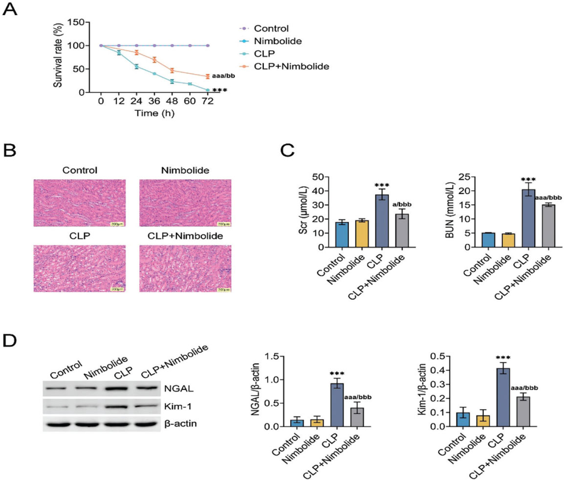
Fig 1.
Nimbolide alleviates CLP-induced renal tissue damage. (A) H&E staining of kidney tissue. (B) Scr levels quantified using a commercial assay kit. (C) Serum BUN levels assessed with a commercially available kit. (D) Western blot analysis of NGAL and Kim-1 in kidney tissue. ***P < 0.001 (vs. Control), aP < 0.05 (vs. Nimbolide), and bbbP < 0.001 (vs. CLP). BUN, blood urea nitrogen; CLP, cecal ligation and puncture; H&E, hematoxylin and eosin; Scr, serum creatinine.
3.2. Nimbolide alleviates CLP-induced renal tissue inflammation
No significant inflammatory response was observed in the serum or kidney tissue of mice in the Control and Nimbolide groups. Serum and kidney tissue levels of the inflammatory cytokines IL-6, IL-1β, and TNF-α were higher in the CLP group than in the Control and Nimbolide groups. On the other hand, IL-6, IL-1β, and TNF-α levels were decreased in the CLP + Nimbolide group (Figure 2), suggesting that Nimbolide could reduce the inflammatory response of septic kidney damage caused by CLP.
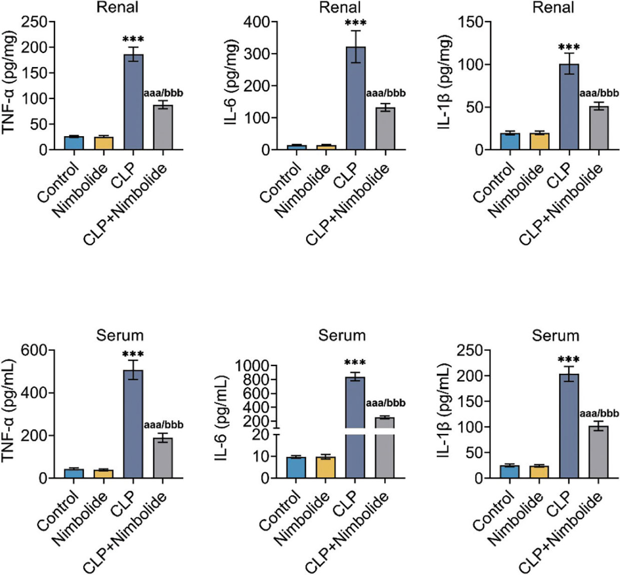
Fig 2.
Nimbolide alleviates CLP-induced renal tissue inflammation. Cytokine levels (IL-6, IL-1β, TNF-α) in patients’ serum and kidney tissues were quantified using commercially available ELISA kits. ***P < 0.001 (vs. Control), aaaP < 0.001 (vs. Nimbolide), and bbP < 0.001 (vs. CLP). CLP, cecal ligation and puncture; IL-6, interleukin-6; IL-1β, interleukin-1β; TNF-α, tumor necrosis factor-α.
3.3. Nimbolide alleviates CLP-induced oxidative stress in kidney tissues
No obvious oxidative stress was observed in the kidney tissues of mice in the Control and Nimbolide groups. In the CLP group, the levels of MDA and ROS, which are markers of oxidative stress, were increased, while the levels of SOD and GSH, markers of antioxidant capacity, decreased in kidney tissues. In contrast, Nimbolide intervention reversed the CLP-induced changes in MDA (Figure 3a), SOD (Figure 3b), GSH (Figure 3c), and ROS levels in mouse kidney tissue. These results indicate that sepsis caused oxidative stress in kidney tissue and that Nimbolide inhibited CLP-induced oxidative stress injury in the kidney.
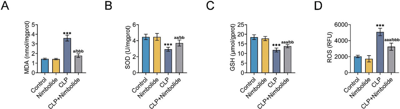
Fig 3.
Nimbolide alleviates CLP-induced oxidative stress in kidney tissue. (A) MDA, (B) SOD, and (C) GSH levels measured in renal tissues using their corresponding kits. (D) ROS content in kidney tissue. ***P < 0.001 (vs. Control), aP < 0.05, aaP < 0.01, aaaP < 0.001 (vs. Nimbolide), bbP < 0.01, and bbbP < 0.001 (vs. CLP). CLP, cecal ligation and puncture; GSH, glutathione; MDA, malondialdehyde; ROS, reactive oxygen species; SOD, superoxide dismutase.
3.4. Nimbolide alleviates CLP-induced cell apoptosis in kidney tissues
No significant tissue cell apoptosis was observed in the kidney tissues of mice in the Control and Nimbolide groups. In the CLP group, TUNEL positivity (Figure 4a) was increased in the kidney tissue of mice, along with increased expression of apoptosis-related proteins Bax and cleaved-caspase 3 and decreased expression of Bcl-2 (Figures 4b,c). Conversely, in the CLP + Nimbolide group, TUNEL positivity was decreased in the kidney tissues of mice, along with reduced expression of Bax and cleaved-caspase 3 and increased expression of Bcl-2. These findings indicate that Nimbolide may inhibit CLP-induced apoptosis in kidney cells.
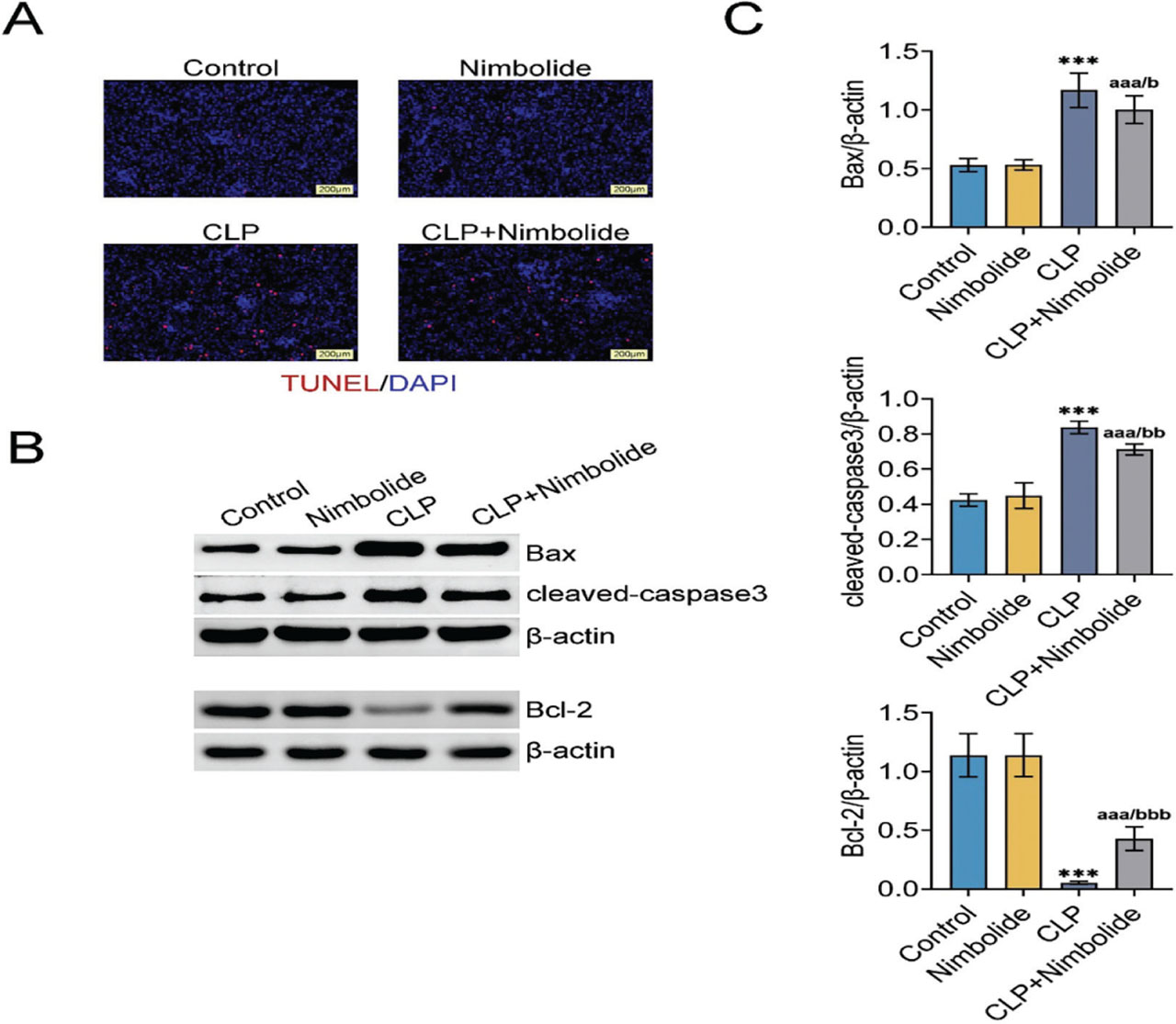
Fig 4.
Nimbolide alleviates CLP-induced cell apoptosis in kidney tissue. (A) Cell apoptosis in kidney tissue assessed using TUNEL staining. (B) Western blot analysis of Bax and Bcl-2 protein expression in renal tissues. (C) Western blot quantification of Bax and Bcl-2 in kidney tissues. ***P < 0.001 (vs. Control), aaaP < 0.001 (vs. Nimbolide), bP < 0.05, and bbP < 0.01 (vs. CLP). CLP, cecal ligation and puncture; DAPI, 4′,6-Diamidino-2-phenylindole; TUNEL, transferase dUTP nick end labeling.
3.5. Nimbolide inhibits the NF-κB/STAT3 pathway
The kidney tissues of mice treated with CLP had higher levels of p-p65/p65 and p-STAT3/STAT3 protein expression (Figure 5a), whereas mice treated with CLP + Nimbolide had lower levels of these proteins, suggesting that Nimbolide could suppress the NF-κΒ/STAT3 pathway activated by CLP in kidney tissues. Rescue experiments were conducted using the STAT3 activator Colivelin, and the results showed that Nimbolide reduced CLP-induced increases in Scr and BUN (Figure 5b), and decreased the expression of Bax and cleaved-caspase 3 proteins (Figure 5c). However, Colivelin reversed the effects of Nimbolide in alleviating kidney injury and renal cell apoptosis.
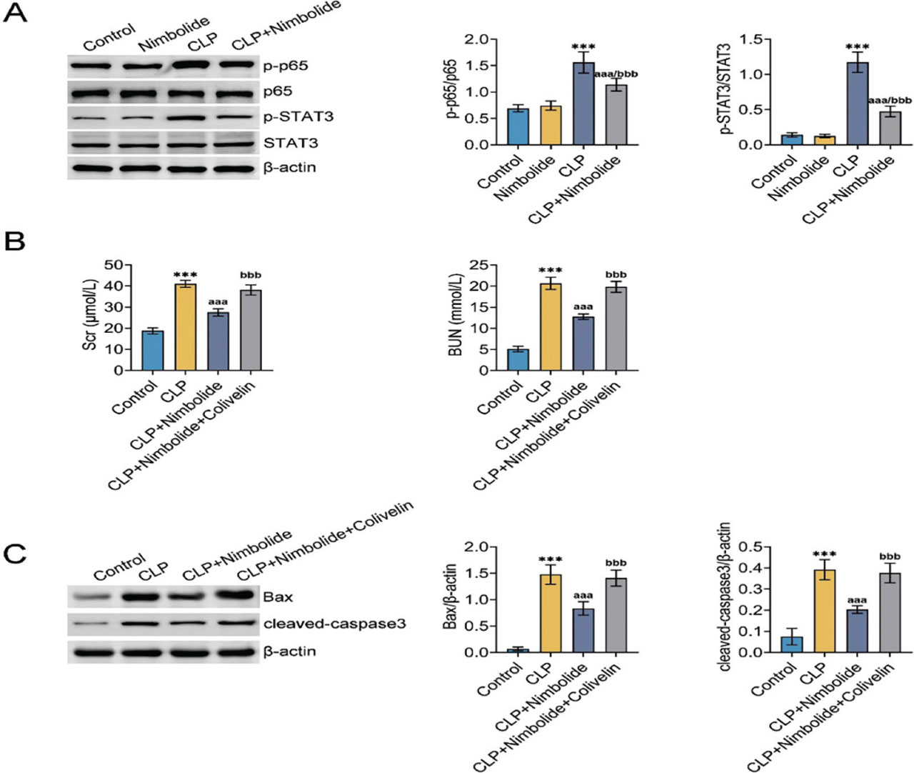
Fig 5.
Nimbolide inhibits the NF-κB/STAT3 pathway. (A) Western blot analysis of p-p65, p65, p-STAT3 and STAT3 protein expression in renal tissues. (B) Scr and BUN levels quantified using a commercial assay kit. (C) Western blot quantification of Bax and cleaved-caspase in kidney tissues. ***P < 0.001 (vs. Control), aaaP < 0.001 (vs. Nimbolide), and bbbP < 0.001 (vs. CLP). BUN, blood urea nitrogen; CLP, cecal ligation and puncture; NF-κB, nuclear factor-κB; Scr, serum creatinine.
4. Discussion
Inflammatory dysregulation is widely acknowledged as a key pathological component of SA-AKI, despite the fact that the pathological mechanisms behind this condition are not entirely understood and there are no particular clinical treatments available. As a result, medication development that targets and modulates inflammatory reactions has grown in importance. Moreover, since natural goods have been shown to possess various benefits with few adverse effects, they have garnered a lot of interest. Herein, our study, which focuses on the limonene compound Nimbolide, provides experimental insights for the development of this natural product as a potential treatment for SA-AKI by demonstrating for the first time its potential in reducing kidney injury in a CLP-induced SA-AKI mouse model.
Previous studies have shown that Nimbolide can effectively improve renal fibrosis in a UUO model by inhibiting the transforming growth factor-β signaling pathway and its downstream EMT/Slug pathway (Annaldas et al. 2019), suggesting its potentially protective role against SA-AKI. This study further confirms that, in a CLP-induced sepsis mouse model, Nimbolide treatment significantly improves renal tissue pathological damage, as manifested by reduced inflammatory cell infiltration and decreased renal tubular vacuolation. Simultaneously, Nimbolide effectively reversed the CLP-induced elevation of Scr and BUN levels. Collectively, these results demonstrate that Nimbolide can effectively alleviate acute kidney injury in the context of sepsis.
In the complex network of mechanisms driving the occurrence and development of SA-AKI, the inflammatory response plays a central driving role (Wang et al. 2025). Besides overactive inflammatory responses, oxidative stress is also a core pathological mechanism in the progression of SA-AKI. Notably, there is a close interaction between inflammation and oxidative stress. Mediators such as nitric oxide (NO), which are produced during the inflammatory response, can directly induce strong oxidative stress, thereby driving renal tubular epithelial cell apoptosis, forming a vicious cycle, and exacerbating kidney damage (Xia et al. 2024). Consequently, SA-AKI may benefit from candidate medications with anti-inflammatory and antioxidant properties. Nimbolide has been shown to successfully reduce oxidative stress and inflammatory reactions in several diseases.
In rats with gestational diabetes, Nimbolide can lower the levels of MDA, increase the activities of SOD, GPx, GST and CAT, and inhibit the expression of TNF-α, IL-1β, and IL-6 (Ma et al. 2022). In mice with acetaminophen-induced acute liver injury, Nimbolide treatment decreased oxidative stress and inflammation (Ba et al. 2025). Consistent with these findings, in mice with CLP-induced acute renal injury, Nimbolide increased the levels of the antioxidant enzymes SOD and GSH, while reducing the levels of the pro-inflammatory factors IL-6, IL-1β, and TNF-α, as well as MDA.
NF-κB plays a pivotal role in the development and progression of sepsis-related kidney injury (Ren et al. 2020). Its activation is a crucial molecular bridge connecting systemic inflammatory responses with localized renal damage. In its resting state, the NF-κB dimer binds to the inhibitory protein IκB and remains in the cytoplasm. During sepsis, upstream signals activate the IκB kinase (IKK) complex, leading to IκB phosphorylation and degradation. NF-κB then dissociates and rapidly translocates into the nucleus, directly initiating the expression of genes encoding pro-inflammatory factors and creates a positive feedback loop (Lou et al. 2025). Conversely, NF-κB activation induces inducible NO synthase to produce large amounts of NO, which reacts with superoxide anions to generate the strong oxidant peroxynitrite (ONOO−), thereby exacerbating oxidative damage to lipids, proteins, and DNA and forming a vicious cycle of “oxidative stress, NF-κB activation, and further ROS production” (Senousy et al. 2022).
STAT3 is a core member of the highly conserved STAT protein family and plays a crucial regulatory role in the pathological processes of various diseases, including sepsis. STAT3 dominates stress and inflammatory responses in the early stages of acute kidney injury and is a central regulator of disease development (Li et al. 2023). In sepsis, PAMPs/DAMPs activate STAT3 through Toll-like receptors (TLRs), leading to its phosphorylation and nuclear translocation, where it directly upregulates the expression of pro-inflammatory cytokine genes such as IL-6 and TNF-α, thereby forming a positive feedback loop that exacerbates local and systemic inflammation in the kidneys (Şahin et al. 2025). Evidence indicates that the protective effect of Nimbolide against streptozotocin-induced diabetic retinopathy in rats is mediated through suppression of the TLR4/NF-κB signaling pathway (Shu et al. 2021). This study revealed that Nimbolide inhibits activation of the NF-κB/STAT3 pathway in kidney tissue induced by CLP. In this study, Nimbolide inhibited both NF-κB p65 and STAT3 phosphorylation in CLP-induced renal injury. Although the direct molecular targets of Nimbolide remain to be fully elucidated, several plausible mechanisms may explain this dual inhibition. Nimbolide may target upstream kinases such as IKK, which is known to regulate both NF-κB activation and STAT3 phosphorylation. Alternatively, given that the CLP model involves TLR4-mediated inflammatory signaling, Nimbolide might interfere with TLR4 or its downstream adaptors. Additionally, since NF-κB drives IL-6 transcription and IL-6 activates STAT3 via JAK, the observed STAT3 inhibition could be a secondary consequence of NF-κB suppression. Further studies are warranted to identify the exact binding targets of Nimbolide.
This study has several limitations. Only male mice were used, and it remains unclear whether the protective effects of Nimbolide apply to female mice. Sex hormones may influence immune responses and susceptibility to sepsis; therefore, future studies should include both male and female mice to assess sex-specific differences.
In summary, this study demonstrates that the natural compound Nimbolide exerts a definitive protective effect against SA-AKI by targeted inhibition of the NF-κB/STAT3 signaling pathway in renal tissue, thereby effectively mitigating sepsis-induced hyperinflammation, oxidative stress damage, and renal tubular cell apoptosis. No kidney damage occurred in mice in the Nimbolide group, and other indicators were not different from those in the Control group, indicating that this dose of Nimbolide was non-toxic to mice. This finding not only provides novel mechanistic insights into the renoprotective role of Nimbolide but also offers important preclinical evidence supporting its potential as a therapeutic strategy for SA-AKI.
Abbreviations
- ANOVA
analysis of variance
- CAT
catalase
- DCFH-DA
2′,7′-dichlorodihydrofluorescein diacetate
- dUTP
2′-deoxyuridine-5′-triphosphate
- ELISA
enzyme-linked immunosorbent assay
- EMT
epithelial-mesenchymal transition
- GPx
glutathione peroxidase
- GST
glutathione-S-transferase
- HDAC-3
histone deacetylase-3
- JAK
Janus kinase
- PAMPs
pathogen-associated molecular patterns
- DAMPs
damage-associated molecular patterns
- STAT3
signal transducer and activator of transcription 3
- UUO
unilateral ureteral obstruction
Acknowledgments
Not applicable.
Notes
[1] Supported by Funding
This work was supported by Zhejiang Provincial Traditional Chinese Medicine Science and Technology Project (No. 2023ZL572), the Medical and Health Research Project of Zhejiang Province (No. 2023RC219), the Construction Fund of Key Medical Disciplines of Hangzhou (No. 2025HZZD02), and the Health Bureau of Zhejiang Province (No. 2023KY433 and No. 2024KY007).
[2] Ethics Approval
Ethical approval was obtained from the Laboratory Animal Management and Ethics Committee of Westlake University (Approval no. HZSY2026001-1).
[3] Contributed by Author Contributions
Yihua Yu—designed the study and carried them out; Yihua Yu, Yangling Li, Ying Gong—supervised the data collection, Yihua Yu, Ying Gong—analyzed the data, Yihua Yu, Yangling Li—interpreted the data, Yihua Yu, Ying Gong—prepared the manuscript for publication and reviewed the draft of the manuscript. All authors have read and approved the manuscript.