
1. Introduction
Bacterial contamination of the fracture site can lead to prolonged sepsis or osteomyelitis, thereby impeding proper bone healing. Furthermore, these infections can cause functional damage and delayed bone repair, which in turn increases hospital stays and medical expenses (Moriarty et al. 2022; Rupp et al. 2024). Despite long-term antibiotic therapy and open fracture debridement, infection can persist, and treatment failure occurs in 10%–30% of cases (Dvorak et al. 2024). Staphylococcus aureus and other coagulase-negative staphylococci, such as Staphylococcus epidermidis, are frequently isolated from open fracture infections (Graan and Balogh 2022). In addition, community-acquired antibiotic-resistant bacteria, including methicillin-resistant Staphylococcus aureus (MRSA) and methicillin-resistant S. epidermidis, are becoming increasingly more (Depypere et al. 2022). Therefore, there is a pressing need to identify and develop novel antimicrobial treatments, particularly for orthopedic applications, due to the growing resistance to conventional antibiotics.
Isovitexin is a flavonoid compound that is naturally present in many edible plants. This compound exhibits a range of pharmacological effects, such as anti-inflammatory, anti-cancer, and antioxidant properties (Abdulai et al. 2021; Cheng 2025). Previous studies have shown that isovitexin can protect mice from MRSA-induced pneumonia, indicating that it possesses certain antibacterial ability (Tian et al. 2022). It may also be administered to people and used as a human angiotensin-converting enzyme 2 (hACE2)-specific treatment drug to prevent COVID-19 infection (Ferdausi et al. 2022). Furthermore, isovitexin has been reported to target Sirtuin3 (SIRT3) to prevent steroid-induced osteonecrosis of the femoral head by regulating mitophagy-mediated ferroptosis (Fan et al. 2025). It has also been demonstrated that isovitexin can prevent methylprednisolone-induced osteopenia through osteogenic and anti-resorptive mechanisms, and can protect against muscle atrophy (Kulkarni et al. 2025). However, further research is necessary to fully understand the function and mechanism of isovitexin in fracture-related infections.
Based on the above considerations, the antibacterial, anti-inflammatory, and osteogenic effects, as well as the underlying molecular mechanisms of isovitexin, were systematically investigated in this study using a model of open midshaft femoral fracture combined with MRSA infection. The findings provide a theoretical and experimental foundation for the development of plant-derived anti-infective and bone repair therapeutics in orthopedic applications.
2. Materials and Methods
2.1. Animals
Twelve-week-old male Wistar rats in good health, weighing between 220 and 250 g, were chosen and kept in a typical animal room with free access to food and water, a 12-h light-dark cycle, and consistent humidity and temperature. The animal experiments were approved by the Ethics Committee of Suzhou Hospital of Integrated Traditional Chinese and Western Medicine, and all procedures were conducted in accordance with relevant animal care guidelines.
The rats were placed in an induction chamber and anesthetized with 4% isoflurane inhalation, followed by maintenance with 2% isoflurane at a flow rate of 0.2 L/min. A longitudinal incision was made on the lateral side of the femur, and the muscles were bluntly separated to expose the midshaft of the femur. The femur was then transected using sterile surgical scissors to establish the fracture model. The fracture site was irrigated with 0.9% sterile saline to remove bone marrow and loose bone fragments, and residual bone chips were carefully removed under magnification to ensure a clean and uniform defect gap. At the same time, the fracture site was injected with 1 × 10⁷ colony-forming unit (CFU)/mL MRSA. The wound was closed using absorbable sutures, and no antibiotics were administered following surgery (Sun et al. 2024).
The rats were randomly divided into four groups (n = 6 per group). The control group received an identical volume of normal saline following the procedure, with a skin incision and exposure that did not result in fracture or infection. For the Model group, a fracture infection model was created, and following surgery, an equivalent volume of regular saline was administered. Vancomycin group: Following modeling, a gavage of Vancomycin (50 mg/kg/day, Sigma-Aldrich; USA) was administered. For the isovitexin group, after modeling, intragastric isovitexin (100 mg/kg/day, Sigma-Aldrich; USA) (Sun et al. 2021) was administered. After 4 weeks, the rats were euthanized by inhalation of excessive isoflurane, and the serum and tissues were taken.
2.2. Hematoxylin and eosin staining
Bone tissues were fixed, embedded in paraffin, and sectioned at a thickness of 5 μm. After deparaffinization in xylene and rehydration through graded ethanol, the sections were stained with hematoxylin for 10 min at room temperature, followed by eosin staining (Solarbio, Beijing, China) for 3 min. The slices were then photographed after being cleaned for 30 s with distilled water.
2.3. Masson staining
Samples of bone tissue were fixed with paraformaldehyde, decalcified with ethylenediaminetetraacetic acid (EDTA), embedded in paraffin, and cut into sections that were 4 μm thick. The local tissue morphology, bone regeneration, and osseointegration were observed using Masson trichrome staining (Solarbio, Beijing, China).
2.4. Immunohistochemistry
At 4 weeks post-surgery, the rats were euthanized, and the femoral tissues were collected. The specimens were fixed in 4% paraformaldehyde for 48 h, decalcified in 10% EDTA for 4 weeks, embedded in paraffin, and sectioned into 4-μm-thick slices. For immunohistochemical staining, the sections were dewaxed in xylene, rehydrated, and subjected to antigen retrieval by heating in sodium citrate buffer (10 mM, pH 6.0) at 95°C for 15 min. Endogenous peroxidase activity was blocked with 3% hydrogen peroxide, and nonspecific binding was blocked with 5% normal goat serum containing 0.1% Triton X-100. The sections were then incubated overnight at 4°C with a primary antibody against CD68 (Abcam, Cat# ab955) at a dilution of 1:200. After washing, the sections were incubated with a horseradish peroxidase (HRP)-conjugated secondary antibody (goat anti-rabbit IgG, 1:500) for 1 h at room temperature. Antibody binding was visualized using DAB substrate, resulting in a brown precipitate. The sections were counterstained with hematoxylin, dehydrated, and mounted. Negative controls were processed without the primary antibody. Images were captured using a light microscope at 100× and 200× magnification, and CD68-positive cells were counted in five randomly selected fields per section using ImageJ software.
2.5. Enzyme-linked immunosorbent assay
According to the manufacturer's instructions, the corresponding enzyme-linked immunosorbent assay (ELISA) kits (R&D Systems) were used to measure the levels of tumor necrosis factor (TNF)-α, interleukin (IL)-1β, IL-6, β-CrossLaps of type I collagen (β-CTX), procollagen type I N-terminal Propeptide (PINP), and bone-specific alkaline phosphatase (BALP) in rat serum.
2.6. Western blot analysis
After the rats were sacrificed, the callus and surrounding bone tissue at the fracture site were immediately collected using pre-cooled sterile instruments. The attached soft tissue was rapidly removed, and the samples were quickly rinsed in pre-cooled phosphate-buffered saline buffer to remove blood and impurities. The cleaned bone tissue was then ground into powder in liquid nitrogen. The tissue homogenate was lysed using RIPA lysis buffer, and the protein concentration was determined using a Bicinchoninic Acid assay protein assay kit (Beyotime, Shanghai, China). Proteins (50 μg per sample) were separated by sodium dodecyl sulfate-polyacrylamide gel electrophoresis (SDS-PAGE) and subsequently transferred onto polyvinylidene difluoride (PVDF) membranes. The membranes were blocked with 5% skim milk for 2 h, followed by incubation with primary antibodies overnight at 4°C. After washing, the membranes were incubated with HRP-conjugated secondary antibodies for 2 h at 37°C. Protein bands were visualized using enhanced chemiluminescence (ECL) chemiluminescence reagent (Roche, Switzerland). Band intensities were quantified using ImageJ software (National Institutes of Health, Bethesda, MD, USA), with glyceraldehyde-3-phosphate dehydrogenase (GAPDH) serving as the internal reference. The primary antibodies used were as follows: BMP2 (1:1000, ab214821, Abcam), Osteopontin (OPN, 1:1000, #66614, CST), RUNX2 (1:1000, # 12556, CST), p-p65 (1:1000, #3033, CST), p65 (1:1000, #8242, CST), Phosphorylated Inhibitor of Nuclear Factor Kappa-B Kinase Subunit Alpha (p-IKB-α) (1:1000, AF2002, Affinity), Inhibitor of Nuclear Factor Kappa-B Kinase Subunit Alpha (IKB-α) (1:1000, AF5002, Affinity), and GAPDH (1:1000, #2118, CST).
2.7. Statistical analysis
The results are expressed as mean ± standard deviation (SD). Comparisons among groups were performed using one-way analysis of variance (ANOVA), and the least significant difference post hoc test was conducted using SPSS 19.0 statistical software (SPSS Inc., Chicago, IL, USA). Statistical significance was defined as P < 0.05.
3. Results
3.1. Isovitexin reduces fracture infection and promotes healing
Figure 1a shows the chemical structure of isovitexin. We used histopathological analysis to assess the therapeutic effects of isovitexin. As shown in Figure 1b, compared with the control group, the femur in the model group exhibited marked inflammatory cell infiltration and bone structural destruction. Although Vancomycin treatment partially improved these changes, the isovitexin treatment group demonstrated a more pronounced repair effect, with significantly improved bone tissue integrity and enhanced bone formation. The quantification of total callus area (TCA, %) based on Masson's trichrome staining is presented in Figure 1c. The model group showed a significantly lower TCA compared with the control group. Isovitexin treatment significantly increased TCA, indicating enhanced new bone formation, whereas Vancomycin treatment resulted in a moderate but incomplete improvement. Figure 1d shows the bacterial load (CFU/mL × 108) at the fracture site. The model group had the highest bacterial count. Both Vancomycin and isovitexin treatments significantly reduced CFU counts, with isovitexin showing a comparable antibacterial effect to Vancomycin. These findings suggest that isovitexin can reduce fracture-related infection and promote bone healing.
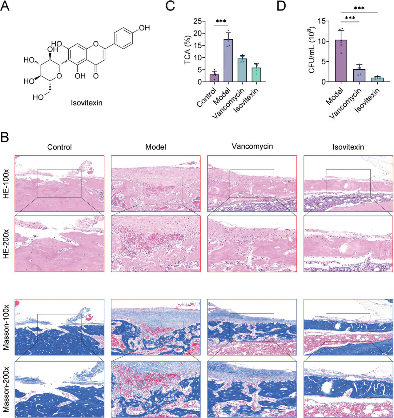
Fig 1.
Isovitexin promotes bone healing and reduces bacterial load in an MRSA-infected femoral fracture model. (A) Chemical structure of isovitexin. (B) Representative histological images of HE staining (top two rows) and Masson's trichrome staining (bottom two rows) at 100× and 200× magnification in the Control, Model, Amoxicillin, and Isovitexin groups. Scale bars are indicated in the images. (C) Quantification of (TCA, %) based on Masson's trichrome staining, showing new bone formation in each group. (D) Bacterial load (CFU/mL × 108) at the fracture site at 4 weeks post-surgery. ***P < 0.001. CFU, colony-forming unit; HE, hematoxylin and eosin; MRSA, methicillin-resistant Staphylococcus aureus; TCA, total callus area.
3.2. Isovitexin reduces inflammation levels
We measured the levels of inflammatory factors in serum to investigate their anti-infective effect. As shown in Figure 2a, the serum levels of TNF-α, IL-6, and IL-10 were assessed at 2 and 4 weeks after surgery. At both time points, the model group exhibited significantly increased levels of pro-inflammatory cytokines TNF-α and IL-6, and decreased levels of the anti-inflammatory cytokine IL-10, compared with the control group. Vancomycin treatment partially reversed these changes. Notably, isovitexin treatment significantly decreased TNF-α and IL-6 levels and increased IL-10 levels at both 2 and 4 weeks, with effects comparable to or greater than those of Vancomycin (Figure 2a). CD68 immunohistochemical staining was performed to evaluate macrophage infiltration (Figure 2b). In the control group, few CD68-positive cells were observed. In contrast, the model group showed abundant CD68-positive brown staining, indicating extensive macrophage infiltration. The Vancomycin group exhibited moderate CD68 staining. In contrast, the isovitexin group showed markedly fewer CD68-positive cells, similar to the control group. These results indicate that isovitexin effectively suppresses macrophage infiltration and local inflammation, thereby promoting a favorable microenvironment for fracture repair (Figure 2b).
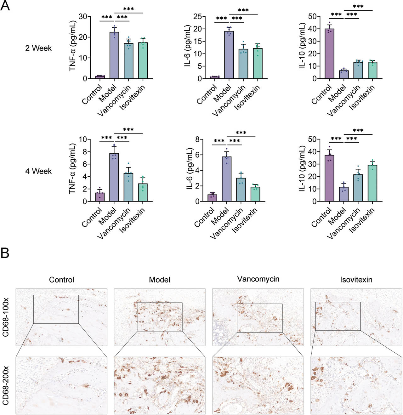
Fig 2.
Isovitexin reduces the level of inflammation. (A) Quantification of serum levels of pro-inflammatory cytokines TNF-α and IL-6, and the anti-inflammatory cytokine IL-10, at 2 and 4 weeks post-surgery in all four groups (Control, Model, Amoxicillin, and Isovitexin). (B) Representative immunohistochemical staining of CD68 (a macrophage marker) at 100× and 200× magnification in femoral tissue sections from each group. Brown staining indicates CD68-positive cells. Data are presented as mean ± SD (n = 6). ***P < 0.001. IL-6, interleukin-6; IL-10, interleukin-10; SD, standard deviation; TNF-α, tumor necrosis factor-α.
3.3. Isovitexin promotes bone metabolism level
Given its anti-inflammatory and healing effects, we further assessed the impact of isovitexin on bone metabolism. The model group exhibited an imbalance in bone metabolism, as indicated by increased levels of the bone resorption marker β-CTX and decreased levels of the bone formation indicators PINP and BALP (Figure 3a). Isovitexin treatment corrected this imbalance and promoted bone formation. Western blot analysis further demonstrated that isovitexin markedly increased the expression of important osteogenic proteins, including BMP2, OPN, and RUNX2 (Figure 3b). These findings indicate that isovitexin not only inhibits bone resorption but also promotes bone formation, thereby providing a biological and molecular basis for accelerated fracture healing.
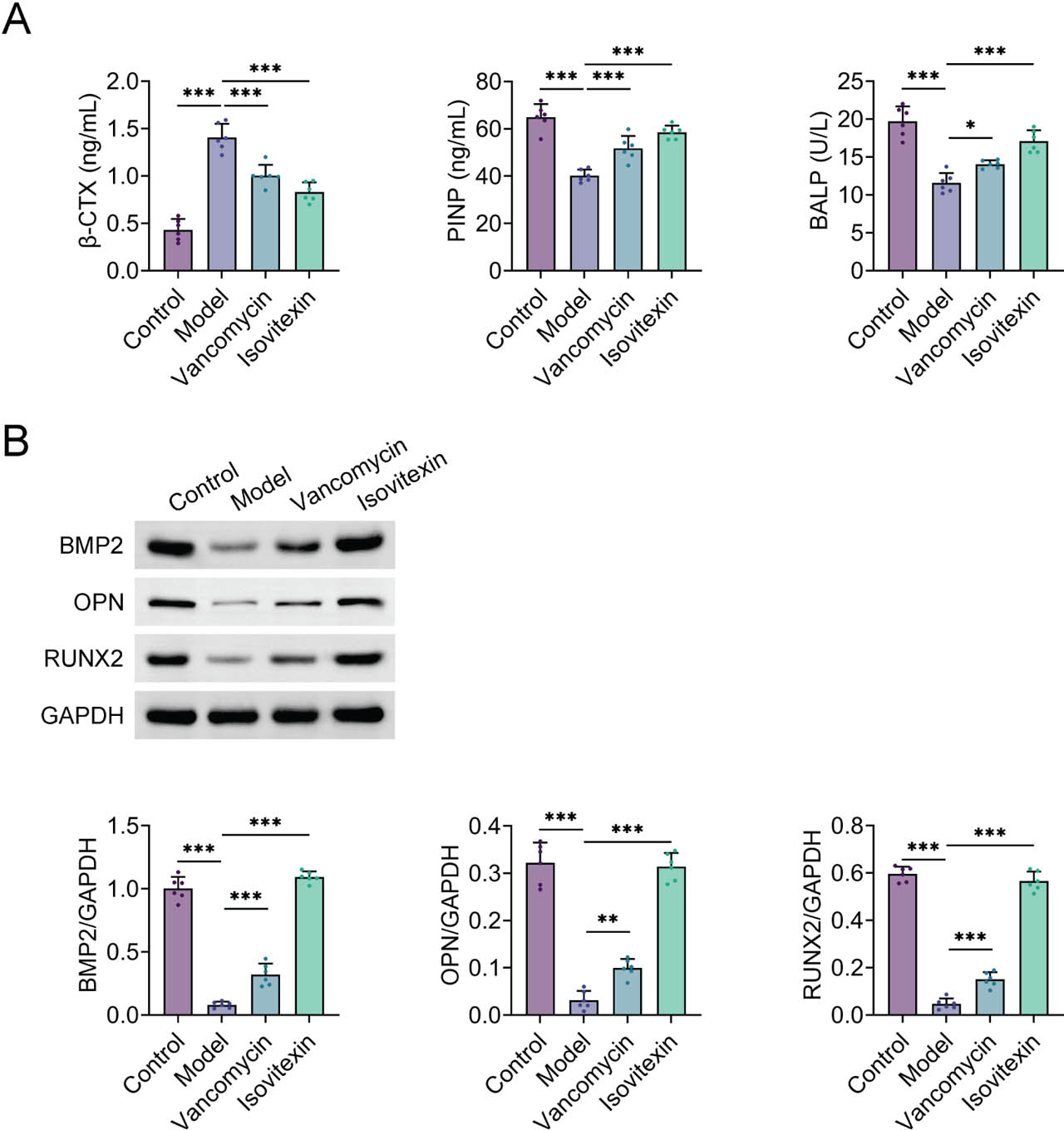
Fig 3.
Isovitexin promotes bone metabolism. (A) Measurement of serum bone metabolism markers β-CTX, PINP, and BALP. (B) The protein expression levels of BMP2, OPN, and RUNX2 in bone tissue were detected by Western blot. Data are presented as mean ± SD (n = 6). ***P < 0.001. BALP, bone-specific alkaline phosphatase; PINP, procollagen type I N-terminal propeptide; β-CTX, β-CrossLaps of type I collagen; SD, standard deviation.
3.4. Isovitexin inhibits the activation of the NF-κB pathway
Lastly, we concentrated on the NF-κB signaling pathway, which plays a central role in inflammation. The phosphorylation levels of IKB-α and p65 were elevated in the model group, indicating an activation of the NF-κB pathway. Isovitexin treatment effectively inhibited pathway activation and reduced the phosphorylation levels of p65 and IKB-α (Figure 4). This finding is consistent with the results described above, suggesting that isovitexin reduces inflammatory damage by suppressing the activation of the NF-κB pathway and subsequently decreasing the production of downstream pro-inflammatory factors.
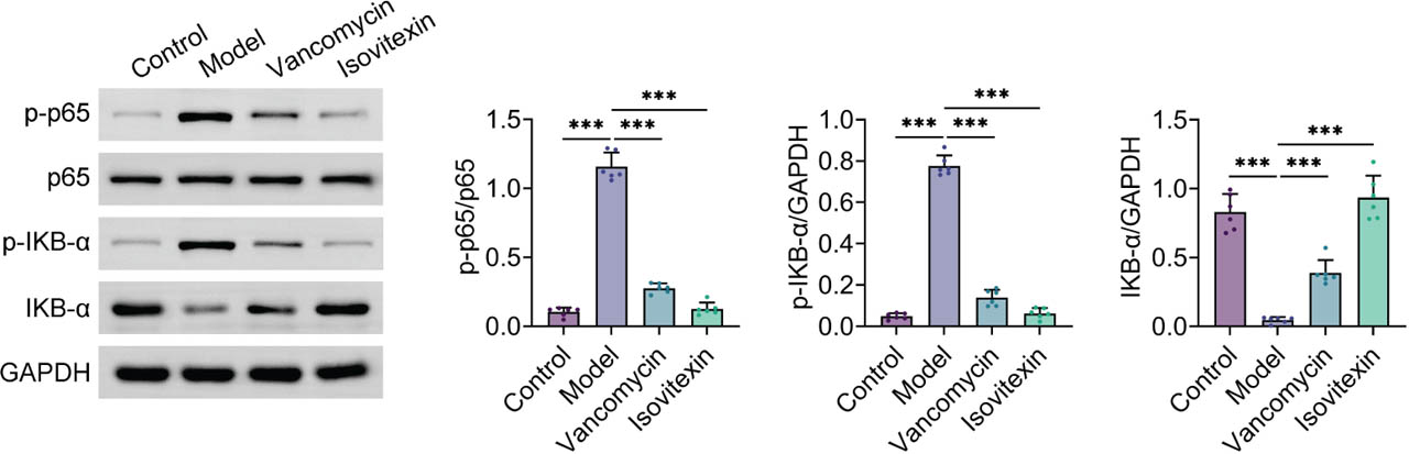
Fig 4.
Isovitexin inhibits the activation of the NF-κB pathway. The protein expression levels of p65, p-p65, IKB-α, and p-IKB-α in bone tissue were detected by Western blot. Data are presented as mean ± SD (n = 6). ***P < 0.001. GAPDH, glyceraldehyde-3-phosphate dehydrogenase. SD, standard deviation.
4. Discussion
The process of bone healing is severely hampered by the complicated inflammatory milieu of fracture-related infection, which provides a significant challenge in the clinical treatment of orthopedics (He et al. 2023). In this study, the therapeutic effects and potential mechanisms of the natural flavonoid isovitexin were thoroughly investigated in a fracture-related infection model. Our findings demonstrate that isovitexin can successfully reduce inflammation while enhancing bone metabolism and promoting bone tissue repair, with inhibition of the NF-κB signaling pathway as a key underlying mechanism. These results offer preclinical insights for the development of isovitexin as a possible treatment medication for fracture-related infection.
The NF-κB pathway is a critical signaling hub that regulates the inflammatory response. After infection and trauma, it is rapidly activated, creating a significant production of pro-inflammatory molecules such as TNF-α and IL-6, resulting in tissue damage (Dai et al. 2024). In the present study, the model group exhibited markedly elevated serum levels of TNF-α and IL-6, accompanied by activation of the NF-κB pathway. It is noteworthy that isovitexin treatment can simultaneously downregulate the levels of downstream pro-inflammatory factors and effectively prevent the activation of the NF-κB pathway, suggesting that isovitexin's suppression of the NF-κB pathway may be the cause of its anti-inflammatory effects. Compared with the bactericidal activity of antibiotics such as Vancomycin, isovitexin appears to regulate excessive host immune responses at the signaling level. This observation is consistent with the concept of adjunctive infection therapy through modulation of host responses proposed in previous studies (Rothe et al. 2019). Also, in the second week, serum TNF-α and IL-6 levels were significantly increased in all infection groups, and only slightly reduced in the isovitexin and Vancomycin treatment groups, reflecting a strong acute-phase response to MRSA infection that may exceed the capacity of early intervention. However, by week 4, isovitexin-treated animals exhibited a marked decrease in pro-inflammatory cytokines and a sustained increase in IL-10, whereas the model group remained in a chronic inflammatory state. These temporal changes indicate that the anti-inflammatory effects of isovitexin are time-dependent and cumulative.
An excessive inflammatory response disrupts the balance between osteogenesis and osteoclast activity, leading to increased bone resorption and reduced bone formation (Matsumoto and Rottapel 2020). According to this study, isovitexin greatly reduced inflammation and enhanced bone metabolism. At the molecular level, isovitexin increased the expression of RUNX2, a key osteogenic transcription factor, and its downstream target proteins, BMP2 and OPN. The anti-inflammatory environment created by isovitexin is likely to facilitate these osteogenic processes. Previous studies have demonstrated that excessive activation of the NF-κB pathway can directly inhibit osteoblast differentiation and function, whereas blocking this pathway can relieve suppression of osteogenic gene expression (Novack 2011). Therefore, we speculate that isovitexin may promote bone healing by inhibiting NF-κB and reducing inflammation, thereby achieving the dual benefits of anti-inflammation and promoting bone formation.
Nevertheless, this study has several limitations. First, as the findings are primarily based on an animal model, further clinical studies are required to validate these results. Second, the optimal dosage and pharmacokinetic properties of isovitexin in vivo require further clarification. Lastly, more research should be done to determine whether isovitexin influences other signaling pathways involved in bone healing.
In conclusion, our study demonstrates that isovitexin can effectively improve bone metabolism and repair while reducing the inflammatory response associated with fracture-related infection. This effect is achieved, at least in part, through inhibition of the NF-κB signaling pathway. These findings provide a theoretical basis for the subsequent development of novel anti-infective bone repair therapies and highlight the potential of isovitexin as a pleiotropic agent for the adjuvant treatment of fracture-related infections.
Acknowledgments
Not applicable.
Notes
[2] Conflicts of interest Competing Interests
The authors state that there are no conflicts of interest to disclose.
[3] Ethics Approval
Ethical approval was obtained from the Ethics Committee of Suzhou Hospital of Integrated Traditional Chinese and Western Medicine (Approval no. 2025038).
[5] Data Availability
The data that support the findings of this study are available from the corresponding author upon reasonable request.
[6] Contributed by Author Contributions
Yaping Gu and Ping Xu—designed the study and carried them out; Yaping Gu and Ping Xu—supervised the data collection, Yaping Gu and Ping Xu—analyzed the data, Yaping Gu and Ping Xu—interpreted the data, Yaping Gu and Ping Xu—prepared the manuscript for publication and reviewed the draft of the manuscript. All authors have read and approved the manuscript.