
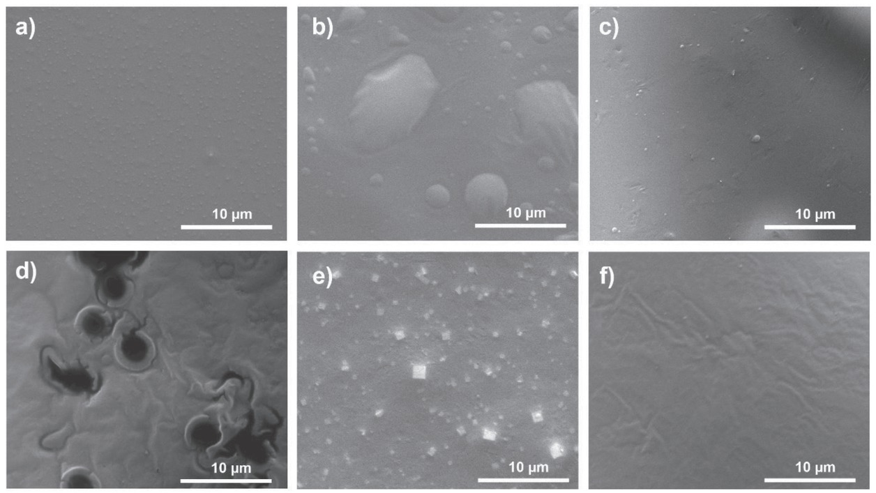
Figure 1
SEM image of self-assembled Gly-His dipeptide films in various solvents: a) 1,1,1,3,3,3-hexafluoro-2-propanol, b) water, c) ethanol, d) acetone, e) chloroform, and f) tetrahydrofuran
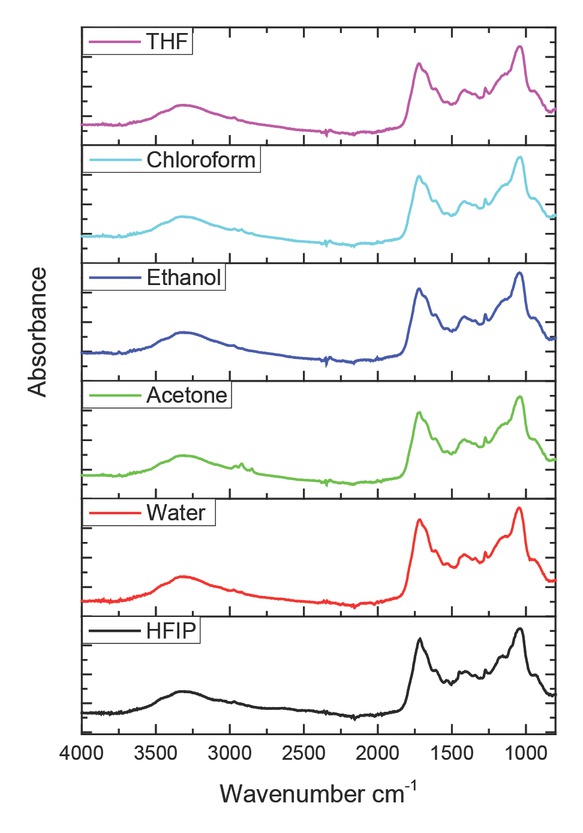
Figure 2
FTIR spectra of Gly-His dipeptide films self-assembled in various solvent media. HFIP – 1,1,1,3,3,3-hexafluoro-2-propanol; THF – tetrahydrofuran
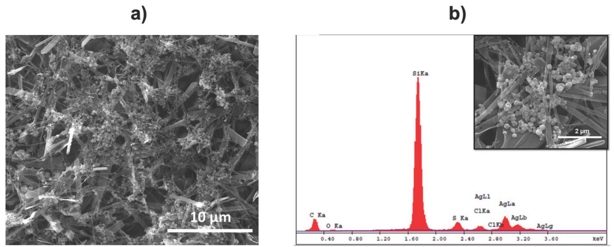
Figure 3
SEM images of (a) morphological changes in Gly-His films formed in AgNO3 solutions for 6 h and (b) zoomed AgNP-decorated Gly-His structure with the EDX spectrum
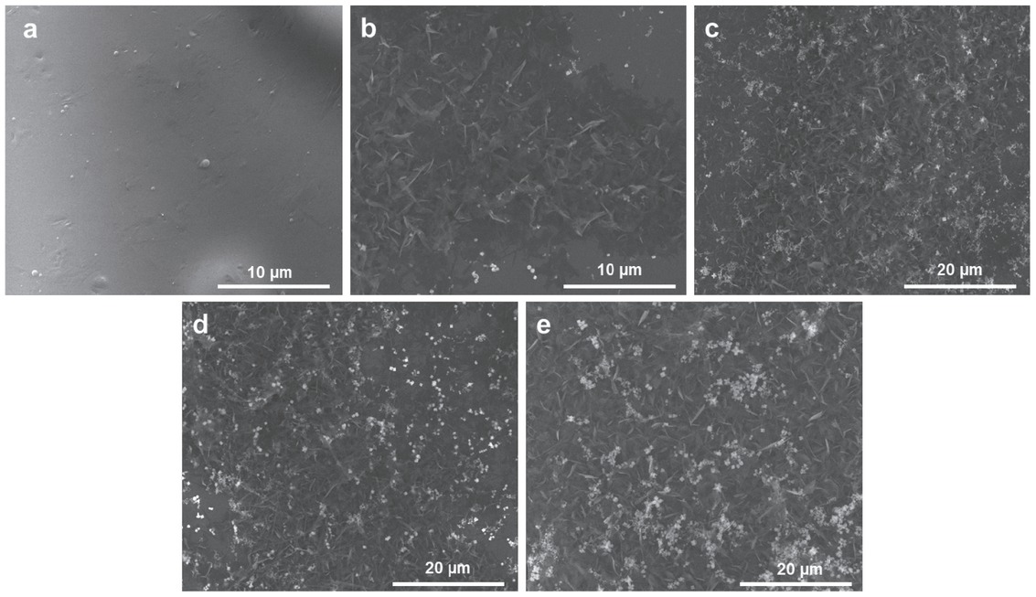
Figure 4
SEM images of Gly-His film structures kept in AgNO3 solution for a) 0 h, b) 3 h, c) 6 h, d) 12 h, and e) 24 h
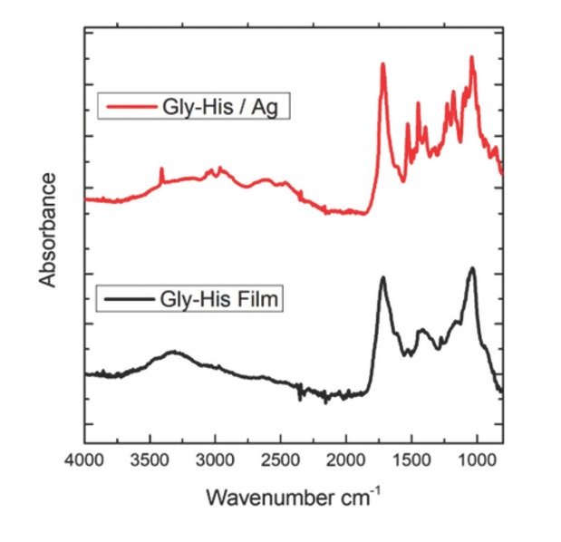
Figure 5
FTIR spectra of Gly-His dipeptide films with and without Ag
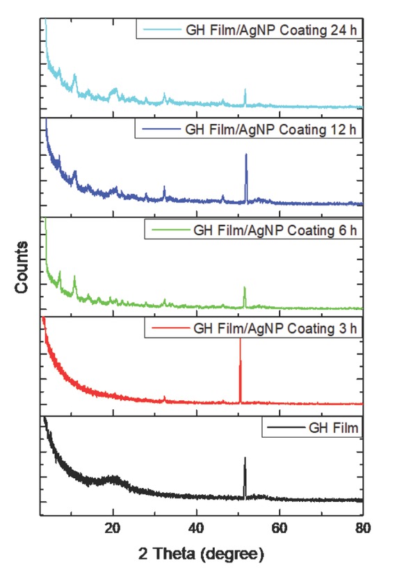
Figure 6
XRD spectra of Gly-His film alone or kept in AgNO3 solution for various times
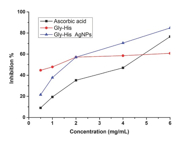
Figure 7
Antioxidant activity of Gly-His films alone and those coated with AgNPs
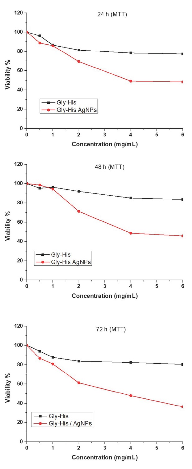
Figure 8
Cytotoxic effects of Gly-His dipeptide with and without AgNPs in L929 cells
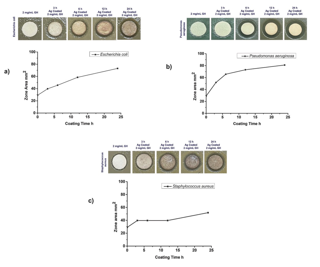
Figure 9
Antibacterial activity of Gly-His dipeptides with or without AgNPs against a) Escherichia coli, b) Staphylococcus aureus, and c) Pseudomonas aeruginosa