
Diabetic retinopathy (DR) is a chronic complication of diabetes mellitus and remains one of the most frequent causes of preventable vision loss in working-age populations. DR affects approximately one-third of diabetic patients [1]. DR is the most common and severe ocular complication of diabetes. Twenty years after diagnosis, it occurs in almost all patients with type 1 diabetes (T1D) and in 60% of people with type 2 diabetes. DR is a factor that negatively affects overall health, vision, and mental status, which reduces quality of life in patients with diabetes [2]. Already in the subclinical stage of DR, functional and microvascular changes occur in the retina and a reduction in the thickness of the inner retinal layers is observed [3]. Hyperglycemia and associated metabolic pathways can lead to neurodegeneration, early microvascular disorders, and neurovascular unit disorders [1]. Increased understanding of the importance of neuronal degeneration and neurovascular unit in DR has led to proposals to include neuronal dysfunction in the assessment of DR severity.
A key element in the medical care of patients with diabetes mellitus is rapid and effective diagnosis before observable symptoms of DR occur. A diagnostic element that fulfils this criterion appears to be retinal functional testing, or electroretinography (ERG). Some of the most important findings from ERG studies in diabetic patients over the past two decades include the finding of early involvement of photoreceptors and bipolar cells and the ability to predict progression of vascular abnormalities.
The aim of this narrative review is to evaluate the available clinical evidence regarding the use of the RETeval™ handheld ERG device, including combined pupillometry, for the detection, staging, and risk assessment of DR across different phases of the disease.
This work was conducted as a narrative literature review aimed at synthesizing current clinical and experimental evidence regarding the use of the RETeval™ handheld ERG device in the context of DR. A search of the PubMed database was performed to identify relevant studies published between January 2010 and March 2024. Search terms included combinations of keywords related to diabetes, DR, ERG, RETeval, flicker ERG, and pupillometry. Only articles published in English were considered.
Studies were included if they met the following criteria: (1) involved human participants with diabetes mellitus; (2) evaluated retinal function using the RETeval™ device; (3) reported outcomes related to DR detection, staging, or risk prediction; and (4) employed observational or interventional study designs. Case reports, conference abstracts, animal studies, and non-peer-reviewed articles were excluded.
Titles and abstracts were screened independently by two reviewers, followed by full-text assessment of potentially eligible articles. Discrepancies were resolved through discussion. Extracted data included study design, population characteristics, RETeval protocol parameters, outcome measures, and key findings.
The methodological quality of included studies was assessed using appropriate tools depending on study design, with particular attention to selection bias, outcome assessment, and reporting transparency.
The RETeval™ handheld device was developed to facilitate objective assessment of retinal function in clinical environments where conventional full-field ERG is impractical. Unlike standard ERG systems that require pupil dilation, dark adaptation, and specialized laboratory settings, RETeval enables rapid testing under photopic conditions without pharmacological preparation.
The system employs a compact, handheld Ganzfeld stimulator that delivers diffuse white-light flicker stimuli generated by integrated light-emitting diodes. Retinal illumination is expressed in Trolands, a unit that incorporates both stimulus luminance and real-time pupil area. Continuous pupillometry allows automatic adjustment of stimulus strength to maintain consistent retinal illuminance throughout the recording, thereby reducing variability related to pupil dynamics.
Electrophysiological signals are acquired using a disposable skin electrode strip positioned along the lower orbital rim. The electrode array integrates active, reference, and ground electrodes within a single adhesive unit. Although skin electrodes produce lower signal amplitudes compared with corneal electrodes, modern signal amplification and frequency-domain analysis enable reliable extraction of flicker ERG responses suitable for clinical interpretation.
Within the DR assessment protocol, the device records flicker responses at multiple stimulus intensities and simultaneously evaluates pupillary reactions to light. Key electrophysiological parameters, including implicit time (IT) and response amplitude, are combined with pupillometric indices and patient age. These variables are processed using an automated algorithm to generate a composite DR risk score intended to support clinical decision-making.
Clinical studies investigating the use of RETeval in diabetic populations consistently demonstrate functional retinal abnormalities in patients without ophthalmoscopically visible retinopathy. Delays in flicker ERG IT and reductions in response amplitude have been observed in individuals classified as having no DR, suggesting early neuroretinal involvement preceding detectable microvascular lesions.
With increasing severity of DR, electrophysiological dysfunction becomes more pronounced. Progressive prolongation of ITs and attenuation of ERG amplitudes have been reported across non-proliferative and proliferative stages. In parallel, pupillometric analysis reveals diminished pupil constriction amplitude, reduced contraction velocity, and impaired dilation dynamics, reflecting diabetes-related autonomic dysfunction.
Several diagnostic accuracy studies have evaluated the performance of the RETeval-derived DR risk score. While sensitivity and specificity for detecting any stage of DR are moderate, higher diagnostic performance has been reported for identifying vision-threatening disease. Importantly, elevated DR risk scores have been associated with an increased probability of disease progression over follow-up periods, indicating potential prognostic relevance rather than purely cross-sectional diagnostic utility.
The RETeval™ device enables objective assessment of retinal function in routine clinical settings where conventional full-field ERG is impractical. Standard ERG requires pupil dilation, dark adaptation, and dedicated laboratory space, whereas the RETeval system facilitates rapid testing under photopic conditions without pharmacological preparation [4].
In the context of DR assessment, the device generates a multifactorial risk score. The DR risk assessment protocol for the device was provided by the manufacturer. The original calculation method was developed based on a multisite study by Maa et al. for screening [5]. Compared with the standard ERG, DR screening, performed in the primary care setting, can reduce subjective errors and make it more useful and common [6]. The test is performed without the administration of eye drops and can be performed by orthoptists, optometrists, or nurses.
The DR RETeval protocol (Figure 1) is a study based on standard International Society for Clinical Electrophysiology of Vision protocols for daytime adaptation and extended protocols, including those developed for diabetic patients [7]. Default normal values for the DR factor range from 7 to 19.9 [7]. In the DR protocol generated in the study, the operator set the reference range from 7.0 to 23.4.

RETeval™ test protocol: DR test. Test result of both eyes in a patient with T1D without DR
Stimulation and pupillometry are integrated to standardize retinal illuminance. The handheld device employs a compact Ganzfeld stimulator with light-emitting diodes to deliver diffuse white-light flicker. Retinal illumination is quantified in Trolands, a unit that accounts for both stimulus luminance and real-time pupil area. Continuous pupillometry allows for automated adjustment of stimulus strength, maintaining consistent retinal illuminance throughout the recording and minimizing variability associated with pupillary dynamics [8]. Electrophysiological signals are acquired via a skin electrode for patient tolerance and clinical efficiency. A single-use, adhesive electrode strip positioned along the lower eyelid margin incorporates active (Figure 2), reference, and ground electrodes (Figure 3). While skin electrodes yield lower signal amplitudes compared to corneal electrodes, the system utilizes specialized amplification and signal-processing algorithms to reliably extract ERG waveforms suitable for clinical evaluation.
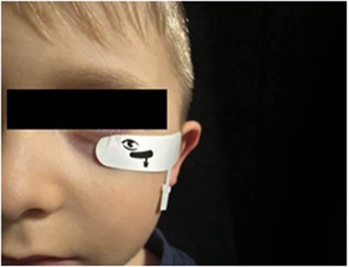
Skin electrode array (Sensor Strip, LKC Technologies, Inc.) placed on the orbital rim, 2 mm from the edge of the lower eyelid
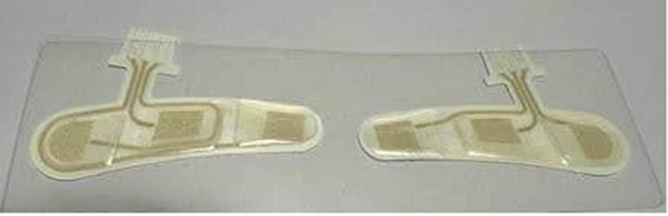
An arrangement of three electrodes: An active (positive) electrode, a reference (negative) electrode, and a ground electrode, all within a single adhesive tape
In a study by Zeng et al. in a group of patients without DR (NDR) there was delayed IT and reduced amplitude compared to the control group (p < 0.01). DR score was significantly higher in the NDR group, while pupil area ratio was significantly lower in the NDR group (p < 0.01) compared to the control group [8]. Smith and Smith found that as DR progressed, DR scores gradually increased, with longer ITs and reduced amplitudes, as well as worse pupil responses. The changes were related to the severity and degree of DR. The amplitude of pupillary response after light stimulation decreases with increasing DR severity, and the impaired pupillary dilation and reflex response to light in diabetes may be due to sympathetic neuropathy or parasympathetic dysfunction. When the pupil is not artificially dilated, measurement of its response to light can act as an independent indicator of DR severity [9]. A study showed that patients with NDR had lower amplitude and longer IT than healthy subjects [8], while a study by Tyrberg et al. showed only longer IT [10]. In Fukuo’s study, both the amplitude and default 8Td-s flash time were not significantly different between the healthy control group and the NDR group, which we suspect may be related to the weaker intensity of light stimulation [4,11]. The flash ERG is the response of the cones, which have a higher density in the fovea; therefore, the test becomes more sensitive and helpful in assessing the efficacy of diabetic maculopathy treatment [12,13]. The sensitivity and specificity in detecting vision-threatening diabetic retinopathy (VTDR) with RETeval were higher than in early DR suggesting that the diagnostic value of the DR test in early DR is not as good as in advanced DR. In Fukuo’s study, the sensitivity and specificity of the optimal cut-off point for any DR diagnosis were 0.70 and 0.81, while the sensitivity and specificity for the diagnosis of severe non-proliferative diabetic retinopathy were 0.85 and 0.85, respectively [4]. In Zeng’s study, the sensitivity and specificity for any DR were 80.2% and 81.7%, while the sensitivity and specificity for VTDR were 94.6% and 88.8%, respectively [14].
Studies have shown that the ERG amplitudes in people with light choroids were higher than in those with dark pigmentation. This would be related to increased melanin-related resistance or reduced effective retinal illumination, resulting in lower ERG amplitudes [15,16]. Iris color should be considered as a factor that may influence the results regardless of the stage of DR.
The DR protocol measures pupillary response in addition to ERG flicker amplitude and IT to generate a DR risk score, which helps assess a patient’s risk of disease progression and guide treatment decisions. Pupil size is dependent on the autonomic nervous system, through the pupil sphincter and dilation muscles [17]. Impaired sympathetic innervation is the cause of the narrower pupil in diabetic patients. Because of the increased sensitivity in diabetic patients to phenylephrine, neuropathic etiology appears to have a greater effect on pupillary diameter abnormalities than myopathy [18,19]. Polaroid pupillometry showed subclinical autonomic neuropathy in children and adolescents with T1D, which was associated with poorer glycemic control, longer duration of diabetes, and other indicators of microvascular disease in these patients [20]. Resting pupil size is significantly reduced in the dark [21,22]. In addition to reduced pupil diameter, reduced pupil response amplitude to light has also been shown in patients with T1D, and patients with proliferative DR have been shown to have significant changes in automatic pupillometry compared to healthy subjects [17,23]. In addition, lower values of pupil contraction amplitude, pupil contraction velocity, and pupil dilation velocity were observed [24].
The study by Davis et al. focused on comparing the most effective methods for assessing patients with NDR and their risk of progressing to VTDR within 48 weeks. Four methods were used to evaluate the status of DR: ERG/pupillometry, color fundus photography, optical coherence tomography angiography, and ultra-widefield fluorescein angiography), and a total of 56 parameters were measured based on these methods. Results have shown that the strongest predictor of progression DR was RETeval DR score ≥26.9, which shows a 5.6 times increased risk of having VTDR [25].
The RETeval DR assessment protocol detects retinal abnormalities resulting from diabetes and creates an objective DR risk score. The combination of retinal function and pupil reactivity analysis takes into account the vascular and neural aspect of ocular pathologies resulting from diabetes. This increases the potential sensitivity and specificity of the test.
The RETeval device can be operated and read after simple training even without the involvement of ophthalmologists. Furthermore, ERG data can be documented for pre- and post-treatment follow-up.
Our own experience allows us to define the limitations of the test. Analysis of the test result should take into account the history of uveitis and retinitis pigmentosa, a history of ocular trauma, high myopia (>−6.0 Dsph), congenital or genetically determined retinal diseases, other eye diseases that interfere with retinal function (e.g., intraocular tumors, intraocular foreign body conditions, conditions after thromboembolic incidents in the retinal vessels), anterior segment diseases preventing pupil dilation, retinal dystrophies, connective tissue diseases, chronic oncological treatment, chronic immunosuppression, hyperbaric chamber treatment, and past surgical procedures. Patients with epilepsy are not eligible for the study. There is a need to establish the dependence of the outcome on the age of the patient and to test on a group of children of different ages.
While the RETeval™ system offers advantages in terms of portability and ease of use, several limitations must be acknowledged. Physiological and demographic factors may affect ERG and pupillometry results. Ocular pigmentation has been shown to influence ERG amplitudes, with lower responses observed in individuals with darker fundus pigmentation. Iris color and baseline pupil size should therefore be considered when interpreting results.
Systemic factors such as duration of diabetes, glycemic control, and presence of autonomic neuropathy may also impact pupillary responses.
Ocular comorbidities, particularly inherited retinal dystrophies or significant media opacities like cataract, directly alter phototransduction and signal amplitude. Systemic factors pose another challenge; diabetic autonomic neuropathy can impair pupillary reflexes. Furthermore, baseline physiological variables – most notably advanced age and differences in fundus pigmentation – inherently modulate ERG parameters and must be contextualized.
The device should not be used in isolation to establish a definitive diagnosis but rather as an adjunct to established screening and diagnostic methods. Appropriate patient selection and awareness of contraindications are essential for accurate interpretation.
Current evidence suggests that RETeval™-based flicker ERG combined with pupillometry provides a useful objective measure of retinal dysfunction associated with DR. The device shows particular promise for identifying patients at higher risk of progression to vision-threatening disease and may serve as a complementary tool in screening and follow-up strategies. Further large-scale prospective studies across diverse populations are needed to refine cut-off values, assess long-term prognostic utility, and establish standardized clinical pathways for implementation.
Diabetic Retinopathy Electroretinography Inner Retinal Layer Implicit Time No Diabetic Retinopathy Troland Seconds Type 1 Diabetes Vision-threatening Diabetic Retinopathy
Sebastian Sirek: Research concept and design, acquisition of data, data analysis and interpretation, preparation, writing, visualization, final proofreading, and approval of the version for publication. Aleksandra Górska: Acquisition of data, data analysis and interpretation, writing, and visualization. Krzysztof Marcinkowski: Acquisition of data, data analysis and interpretation, preparation, writing, and visualization. Dorota Pojda – Wilczek: Research concept and design, supervising the project, final proofreading, and approval of the version for publication.
Sebastian Sirek https://orcid.org/0000-0002-3138-3011
Aleksandra Górska https://orcid.org/0000-0002-6851-5065
Krzysztof Marcinkowski https://orcid.org/0000-0002-6851-5065
Dorota Pojda-Wilczek https://orcid.org/0000-0002-7579-2546
The authors have no potential conflicts of interest to declare.