
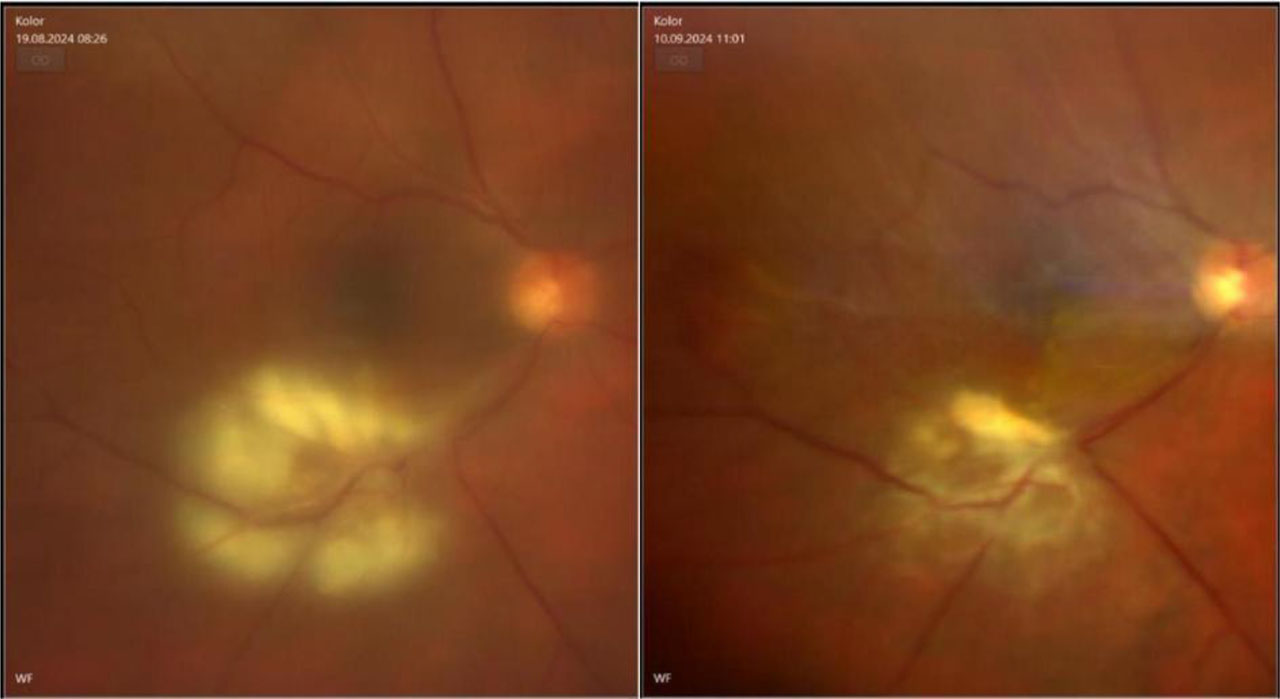
Figure 1.
Fundus photo of the right eye with inflamatory chorioretinal changes –the left side pre-treatment and on the right site there’s an image made after the treatment
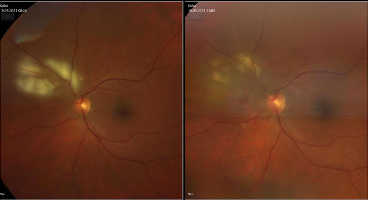
Figure 2.
Fundus photo of the left eye with inflamatory chorioretinal changes – on the left side pre-treatment and on the right side there’s an image made after the treatment
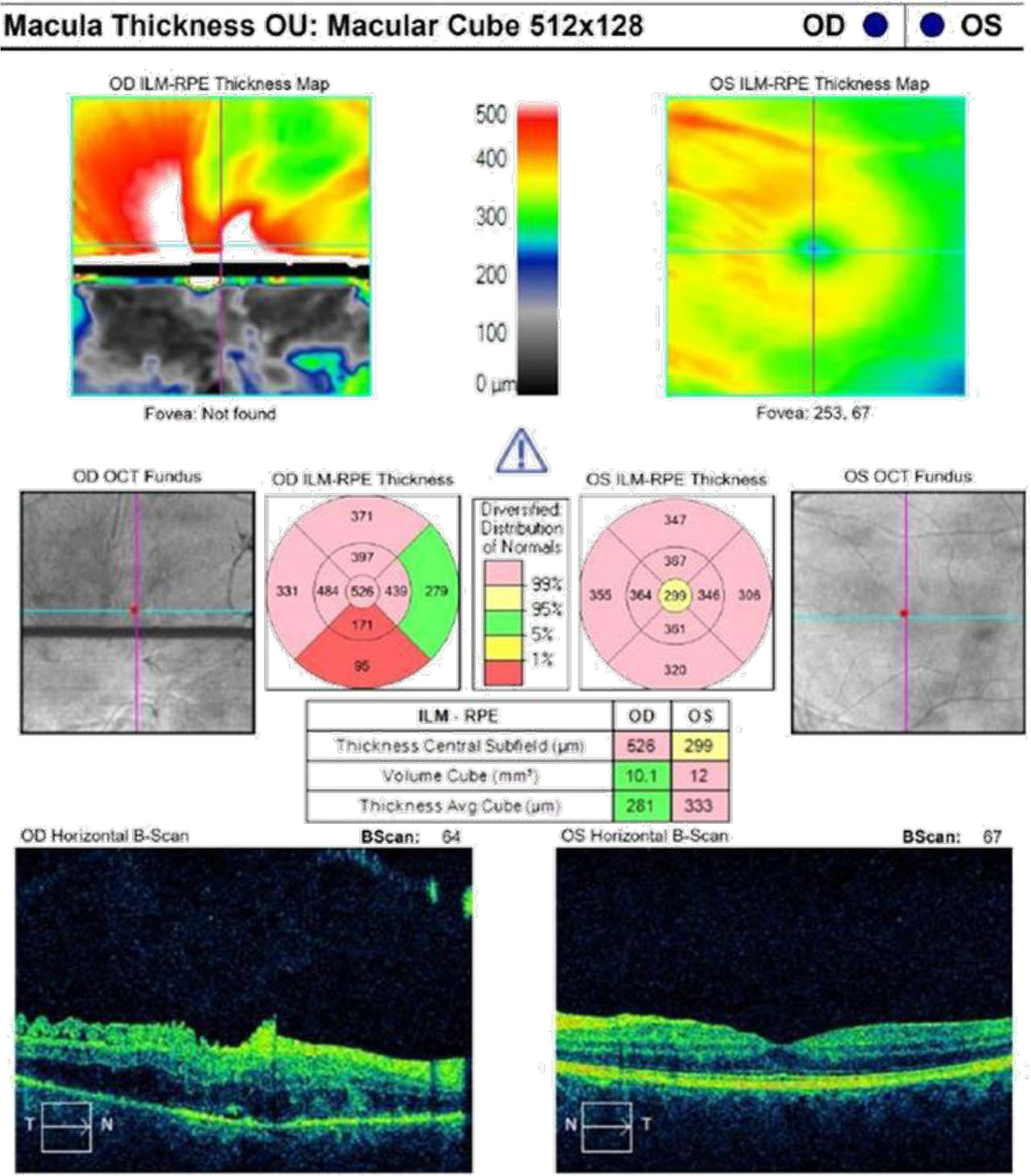
Figure 3.
OCT through inflammatory changes of the right eye: RPE preserved, with visible edema of the retinal layers and hyperreflectivity of the inner retinal layers and the left eye: normal RPE
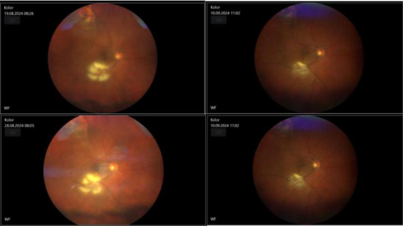
Figure 4.
Fundus photo of the right eye shows changes of inflammation before the treatment and after it
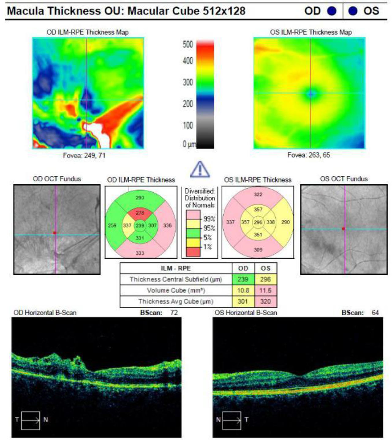
Figure 5.
OCT of the macula was performed, revealing RPE thinning and ERM with irregular surface the right eye, while the foveal morphology in the left eye remained preserved