
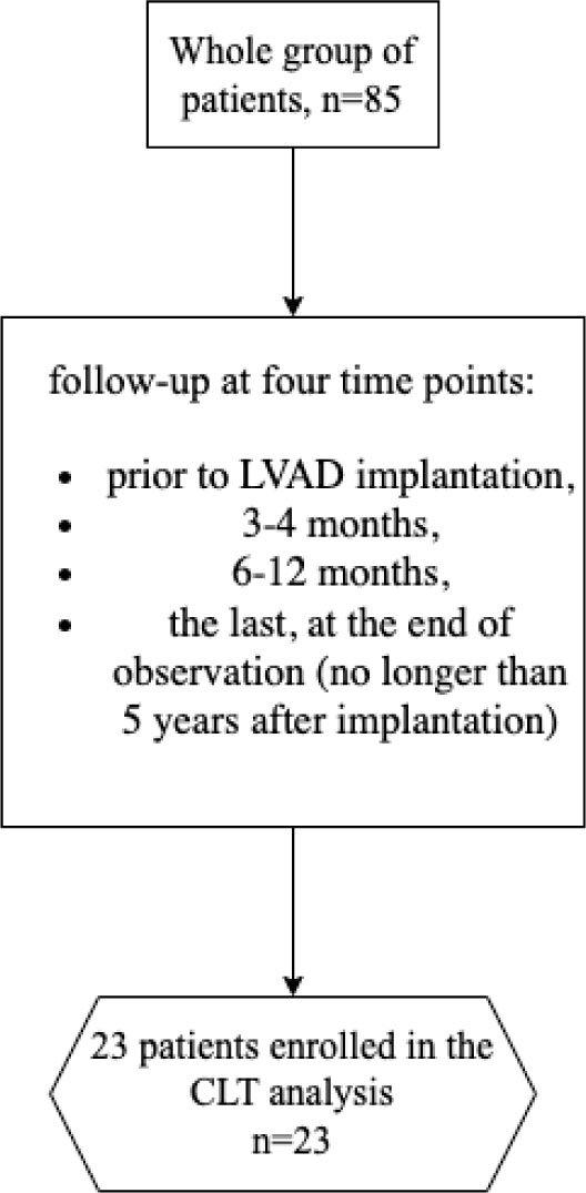
Figure 1.
Flowchart of the study
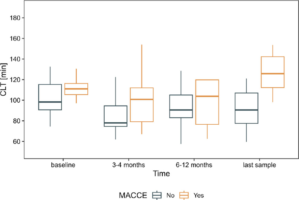
Figure 3a.
Comparison between patients with and without MACCE in the subgroup of patients who completed four observations (n=23). Whiskers presents range, box presents interquartile range (IQR) and the bar within the box displays median.
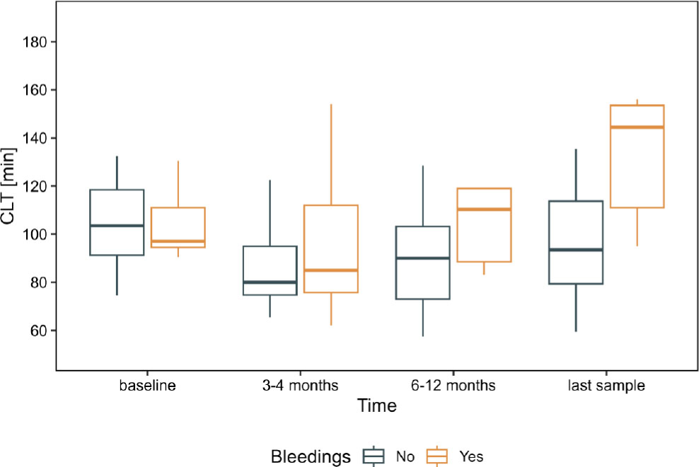
Figure 3b.
Comparison between patients with and without bleeding in the subgroup of patients who completed four observations (n=23). Whiskers present range, the box presents interquartile range (IQR), and the bar within the box displays median
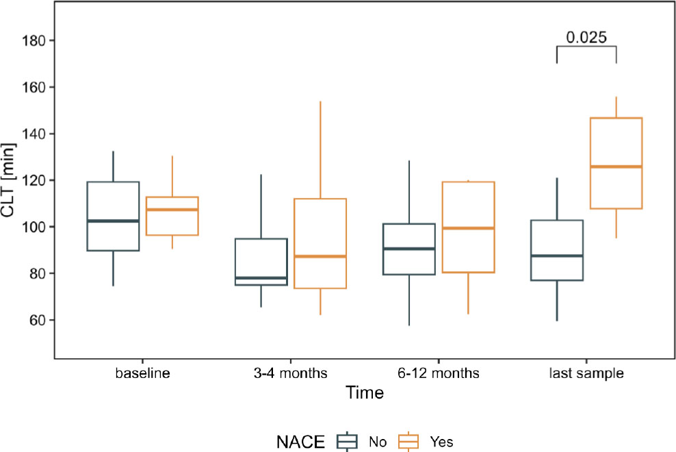
Figure 3c.
Comparison between patients with and without NACE in the subgroup of patients who completed four observations (n=23). Whiskers presents range, box presents interquartile range (IQR) and the bar within the box displays median. The square bracket (top-right) shows statistically significant difference between groups with the p-value after Holm-Bonferroni correction
Table 1.
Baseline characteristics of LVAD patients (prior to LVAD implantation), 85 patients
| Variable | |
| Age at the time of implantation, years | 58.2 [48.6–63.8] |
| NYHA class | 4[3.5–4] |
| INTERMACS | 3[2–4] |
| LVEF, % | 15[11–17] |
| LVEDD, mm | 75[70–82] |
| Prior stroke | 13 (17.6) |
| Hypertension | 38 (44.7) |
| Diabetes mellitus | 35 (41.2) |
| eGFR< 60ml/min/1.73m2, | 37 (45.1) |
| Medications, n (%) | |
| Beta blockers | 65 (76.5) |
| ACEI | 44 (51.8) |
| ARB | 12 (14.1) |
| Aldosterone antagonists | 74 (87.1) |
| Aspirin | 24 (28.2) |
| Clopidogrel | 5(5.9) |
| Statin | 51 (60) |
| Vitamin K antagonists | 38 (44.7) |
Table 2.
Laboratory investigations in LVAD patients who completed the whole study, n=23. Patients who died prior to the end of observation and incomplete observations were excluded from the further analyses
| Laboratory parameters | baseline, N = 23 | 3–4 months, N = 23 | 6–12 months, N = 23 | last sample, N = 23 | p-value1 | q-value2 | Effect size | Effect size (without baseline) |
|---|---|---|---|---|---|---|---|---|
| White blood cell count, 103/µL | 9.93 (7.52–11.05) | 7.70 (6.28–9.17) | 7.31 (5.88–8.76) | 7.80 (6.89–9.41) | 0.066 | 0.078 | 64.3% | 21.1% |
| Hemoglobin, mmol/L | 7.70 (7.15–9.00) | 8.10 (7.30–8.45) | 8.30 (7.50–8.90) | 8.80 (7.85–9.45) | 0.015 | 0.022 | 48.5% | 53.6% |
| Platelet count, 103/µL | 194 (146–254) | 245 (199–261) | 211 (171–257) | 185 (172–228) | 0.003 | 0.008 | 47.4% | 56.2% |
| Bilirubin, µmol/L | 18 (8–33) | 8 (7–11) | 11 (9–13) | 11 (8–15) | <0.001 | 0.004 | 70.0% | 55.1% |
| Albumin, g/L | 40.0 (35.0–42.0) | 44.0 (41.5–47.5) | 45.0 (43.0–49.0) | 46.0 (42.0–47.5) | <0.001 | <0.001 | 81.2% | 17.0% |
| Creatinine, µmol/L | 125 (110–184) | 100 (81–125) | 110 (93–131) | 111 (95–154) | 0.001 | 0.005 | 58.4% | 35.3% |
| hsCRP, mg/L | 8 (4–25) | 8 (3–18) | 6 (4–10) | 4 (2–13) | 0.2 | 0.2 | 38.7% | 28.4% |
| NT-proBNP, pg/mL | 11259 (5521–20366) | 2244 (1230–3273) | 1854 (895–2995) | 1992 (1138–3005) | <0.001 | <0.001 | 89.0% | 10.1% |
| Fibrinogen, g/L | 479 (380–542) | 424 (359–476) | 394 (346–428) | 368 (321–453) | 0.010 | 0.017 | 62.7% | 34.4% |
| D-dimer, ng/mL | 1.66 (1.00–3.46) | 1.79 (1.43–2.46) | 1.25 (0.97–1.66) | 1.11 (0.69–1.53) | 0.001 | 0.005 | 62.7% | 77.9% |
| CLT, min | 104 (93–125) | 82 (75–104) | 91 (79–113) | 98 (86–117) | 0.022 | 0.027 | 56.6% | 41.0% |
| Von Willebrand Factor, % | 290 (210–368) | 168 (140–215) | 177 (139–212) | 174 (131–288) | 0.009 | 0.017 | 77.3% | 17.9% |
| Factor VIII, % | 281 (232–349) | 200 (171–229) | 197 (131–226) | 174 (114–226) | 0.007 | 0.015 | 83.4% | 28.0% |
| Antitrombin, % | 87 (73–94) | 93 (87–99) | 97 (92–104) | 104 (90–108) | 0.013 | 0.020 | 76.0% | 52.6% |
| International Normalized Ratio, % | 1.53 (1.21–1.89) | 2.29 (2.06–2.37) | 2.24 (2.01–2.65) | 2.31 (2.17–2.57) | <0.001 | <0.001 | 90.0% | 17.4% |
| Thrombin Time, s | 16.90 (15.85–18.05) | 16.00 (15.70–16.60) | 15.80 (15.20–16.38) | 16.60 (15.85–17.30) | 0.017 | 0.023 | 66.7% | 63.7% |
| Activated Partial Thromboplastin Time, s | 43 (33–46) | 45 (40–50) | 41 (37–45) | 38 (36–42) | 0.003 | 0.008 | 56.7% | 71.0% |
| ASPI, AU × min | 590 (362–1,058) | 431 (276–705) | 331 (239–754) | 533 (312–880) | 0.2 | 0.2 | 37.2% | 22.4% |
| ADP, AU × min | 364 (274–658) | 466 (223–621) | 408 (268–655) | 461 (341–653) | 0.10 | 0.11 | 6.9% | 7.3% |
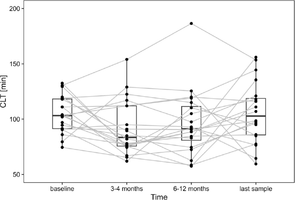
Figure 2.
CLT in the subgroup of patients during follow-up, n=23. Dots represent individual patients. The gray line connects the same patient at consecutive time points. The box presents an interquartile range (IQR), and the bar within the box displays the median
Table 3.
Major adverse cardiac and cerebrovascular events (MACCE), bleeding events and Net Adverse Clinical Events (NACE) in the whole group of patients (n=85)
| 0 to 3 months | 3–4 months | 6–12 months | Last available (median 21 months;IQR:10–29 months) | |
|---|---|---|---|---|
| MACCE, pts. (%) | 11(12.9) | 4(5.1) | 10(12.9) | 9(13.4) |
| Bleeding events, pts. (%) | 3(3.5) | 8(10.3) | 5(6.5) | 6(8.9) |
| NACE, pts (%) | 14(16.5) | 12(15.4) | 14 (18.2) | 15(22.4) |
| Deaths, pts (%) | 7(8.2) | 1(1.3) | 10(12.9) | 9(13.4) |
Table 4a.
Comparison of CLT according to Major Adverse Cardiac and Cerebrovascular Events (MACCE) in the course of time, subgroup of patients with completed four observations (n=23). In the analyzed subgroup of patients MACCE occurred in 7 patients
| Clot Lysis Time [min] | baseline | 3–4 months | 6–12 months | Last available |
|---|---|---|---|---|
| Patients with MACCE during the whole observation period, n=6 | 111 (105.4–116.3) | 100.8 (79.2–112) | 103.8 (76.5–119.8) | 125.8 (112.33–142.2) |
| Patients without MACCE during the whole observation period, n=17 | 103 (91–129.5) | 78 (74.5–94.5) | 90.5 (83–105) | 90.5 (77.5–105) |
Table 4b.
Comparison of CLT according to bleeding events in the course of time. Subgroup of patients with completed four observations (n=23). In the analyzed subgroup of patients, bleedings occurred in 5 patients
| Clot Lysis Time [min] | baseline | 3–4 months | 6–12 months | Last available |
|---|---|---|---|---|
| Patients with bleedings during the whole observation period, n=5 | 90.5 (94.5–111) | 85 (75.6–112) | 110.3 (88.5–119) | 144.5 (111–153.5) |
| Patients without bleedings during the whole observation period, n=18 | 107.5 (94.1–127.1) | 80 (74.8–94.9) | 110.3 (88.5–119) | 144.5 (111–153.5) |
Table 4c.
Comparison of CLT according to Net Adverse Clinical Events (NACE) events in the course of time. Subgroup of patients with completed four observations (n=23). In the analyzed subgroup of patients, NACE occurred in 8 patients
| Clot Lysis Time [min] | baseline | 3–4 months | 6–12 months | Last available |
|---|---|---|---|---|
| Patients with NACE during the whole observation period, n=8 | 107.3 (96.4–112.8) | 87.3 (73.6–112) | 99.4 (80.4–119.3) | 125.8 (107.8–146.7) |
| Patients without NACE during the whole observation period, n=15 | 104 (91.3–131) | 78 (75–94.8) | 90.5 (79.5–101.3) | 87.5 (77–102.8) |