
INTRODUCTION
Wound healing is a complex natural process that occurs when the skin barrier is damaged by physical, chemical, or biological stressors. This dynamic process involves multiple biological responses and occurs in four harmonious, interrelated, and highly organized stages that facilitate the restoration of the skin barrier and the regeneration and repair of damaged tissues (Yang et al., 2021). The first stage of wound healing is hemostasis, which begins immediately after a skin injury to minimize bleeding at the wound site. During this stage, vascular constriction and clot formation occur. This is followed by the inflammatory phase, characterized by the infiltration and recruitment of immune and pro-inflammatory cells. These cells secrete various pro-inflammatory cytokines and proteolytic enzymes to eliminate antigens, foreign particles, and cellular debris from the wound site. They also induce oxidative stress and promote the production of reactive oxygen species to protect the damaged tissue from microbial infections (Deng et al., 2021). Once the inflammatory phase is complete, the proliferation phase begins. During this stage, epithelialization, fibroblast migration, angiogenesis, and the deposition of extracellular matrix are accelerated at the wound site. The final stage is the remodeling phase, during which the newly generated tissue increases its tensile strength through the reorganization of collagen fibers (Ellis et al., 2018; Singh et al., 2017).
The effective healing of wounds requires a sequential and well-coordinated progression through these phases. In fact, the prolonged bleeding, inflammation, excessive production of free radicals, persistent oxidative stress, and microbial infections at the wound site can delay the healing process and affect the properties and functions of the generated tissues (Deng et al., 2021; Guo & DiPietro, 2010; Schilrreff & Alexiev, 2022). Consequently, there is a growing interest in the pharmaceutical and cosmetic industries in developing new wound-healing agents that have antioxidant, anti-inflammatory, antimicrobial, hemostatic, and regenerative activities (Ghuman et al., 2019). These qualities promote optimal wound repair and support tissue regeneration. Additionally, the antioxidant and antimicrobial activities improve the stability of pharmaceutical formulations.
Medicinal plants remain a vital source of bioactive compounds useful for discovering and developing new drugs and treatments (Shedoeva et al., 2019). The genus Linaria (Scrophulariaceae) is one of the most used taxa in folk medicine due to the richness of its plants in diverse classes of bioactive molecules (Cheriet et al., 2015). Among its members, the species Linaria scariosa Desf. is an annual plant employed traditionally in North Africa for the treatment of acne and infected wounds. Previous reports on this species confirmed its potential in vitro biological activities, including photoprotective, anti-denaturant, hemostatic, anticholinesterase, antibacterial, and antioxidant (Ahmed-Chaouch et al., 2021; Mouffouk et al., 2020). However, no prior reports regarding its in vivo pharmacological properties were published. Therefore, the present work aims to develop a herbal cream and to assess the in vivo wound healing and in vitro biological effects of Linaria scariosa species, to validate its traditional application.
MATERIAL AND METHODS
Plant material
Linaria scariosa Desf. species was collected during its flowering period in May 2023, from Hidoussa region (geographic coordinates in decimal degrees (WGS84): latitude 35.494, longitude 5.906, Algeria), and was identified by botanists from the Department of Agronomy of the University of Batna 1. A reference specimen was deposited under reference number 826/LCCE.
Chemicals and reagents
All solvents used in the experiments (methanol, acetone, hexane, and DMSO) were purchased from PROLAB, MERK EUROLAB. Cera alba (White beeswax), paraffin oil, eumulgin B2, and cutina GMS were purchased from DZAIR MATIERE PREMIERE. 2,2-diphenyl-1-picrylhydrazyl (DPPH), potassium ferricyanide (K3Fe[CN]6), trichloroacetic acid (CCl3COOH), ferric chloride (FeCl3), Folin-Ciocalteu reagent, sulfuric acid (H2SO4), trichloroaluminum (AlCl3), vanillin (C8H8O3), sodium carbonate (Na2CO3), dipotassium phosphate (K2HPO4), monopotassium phosphate (KH2PO4), sodium chloride (NaCl), potassium permanganate (KMnO4), and ammonium molybdate (NH4)2MoO4 were purchased from Sigma-Aldrich Chemie and Fluka Chemie. EDTA (C10H14N2Na2O8), ferrous chloride (FeCl2), ferrozine (C20H13N4NaO6S2), butylate hydroxyanisole (BHA; C11H16O2), ascorbic acid (C6H8O6), sodium tetraborate decahydrate (Na2B4O7 · 10H2O), and Mueller-Hinton were purchased from Sigma-Aldrich Co., ST Lowis, Mo.
Instruments
Spectrophotometric measurements were conducted using a SHIMADZU UV-1700 UV-Vis spectrophotometer. The pH measurements of the formulation were determined with a digital pH meter (HANNA instruments, model HI 2210). Color parameters were assessed using the KONICA MINOLTA CR-10 color reader (Konica Minolta, Japan) operating with a CIELAB color system, and viscosity measurements were performed by a Discovery HR 30 rheometer with a 40 mL parallel plate geometry at a 1000 μm gap to evaluate the rheological behavior of the cream formulations.
Extraction procedure
Three hundred grams (300 g) of the entire plant Linaria scariosa, including roots, stems, leaves, and flowers were collected; dried in a dark and dry place; and subsequently ground into a fine powder using an electric grinder. Extraction was carried out by maceration in 3 L of methanol for 48 hours at room temperature under continuous stirring. The extraction was repeated twice under identical conditions using fresh solvent to optimize the yield of extraction. After each extraction cycle, the extract was filtered, and the filtrates were combined before being concentrated under reduced pressure at 35 °C using a rotary evaporator to obtain 3.69 g of the methanolic extract.
Total bioactive contents
To determine the total phenolic content, a volume of 200 μL of crude extract was mixed with 1 mL of Folin-Ciocalteu solution (10%). After 4 minutes of reaction, 800 μL of a saturated sodium carbonate solution (Na2CO3, 75 g/L) was added. The obtained solution was incubated in a dark place at room temperature for 2 hours. The absorbance was then measured at 765 nm (Aichour et al., 2023). The total phenolic content was determined using a gallic acid calibration curve (25–400 μg/mL, y = 0.0022x + 0.1497, R2 = 0.9901). Results were expressed as microgram equivalents of gallic acid per milligram of extract (μg GAE/mg).
To quantify the total flavonoid content, 1 mL of trichloroaluminum solution (2%) was added to 1 mL of the plant extract. The mixture was vigorously stirred and incubated at room temperature for 10 minutes. The absorbance of the resulting solution was then measured at 430 nm (Aichour et al., 2023). The total flavonoid content was determined using a calibration curve established with quercetin (25–600 μg/mL, y = 0.0018x + 0.0343, R2 = 0.9977). Results were expressed as micrograms equivalents of quercetin per milligram of crude extract (μg QE/mg).
The total terpenoid content was resolved by mixing 150 μL of LS extract with 500 μL of vanillin solution (8%; w/v) and 500 μL of sulfuric acid (72%; w/v). The resulting solution was heated in a water bath at 60°C for 10 minutes and then cooled in ice water for 15 minutes. The absorbance of the obtained solution was measured at 538 nm. The total terpenoid content was calculated using a calibration curve established with ursolic acid (4–160 μg/mL, y = 0.0079x + 0.3145, R2 = 0.9906). Results were presented as microgram equivalents of ursolic acid per milligram of extract (μg UAE/mg) (Hiai et al., 1976).
Total lycopene and carotenoid content was measured using the method described by Barros et al. (2008). This method is based on calculating the absorbance of the plant extract dissolved in a mixture of acetone and hexane (6:4, v/v) at wavelengths of 453, 505, and 663 nm. Total β-carotene and lycopene content was calculated using the following equations (1 and 2):
Antimicrobial activity
The inhibitory potential of L. scariosa cream on the growth of microbial strains was evaluated in vitro using the solid-state diffusion method (Dekkiche et al., 2021). This activity was tested on a yeast (Candida albicans ATCC 90029) and several bacterial strains, including five reference strains from the American Type Culture Collection (Staphylococcus aureus ATCC 25923, Escherichia coli ATCC 25922, Klebsiella pneumoniae ATCC 70063, Morganella morganii ATCC 25830, Pseudomonas aeruginosa ATCC 27853, and Streptococcus pneumoniae ATCC 49619) and two pathogenic isolates (Streptococcus D and Enterococcus sp.). All these strains were kindly provided by the Bacteriology Laboratory of the University Hospital Center of Batna (CHU). The growth of the tested microbial strains was optimized by inoculating the bacteria into Mueller-Hinton agar plates and incubating them at 37°C for 24 hours. After, many well-isolated colonies were picked using a platinum loop and transferred to a tube containing 5 mL of nutrient broth. The mixture was then homogenized and incubated for 10–24 hours at 37°C. The turbidity of each 1 mL microbial suspension was adjusted to match a McFarland 0.5 standard by measuring the optical density at 626 nm. A McFarland 0.5 standard corresponds to an OD value of 0.08–0.1 at 625 nm, equivalent to approximately 1–2 × 10⁸ CFU/mL, thereby ensuring a standardized inoculum density for all antimicrobial assays.
Petri dishes containing Mueller-Hinton medium were inoculated with the microbial suspensions using the swabbing technique. Whatman paper discs impregnated with 10 μL of samples (extract and antibiotics) at different concentrations were carefully placed on the inoculated agar surface using sterile forceps. The Petri dishes were then incubated for 24 hours at 37 °C. The tests were performed in triplicate. The lowest concentration that inhibited visible bacterial growth was recorded as the minimum inhibitory concentration (MIC).
In vitro anti-inflammatory activity
The anti-inflammatory effect of the LS-cream was evaluated by studying the membrane stability of red blood cells; 4 mL of blood was collected from a healthy adult in tubes containing sodium oxalate. The samples were stored at 4°C for 24 hours before use, then centrifuged (5 min at 2500 rpm), and the supernatant discarded. The aliquot was washed with sterile saline and then centrifuged (2500 rpm for 5 min). Finally, a 40% (v/v) suspension was prepared in phosphate-buffered saline and used in the heat-induced hemolysis test (Mouffouk et al., 2023).
Antioxidant activity
A volume of 1 mL of potassium permanganate solution prepared in methanol (0.02 M) was added to 1 mL of the samples at different concentrations. The obtained solutions were shaken and incubated in the dark at room temperature for 30 minutes. The absorbances were measured at 514 nm. The percentages of scavenging activity were calculated using the following equation (3):
The chelation of ferrous iron was estimated using the method of Le et al. (2007). Indeed, 500 μL of the samples (LS-cream and EDTA) were mixed with 100 μL of ferrous chloride (0.6 mM) and 900 μL of methanol. After 5 min, 100 μL of ferrozine solution (5 mM) was added to the mixture. The performing tubes were agitated and incubated for 10 min at room temperature, after which the absorbance was measured at 560 nm. The results of ferrous iron chelation were expressed as a % of chelation, calculated using equation (4):
The antioxidant activity of the samples was assessed using the DPPH radical scavenging assay. A freshly prepared DPPH solution in methanol was used for the analysis. Briefly, 100 μL of the samples at different concentrations was mixed with 900 μL of the DPPH solution. The reaction mixture was agitated and incubated in the dark at room temperature for 30 min. A control was prepared by replacing the samples with methanol under the same experimental conditions. After incubation, the absorbance was measured at 517 nm. The ability of the samples to scavenge DPPH radicals was expressed as percentage of inhibition and calculated according to equation (3).
The total antioxidant potential of the tested samples was evaluated using a colorimetric method. A volume of 100 μL of the formulation at a concentration of 1 g/mL was added to a tube containing 900 μL of a reagent [H2SO4 (0.6 M), Na2PO4 (28 mM), and ammonium molybdate (4 mM)]. All tubes were incubated at 95 °C for 90 minutes. After incubation, the mixtures were cooled, and the absorbances were measured at 695 nm. The results are expressed as μg equivalents of ascorbic acid per g of cream (μg eq AA/g), using the calibration curve established with ascorbic acid (0.25–3 μg/mL, y = 47.071x + 0.0279, R2 = 0.9978).
The ability of cream to reduce ferric ions was measured by spectrophotometry. Indeed, 100 μL of samples at a concentration of 1 g/mL was mixed with 500 μL of phosphate buffer (pH 6.6; 0.2 M) and 500 μL of a 1% ferrocyanide solution. The mixtures were incubated at 50 °C for 20 minutes. After incubation, 500 μL of a 10% trichloroacetic acid solution was added, and the resulting mixtures were centrifuged at 3000 rpm for 10 minutes. After centrifugation, 500 μL of the supernatant was diluted in an equal volume of double-distilled water, and then, 125 μL of 0.1% FeCl3 was added. The absorbance of each sample was measured at 700 nm, and the reducing potential was determined using a calibration curve established with ascorbic acid (25–300 μg/mL, y = 7.77086x + 0.1209, R2 = 0.9961). The results are expressed as μg equivalents of ascorbic acid per g of cream (μg eq AA/g)
Herbal cream preparation
The herbal cream was formulated using L. scariosa extract and a water/oil emulsion following an adapted method and commonly used ingredients of cold cream preparation (Table 1) (Sundar & Lingakumar, 2023). First, the oil phase was prepared by melting Cera alba (white beeswax), Paraffin oil, Eumulgin B2, and Cutina GMS on a water bath at about 70–75°C. The mixture was stirred until a clear, homogeneous melt was obtained. Then, the aqueous phase was prepared by dissolving both the extract and borax in purified water with gentle heating up to 50 °C. Emulsification was initiated by slowly adding the aqueous phase to the melted oil phase under continuous mechanical stirring for 5 minutes, until a smooth and semi-solid consistency was achieved. The prepared herbal cream was then transferred into sterile, opaque, low-density containers and stored in a refrigerator at 4°C until use.
Table 1.
Composition of the herbal cream prepared from Linaria scariosa extract
| Ingredient | Quantity for 100 g | Function |
|---|---|---|
| Linaria scariosa extract | 200 mg | Active ingredient |
| Cera alba (White beeswax) | 18 g | Thickening agent and consistency enhancer |
| Paraffin oil | 50 g | Oil phase, emollient |
| Eumulgin B2 | 0.3 g | Non-ionic emulsifying agent |
| Cutina GMS | 0.8 g | Co-emulsifier and consistency enhancer |
| Borax (Sodium tetraborate decahydrate) | 0.05 g | Emulsion stabilizer and pH-adjusting agent |
| Distilled water | q.s. to 100g | Aqueous phase |
Physicochemical, rheological, and microbiological examination of the herbal cream
Samples of the cream were used to verify its rheological, physicochemical, and microbiological stability. Organoleptic properties, including color, odor, and texture, as well as phase separation, were physically assessed. The color of the cream was determined using a KONICA MINOLTA CR-10 color reader (Konica Minolta, Japan) operating with a CIELAB color system. The color parameters evaluated include L*, a*, and b*, where L* represents lightness on a scale ranging from 0 (black) to 100 (white), a* represents the green (−60) to red (+60) axis, and b* represents the blue (−60) to yellow (+60) axis. The spreading time was evaluated by placing 0.5 g of the prepared cream between two cleaned microscopic slides. To ensure a uniform spread, a 100 g weight was placed on the slides. The time required for slide separation was recorded, and the following equation (5) was used to report the spreadability coefficient (S):
where M = weight tied to upper slide (g), L = length of glass slide covered (cm), and T = time (sec).The pH of each cream (1 g of cream dissolved in 10 mL of distilled water) was measured with a digital pH meter. The viscosity measurement of the formulated cream was conducted using a Discovery HR30 rheometer, equipped with a 40-mm stainless-steel parallel-plate geometry at a gap of 1000 μm. Measurements were performed at 25 °C over a shear-rate of 0.0069–1000 s−1. The rheological data were analyzed using the Cross Model. Other physicochemical properties of the formulated cream were evaluated using standard analytical procedures, including ash content using the incineration method and moisture and dry matter contents using the oven-drying method. Additionally, the density of the cream was measured using a densimeter.
The microbiological stability of the LS-cream was assessed in accordance with applicable ISO standards for cosmetic products. Five samples of the prepared herbal cream (E1 to E5) were submitted for microbiological analysis to verify compliance with the acceptance criteria established for topical products. Total aerobic mesophilic count (TAMC) was determined in accordance with ISO 21149. Yeast and mold testing was performed in accordance with ISO 16212. The presence of specified microorganisms was also investigated, including coagulase-positive Staphylococcus aureus according to ISO 22718, Escherichia coli according to ISO 21150, Pseudomonas aeruginosa according to ISO 22717, and Candida albicans according to ISO 18416.
In vivo wound-healing effect
After assessing the in vitro antioxidant, antimicrobial, and anti-inflammatory effects of the herbal cream, its in vivo wound-healing activity was evaluated using the excision wound model, in accordance with institutional ethical guidelines for the care and use of laboratory animals. The study design, animal handling, outcome assessment, and reporting followed the ARRIVE 2.0 guidelines to promote transparency, animal well-being, and reproducibility. Initially, five groups (n = 6) of healthy, well-adapted adult male albino Wistar rats, weighing an average of 150–160 g, were purchased from the Pasteur Institute of Algeria and randomly combined. The back of tranquilized rats was clipped and carefully shaved in aseptic conditions. Then, all rats were anesthetized with 45 mg/kg sodium thiopental, and a full-thickness wound (1.5 × 1.5 cm) was created on their shaved dorsal areas. The area of excised skin of different groups was treated topically for 23 days as follows:
Group 1: treated once with the prepared herbal cream
Group 2: treated twice with the prepared herbal cream.
Group 3: treated with commercial BETADINE® (positive control group)
Group 4: treated with the same prepared water/oil emulsion without the extract (vehicle group)
Group 5: does not receive any treatment (control group)
The wound areas were measured every 48 hours by placing transparent tracing paper over the wound and tracing it out. The tracing papers were then scanned, and the surfaces were estimated using ImageJ software (Schneider et al., 2012), and the area was recorded. The morphological, macroscopic, and histological parameters were evaluated. The percentage of wound contraction was calculated as follows (6):
On day 23, the animals were sacrificed, and tissue samples were harvested from the wound site of each animal and stored separately in 10% formalin solution for 72 hours. After undergoing the routine histopathological treatment and H&E staining described by Suvarna et al. (2019), the prepared sections were submitted to microscopic evaluation, and some wound-healing parameters, including the degree of epithelialization, differentiation, amount of granulation tissue, inflammation, orientation of collagen fibers, and neovascularization, were estimated.
Statistical analysis
Means, standard error of measurement, and standard deviations of the different assays were calculated using a one-way ANOVA test followed by Tukey's post hoc tests to determine the inner group differences. Data were analyzed using Minitab Statistical Software 22 on Windows, and p value < 0.05 was set for all analyses.
RESULTS AND DISCUSSION
Total bioactive contents
The quantities of major bioactive compounds found in the LS-extract derived from the species L. scariosa were determined through the standardized spectrophotometric assays, and the results are reported in Table 2. According to the results, the crude extract of L. scariosa contains carotenoids (0.57 mg/100 mL) and lycopene (0.36 mg/100 mL). These natural lipophilic pigments are responsible for a large range of the pharmaceutical and nutritional properties and exert excellent antiaging, photoprotective, anti-inflammatory, immunomodulatory, and antioxidant activities (Medina-García et al., 2025; Zhu et al., 2025). No previous reports have quantified carotenoids or lycopene in plants of the genus Linaria. Therefore, no direct comparisons with literature have been made. However, the reported values are considered low compared with several culinary herbs and medicinal plants across multiple taxa (Daly et al., 2010; Othman et al., 2017). Additionally, this extract possesses moderate levels of flavonoids and phenolic compounds. This finding is consistent with the results of many recent studies on species belonging to the Linaria taxa. In fact, low to moderate phenolic and flavonoid contents were estimated in Linaria triphylla, L. tarhunensis, L. corifolia, L. aegyptiaca, and L. reflexa organic extracts (Al-Awjali et al., 2025; Atayalla et al., 2025; Bekhaoua et al., 2019; Utlu & Ercil, 2024). However, a low amount of terpenoids (0.14 ± 0.15 μg EUA/mg) was estimated in this extract. Despite the existence of several classes of terpenoids in Linaria species, such as pentacyclic triterpenes and iridoids isolated from L. alpine L. corifolia, L. aegyptiaca, and L. tingitana organic extracts (Ferhat et al., 2010; Frezza et al., 2019; Hanfer et al., 2017; Utlu & Ercil, 2024).
Table 2.
Total phenolic, flavonoid, terpenoid, carotene, and lycopene contents of L. scariosa extract
| Classes of bioactive compounds | LS extract |
|---|---|
| Total phenolic content (μg EAG/mg) | 108.98 ± 0.67 |
| Total flavonoid content (μg EQ/mg) | 78.77 ± 0 .18 |
| Total terpenoid content (μg EUA/mg) | 0.14 ± 0.15 |
| Total carotene content (mg/100 mL) | 0.57 ± 0.12 |
| Total lycopene content (mg/100 mL) | 0.36 ± 0.08 |
Herbal cream formulation
The prepared cream was a water/oil emulsion, characterized by a homogeneous texture and emollient properties with a pH of 6.8 ± 0.09 compatible with normal skin physiology. The colorimetric analysis of the herbal cream revealed values of L*= 31.9, a*=−11.0, and b*=+19.1, indicating that the tested cream had a dark yellowish-green color. The cream kept its physical characteristics and organoleptic properties after its storage. Indeed, no color, odor, or phase separation was observed. It had a non-greasy feel and was easily removed with warm water after application. The physicochemical properties of the formulated cream, including ash content (0.02% ± 0.087), humidity (18.24% ± 0.10), dry matter content (81.75% ± 0.12), and density (1.001±0.02 g/mL), are consistent with acceptable standards for cold creams, which indicates the stability and suitability of the formulated cream for topical application (Table 3).
Table 3.
Physicochemical, rheological, organoleptic and microbiological properties of the herbal cream
| Parameters | Physical observations after 6 months | |
|---|---|---|
| Organoleptic properties | Color | Dark yellowish-green color (L*= 31.9, a*=−11.0 and b*=+19.1) |
| Odor | Strong herbal odor | |
| Homogeneity | Homogenous | |
| Texture | Viscous, non-greasy and smooth | |
| Stability | Stable | |
| Physicochemical properties | pH | 6.8 ± 0.09 a |
| Washability | Warm water | |
| Spreading time | 14.91 ± 1.72 a S | |
| Spreadability coefficient (S) | 50.07± 5.94 g·cm/s a | |
| Density | 1.001±0.02 g/ml a | |
| Moisture content | 18.24% ± 0.10 a | |
| Dry matter content | 81.75% ± 0.12 a | |
| Ash content | 0.02% ± 0.087 a | |
| Microbiological stability | Total mesophilic aerobic microorganisms | ≤100 |
| Coagulase-positive Staphylococcus aureus | Complete absence in 1 g | |
| Escherichia coli | Complete absence in 1 g | |
| Pseudomonas aeruginosa | Complete absence in 1 g | |
| Candida albicans | ≤100 | |
| Rheological properties | Zero-shear viscosity (η0) | 1332 Pa·s |
| Flow behavior index (n) | 0.7825 | |
| Coefficient of determination (R2) | 0.998 |
The formulation demonstrated a promoting safety profile, with minimal instances of skin irritation or allergic sensitization. Moreover, all formulations successfully passed rigorous microbial limit tests, including total bacterial and fungal counts after the microbiological stability evaluation following relevant ISO standards for cosmetics. No pathogenic microorganisms, such as Coagulase-positive Staphylococcus aureus, Escherichia coli, Pseudomonas aeruginosa, or Candida albicans, were found in 1g of product. Additionally, no yeasts or molds were detected. Tests on samples E1 to E5 revealed that total mesophilic aerobic microorganisms were below the ISO 21149 limit (≤ 100 CFU/g). These findings confirm that the herbal cream complies with microbiological stability throughout the testing period and after six months of storage.
The rheological analysis of the herbal cream revealed that it exhibited non-Newtonian pseudoplastic (shear-thinning) behavior, confirmed by the flow index value (n=0.7825 (<1)) and characterized by a marked decrease in viscosity with increasing shear rate, indicating that the cream becomes progressively easier to spread upon application. These results correlate with a good spreadability coefficient estimated at 50.07 g·cm/s. The high Zero-shear viscosity (η0 = 1.33238 × 10⁷ Pa⋅s) indicates good structural integrity and strong resistance to flow under near-static conditions. On the other hand, the low infinite-shear viscosity (0.101 Pa·s) suggests that the tested cream becomes considerably less viscous when shear is applied facilitating spreading and application under shear. Additionally, the high coefficient of determination (R2 = 0.998) indicated that the rheological behavior of the herbal cream was successfully described by the Cross model, supporting the reliability of the calculated rheological parameters. Overall, these results suggest that the prepared herbal cream possesses favorable rheological characteristics, combining high viscosity at rest with reduced viscosity during application, which are desirable characteristics for topical formulations (Figure 1).
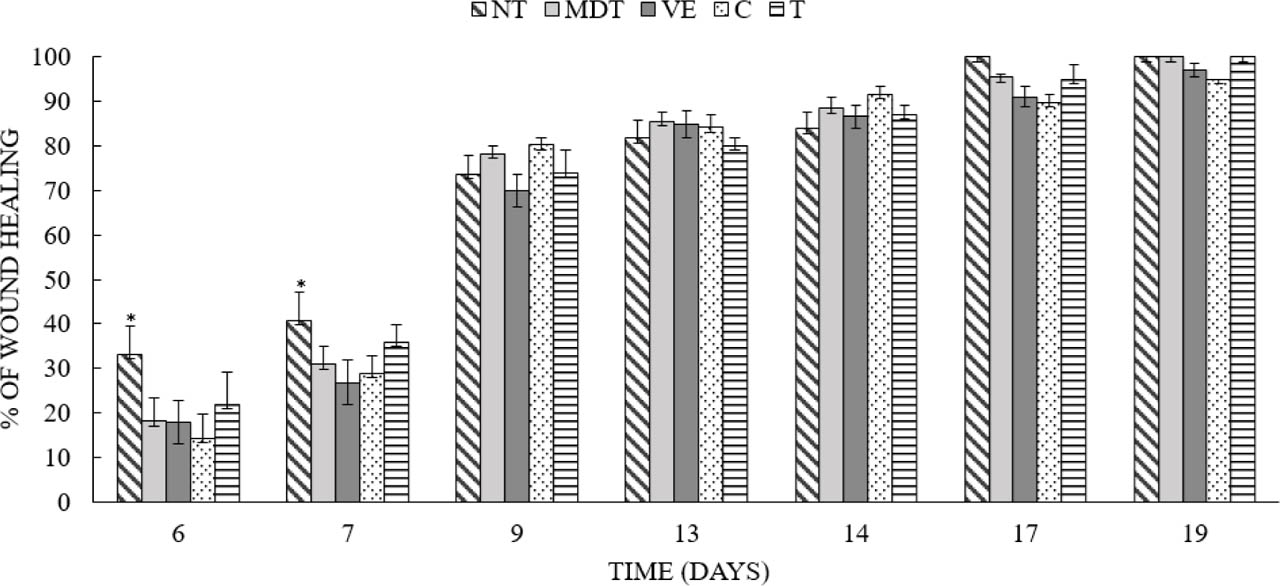
Figure 1.
Percentage wound closure for different treated groups over time. C: Control; MDT: Medicament; VE: Vehicle; T: Single dose of the herbal cream; NT: Double dose of the herbal cream.
Antioxidant activity
The antioxidant properties of the LS-cream were tested by five different spectrophotometric methods, including DPPH radical scavenging assay, ferrous iron chelation method, total antioxidant capacity, and ferric- and permanganate-reducing activities. The results are summarized in Table 4. According to the obtained results, the LS-cream exerted radical scavenging, reducing, and chelating abilities that varied in a dose-dependent manner. However, the reported activities were less efficient than those of ascorbic acid, BHA and EDTA, used as reference molecules. Indeed, the LS-cream showed a moderate DPPH radical scavenging effect (IC50 = 87.63 ± 0.78 μg/mL). This activity was comparable to several previous reports, where the ethyl acetate extracts prepared from both L. scariosa and L. tingitana exerted moderate scavenging properties with values of IC50 estimated at 71.64 ± 1.13 and 82.40 ± 16 μg/mL, respectively (Ahmed-Chaouch et al., 2021; Hanfer et al., 2016). Furthermore, the organic extracts prepared from L. reflexa, L. vulgaris, L. corifolia, and L. genistifolia and the essential oil derived from L. corifolia showed weak DPPH scavenging effects compared to the present study (Cheriet et al., 2017; Vrchovská et al., 2008; Gul, et al., 2017; Zengin et al., 2016). On the other hand, the LS-cream showed interesting reducing effects on both ferric and permanganate ions and strong total antioxidant capacity (EC50 = 3.41 ± 0.27 μg/mL). These activities were superior than those reported previously on the crude extracts prepared from the same plant and collected from different regions (Ahmed-Chaouch et al., 2021; Mouffouk et al., 2020). The differences in antioxidant activity results for this species can be attributed to several factors, such as geographical origin, extraction techniques, and testing methods. This variability reflects the sensitivity of the species' chemical composition to environmental factors such as altitude, climate, temperature, and region, as well as to the extraction process itself, particularly the solvent used water, ethanol, etc. The strong total antioxidant capacity of the LS-cream could be likely associated to the presence of fatty acids and steroids. Several fatty acids and steroids, such as α-linolenic acid, palmitic acid, and β-sitosterol, have already been quantified from this species (Ahmed-Chaouch et al., 2021). These compounds are well-documented for their ability to withstand extreme conditions and to react in buffered mixtures. Moreover, the formulation of the crude extract may increase its total antioxidant capacity by improving its stability under incubation conditions. In addition, the LS-cream exerted iron-chelating effect. Indeed, these results were predictable; prior investigations highlighted the considerable reducing and chelating effects against iron and copper ions exerted by various organic extracts of the same tested plant (Ahmed-Chaouch et al., 2021), and several plants which belong to the same genus, such as L. corifolia (Utlu & Ercil, 2024), L. triphylla (Al-Awjali et al., 2025), and L. pinifolia (Zakaria, 2023). The antioxidant potential of Linaria scariosa may be linked to its chemical composition. Many secondary metabolites, including flavonoids and iridoids, have been isolated from the same plant. In fact, several studies have reported that high levels of pectolinarigenin and pectolinarin, which are found in the crude extracts of Linaria species and were previously isolated from the species Linaria scariosa, are considered as strong antioxidant molecules (Cheriet et al., 2020).
Table 4.
Antioxidant potential of LS-herbal cream
| Sample | Iron chelating power EC50 (μg/mL)a | DPPH radical scavenging assay IC50 (μg/mL)a | Permanganate-reducing activity IC50 (μg/mL)a | Ferric-reducing antioxidant power μg eq AA/mg | Total antioxidant capacity μg eq AA/mg |
|---|---|---|---|---|---|
| LS-cream | 3.41 ± 0.27 | 87.63± 0.78 | 10.38 ± 0.4 | 0.528 ± 0.02 | 0.144± 0.01 |
| Ascorbic acid b | NT | 3.8 ± 0.08 | 0.42 ± 0.22 | NT | NT |
| BHA | NT | 6.73 ± 0.5 | NT | 0.62 ± 0.09 | 0.16 ± 0.07 |
| EDTAb | 0.63 ± 0.07 | NT | NT | NT | NT |
In vitro anti-inflammatory activity
The anti-inflammatory activity of the LS-cream was tested by the heat-induced lysis inhibition method. The results are shown in Table 5. The results indicated that the herbal cream was able to protect erythrocyte membrane against lysis induced by heat in a dose-dependent manner, with a value of IC50 estimated at 338.01 ± 0.25 μg/mL. This activity was better than that exhibited by the reference molecule (Diclofenac), which was less efficient (IC50 = 1.96 ± 0.65 mg/mL). Our previous report on the species L. scariosa showed a moderate in vitro anti-inflammatory activity tested by ovalbumin denaturation assay (Mouffouk et al., 2020). Nonetheless, plants belonging to the genus Linaria are well-documented for their anti-inflammatory properties. Several species, including L. tingitana, L. japonica, L. grandiflora, L. genistifolia, L. confertiflora, L. reflexa, and L. aucheri, demonstrated interesting anti-inflammatory activities. Their ability to modulate inflammation was proved through different mechanisms of action, namely nitric oxide trapping, the inhibition of albumin denaturation, the inhibition of hemolysis induced by hypotony, and the inhibition of rat paw edema induced by carrageenin (Akkol & Ercil, 2009; Cheriet et al., 2021; Hanfer et al., 2017; Jung et al., 2007). Additionally, a recent study reported that Liriodendrin isolated from Linaria vulgaris exerts strong anti-inflammatory and antioxidant properties due to its potential to downregulate the release of numerous inflammatory mediators (Li et al., 2025).
Table 5.
Anti-inflammatory activity of the LS cream
| % of inhibition of RBC hemolysisa | ||
|---|---|---|
| Concentration (mg/mL) | LS-cream | Diclofenacb |
| 0.25 | 44.03 ± 0.98 | 17. 44 ± 1.29 |
| 0.5 | 52.69 ± 0.24 | 24.98 ± 0.72 |
| 1 | 61.62 ± 0.6 | 37.77 ± 0.69 |
| 2 | 68.53 ± 1.42 | 58.96 ± 0.95 |
| 4 | 87.52 ± 0.85 | 73.51±1.09 |
Many factors may explain the greater potential of the tested cream to prevent heat-induced hemolysis compared with the reference non-steroidal anti-inflammatory drug. In fact, water in oil and lipid-based emulsions have been reported to influence erythrocytes' stability under stress conditions due to their multiphase structure and lipid-rich composition, which reduces the effective heat transfer, thereby attenuating thermal stress. In addition, the lipid phase may mitigate hemolysis by directly modulating erythrocyte membrane stability, as lipids are known to influence plasmatic membranes' fluidity. Furthermore, amphiphilic components present in this formulation can also modulate membrane tension and alter permeability responses under stress through their interfacial localization. Consistent with this interpretation, previous studies reported a reduction in hemolytic activity of various bioactive molecules and drugs following their incorporation into lipid-based or emulsified systems (Jaromin et al., 2006; Jeswani et al., 2015). Furthermore, the reference drug used in this study was an injectable diclofenac formulation rather than analytical-grade diclofenac sodium; therefore, the potential influence of formulation excipients and solubility-related factors on the assay outcome cannot be completely excluded. Under the experimental conditions, these formulation components may have altered the physicochemical environment of the reaction mixture, thereby affecting erythrocyte membrane stability and, consequently, the apparent inhibitory activity of diclofenac.
Antimicrobial activity
The antimicrobial activity of the LS-cream was tested using the solid medium diffusion method against a yeast and seven bacterial strains belonging to different types, and the results are represented in Table 6. According to the obtained data, L. scariosa herbal cream exhibited significant antimicrobial effects against all the tested strains with inhibition zones ranging from 8.5 to 15.17 mm, and low minimum inhibitory concentrations (from 6.25 to 25 μg/mL), except the bacterium M. morganii ATCC25830, which resisted all the concentrations of the tested samples. However, the exerted antimicrobial activity was lower than that offered by both reference antibiotics (penicillin and gentamicin). It is important to note that adding borax to the formulation may partially increase the antimicrobial effectiveness of the Linaria scariosa-based cream, as previous reports have indicated a moderate antimicrobial effect of this plant (Ahmed-Chaouch et al., 2021). Indeed, the sensitive strains including S. aureus, E. coli, P. aeruginosa, Enterobacter spp, and C. albicans are documented for being responsible for nosocomial and surgical wound infections (Alharbi, 2022; Surucuoglu et al., 2005) and burn wound contamination (Tchakal-Mesbahi et al., 2021), and they are associated with a wide range of serious hospital-acquired infections and postoperative complications. Additionally, these pathogens are considered resistant to several common and efficient antibiotics; thus, their inhibition by the herbal cream may have a great impact on the cutaneous wound-healing process. In fact, essential oils and organic extracts prepared from species belonging to the taxa Linaria have been documented for their ability to possess pronounced bacteriostatic and bactericidal effects against the tested microbial strains among others including: L. corifolia (Gul et al., 2017), L. tingitana (Cheriet et al., 2015), L. ventricose (Nafis et al., 2021), L. vulgaris (Bombela et al., 2022), and L. reflexa (Cheriet et al., 2021). Several compounds isolated from L. cretacea and L. purpurea showed strong antibacterial and antifungal activities (Bakytzhanova et al., 2025; Frezza et al., 2019). However, some other species have no antimicrobial effects including L. tarhunensis (Atayalla et al., 2025).
Table 6.
The in vitro antimicrobial activity of L. scariosa herbal cream
| Inhibition zones (mm) and minimum inhibitory concentrations | ||||||
|---|---|---|---|---|---|---|
| Gram-negative strains | ||||||
| Samples | Concentration (μg of extract/g of herbal cream) | E. coli ATCC 25922 | Pseudomonas aeruginosa ATCC27853 | Klebsiella pneumoniae ATCC70063 | M. morganii ATCC25830 | |
| LS-cream | 2000 | 15.17 ± 1.18 | 10.5 ± 2.121 | 11 ± 1.41 | - | |
| 1000 | 13.42 ± 1.53 | 9 ±1.414 | 10 ± 0.94 | - | ||
| 500 | 12.10 ± 2.26 | 8 ± 0.001 | 6.5 ± 0.71 | - | ||
| 250 | 11.05 ± 2.05 | 6.5 ± 0.707 | 6 ± 0.71 | - | ||
| 125 | 9.00 ± 1.41 | - | - | - | ||
| 62.5 | - | - | - | - | ||
| MIC (μg/mL) | 125 | 250 | 250 | - | ||
| Penicillin (10 μg/mL) | 22.50 ± 0.70 | 25.00 ± 0.00 | 23.50 ± 0.70 | - | ||
| Gentamicin (10 μg/mL) | 38.00 ± 0.5 | 39.00 ± 0.1 | 27.00 ± 0.5 | - | ||
| Gram-positive and fungal strains | ||||||
| Samples | Dilutions | Staphylococcus aureus ATCC 25923 | Streptococcus D | Enterococcus spp | Candida albicans ATCC 90029 | |
| LS-cream | 1/1 | 15 ± 1.18 | 13.5 ± 0.12 | 8.5± 0.23 | 11 ± 0.03 | |
| 1/2 | 14 ± 1.18 | 12.5 ± 2.12 | 7.5 ± 0.71 | 10.5 ± 0.71 | ||
| 1/4 | 11 ± 0.71 | 12.5 ± 0.54 | 7.3 ± 0.65 | 10 ± 0.46 | ||
| 1/8 | 10 ± 2.59 | 12 ± 0.71 | 7 ± 0.17 | 9.5 ± 1.41 | ||
| 1/16 | 11 ± 0.01 | - | - | - | ||
| 1/32 | 8 ± 1.65 | - | - | - | ||
| MIC (μg/mL) | 62.5 | 250 | 250 | 250 | ||
| Penicillin (10 μg/mL) | 26.00 ±1.4 | 21.00 ±1.41 | 16 ± 0.7 | 22 ± 0.8 | ||
| Gentamicin (10 μg/mL) | 34 ± 0.2 | 27.4 ± 0.32 | 36.00 ± 1.4 | 39 ± 0.2 | ||
In vivo wound-healing effect
According to the results, the daily application of LS cream twice on excision wounds causes a significant reduction in the wound area and strong wound-healing effect during the first week of the treatment compared to all the groups (Figure 1). Indeed, the rate of wound closure in the group treated with herbal cream twice was much faster than that in the control group. A complete wound closure was observed in the group treated with the prepared cream on day 17 (16.33 ± 1.22 day), whereas it took about 22 days in the control group (21.33 ± 0.92 day). The untreated and vehicle-treated groups (21.16 ± 2.30 day) showed no significant differences in wound healing (p > 0.05), thus suggesting that the vehicle used to prepare the cream does not contribute to the healing process. Additionally, animals treated with betadine showed complete wound healing on day 18 (17.70 ± 1.17 day). The microscopic wound-healing parameters, such as the degree of epithelialization, differentiation, amount of granulation tissue, inflammation, orientation of collagen fibers, and neovascularization, were converted into scores according to the modified scoring system adapted by Gupta and Kumar (2015) (Table 7). Mean scores of histological parameters are represented in Table 8. In general, histological results complemented the clinical results of this study. In fact, the groups treated by the herbal cream prepared from L. scariosa have shown a highly significant difference in terms of total histological score compared to the other groups (p<0.01). This enhancement of healing was mainly observed in terms of epithelialization where treated groups with single and double doses of LI Cream (T and NT) and drug group (MDT) have shown marked epithelialization compared to a moderate epithelialization in control (C) and Vehicle (VE) groups (p<0.01) (Figure 2). Also, the plant reduced the inflammation process since the treated groups have both shown weak inflammation process compared to MDT and C groups that have shown moderate and severe inflammation process, respectively (p<0.01) (Figure 2). Furthermore, collagen fiber orientation was horizontal for the treated groups with a marked angiogenesis compared to a mixed collagen fiber orientation and low angiogenesis for C and MDT groups (p<0.01) (Figure 2). Additionally, highly significant differences have been noted between treated groups (NT and T) and the VE group in all studied parameters except the differentiation (p<0.01), which confirms that the observed effect is truly related to the plant. Finally, no difference has been observed between T and NT groups, since they have recorded the same scores for each histological parameter; however, more skin annexes (sweat glands and hair follicles) have been noticed in NT sections. The obtained results confirm that Linaria scariosa cream enhances wound healing. In fact, this beneficial effect is mainly related to a control of the inflammatory phase of wound healing by important anti-inflammatory, antioxidant, and antibacterial activities. In addition, the plant has enhanced the proliferation phase of wound healing by promoting collagen synthesis and angiogenesis, which are the two main processes of this phase. The observed collagen fiber organization may be consistent with tissue remodeling and wound repair. However, additional quantitative immunochemical, histological, and molecular analyses are required to confirm collagen synthesis and maturation, as well as to demonstrate angiogenesis and the regulation of the inflammatory process at the wound site. Thus, further studies are needed to determine the exact mechanism by which the plant promotes wound healing. However, the proven antioxidant, anti-inflammatory, and antimicrobial activities of the plant extract may indicate its possibility to modulate chronic inflammation, oxidative stress, and microbial infections in the injured wound. In addition, Linaria species are well-documented to promote hemostasis, which is considered the first stage of the wound-healing process (Mouffouk et al., 2020). These astringent and coagulant properties reflect their ability to promote vasoconstriction, platelet plug, and fibrin clot formation, which aids in wound management. In fact, the antiplatelet effects of L. scariosa, L. reflexa, and L. vulgaris have been linked to the presence of several classes of secondary metabolites, including flavonoid glycosides, phenolic acids, and iridoids (Cheriet et al., 2021; Konarska, 2023; Schneider et al., 2011).
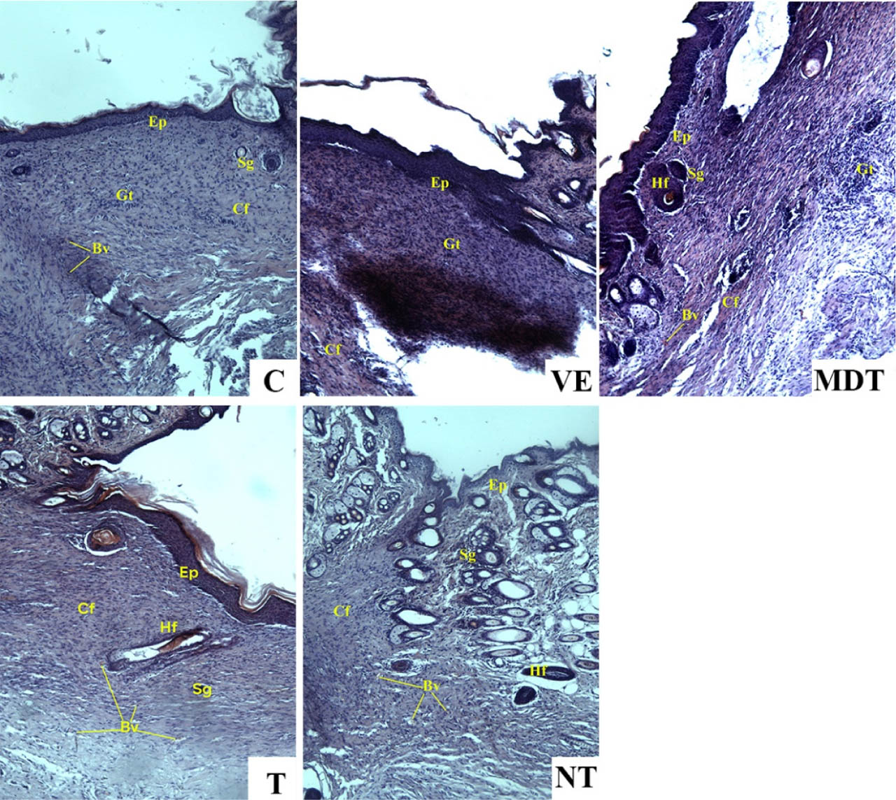
Figure 2.
Histological sections with H&E staining showing better healing in T and NT groups compared to other groups of the study, X400. C: Control; VE: Vehicle; MDT: Medicament; T: Single dose of the herbal cream; NT: Double dose of the herbal cream. Bv: Blood vessel, Cf: Collagen fiber, Ep: Epithelialization, Gt: Granulation tissue, Hf: Hair follicle, Sg: Sweat gland.
Table 7.
Modified histological scoring system adapted by Gupta and Kumar (2015) [44]
| Histological parameters | Score | ||
|---|---|---|---|
| 1 | 2 | 3 | |
| Epithelialization | Absent | Moderate | Marked |
| Differentiation | Absent | Present | / |
| Amount of granulation tissue | Profound | Moderate | Absent |
| Inflammation | Severe | Moderate | Weak |
| Collagen fiber orientation | Vertical | Mixed | Horizontal |
| Angiogenesis | <5/HPF | 6-10/HPF | >10/HPF |
Table 8.
Mean scores ± SD of histological parameters in all treated groups
| Histological parameters | Groups | |||||
|---|---|---|---|---|---|---|
| C | MDT | VE | T | NT | P value | |
| Epithelialization | 1.60 ± 0.54c | 2.60 ± 0.54ab | 1.80 ± 0.44bc | 2.80 ±0.44a | 3.00 ± 0.00a | 0.00 |
| Differentiation | 1.40 ± 0.54a | 2.00 ± 0.00a | 1.60 ± 0.54a | 2.00 ± 0.00a | 2.00 ± 0.00a | 0.03 |
| Amount of granulation tissue | 2.60 ± 0.54a | 1.20 ± 0.44b | 1.60 ± 0.89b | 3.00 ± 0.00a | 3.00 ± 0.00a | 0.00 |
| Inflammation | 1.40 ± 0.54c | 2.20 ± 0.44b | 2.40 ± 0.54ab | 3.00 ± 0.00a | 3.00 ± 0.00a | 0.00 |
| Collagen fiber orientation | 1.60 ± 0.54c | 2.40 ± 0.54ab | 2.20 ± 0.44bc | 3.00 ± 0.00a | 3.00 ± 0.00a | 0.00 |
| Angiogenesis | 1.60 ± 0.54c | 2.60 ± 0.54ab | 2.00 ± 0.70bc | 3.00 ± 0.00a | 3.00 ± 0.00a | 0.00 |
| Total histological score | 10.2 ± 1.09c | 13.00 ± 0.70b | 11.60 ± 1.14bc | 16.80 ± 0.44a | 17.00 ± 0.00a | 0.00 |
CONCLUSION
The present study reported the formulation and biological evaluation of a herbal cream based on Linaria scariosa, a medicinal plant acknowledged for its potential to treat infected wounds. According to the obtained results, the species L. scariosa is rich in several classes of bioactive molecules exerting interesting pharmacological properties. The prepared herbal cream was a water-in-oil emulsion with a safe microbiological profile and acceptable physicochemical and rheological properties after storage. Topical application of the prepared herbal cream to excision wounds resulted in a significant wound-healing effect associated with improved epithelialization and favorable histological features consistent with tissue repair. Also, the prepared herbal cream presents interesting antioxidant, anti-inflammatory, and antimicrobial effects, which confirm its traditional use for the treatment of festering wounds. It can be concluded that Linaria scariosa herbal cream exerts interesting pharmacological applications, but more in-depth in vivo studies are needed in order to confirm its safety and to determine the exact mechanism by which the plant promote wound healing.
ACKNOWLEDGMENTS
The authors thank the DGRSDT and the Algerian Ministry of Higher Education and Scientific Research for their support within the framework of the PRFU projects (B00L01UN050120230002) and (B00L01UN050120230001).
They also thank the Scientific and Technical Research Center in Physico-Chemical Analysis (CRAPC-Batna) and Dr. Souad Khebri for providing instrumental support and help with running the rheological parameters.
Notes
[9] ETHICAL STATEMENT
The experimental protocol used for the assessment of wound-healing effect was conducted in accordance with ethical standards adopted by the national regulations for the use of laboratory animals and complied with the ARRIVE guidelines (Animal Research: Reporting of In Vivo Experiments), ensuring that the rights of animal subjects were fully respected.
Notes
SUPPLEMENTARY MATERIAL
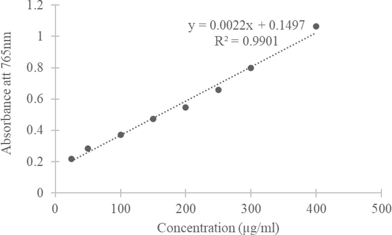
Figure S1.
Straight calibration curve of gallic acid for total phenolic content quantification
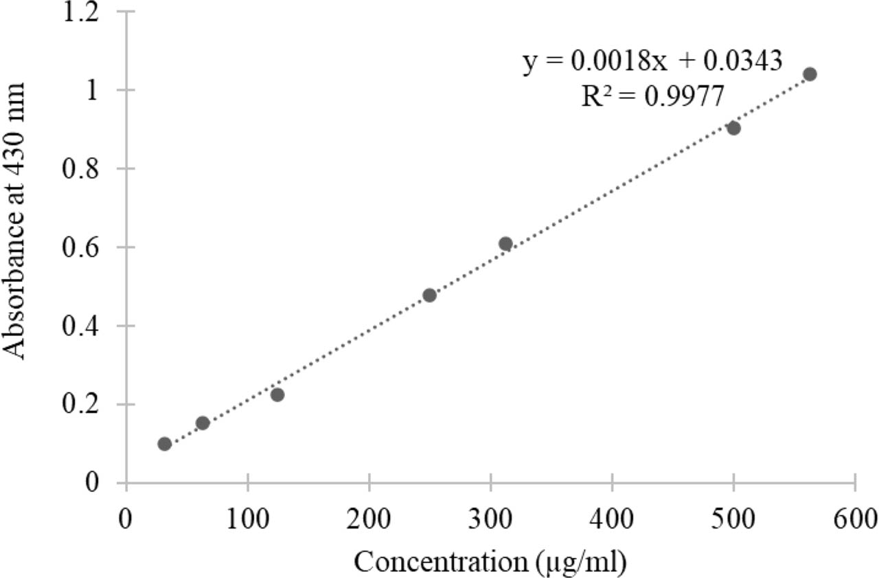
Figure S2.
Straight calibration curve of quercetin for flavonoid content quantification
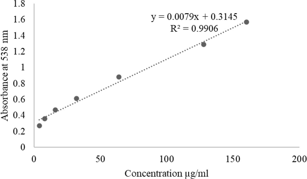
Figure S3.
Straight calibration curve of ursolic acid for total terpenoids content quantification
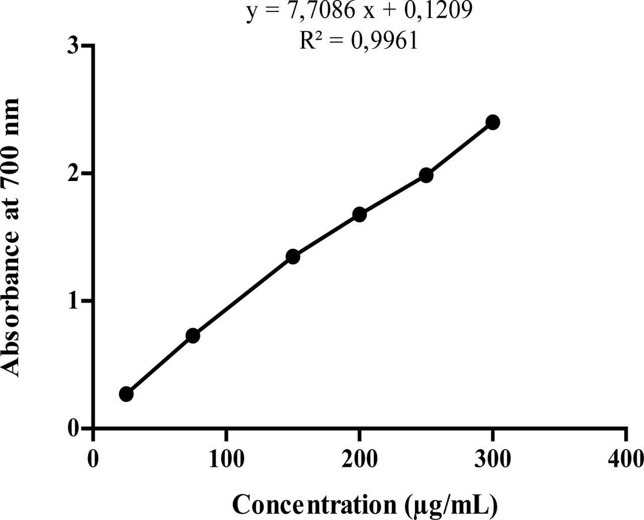
Figure S4.
Straight calibration curve of ascorbic acid for FRAP assay.
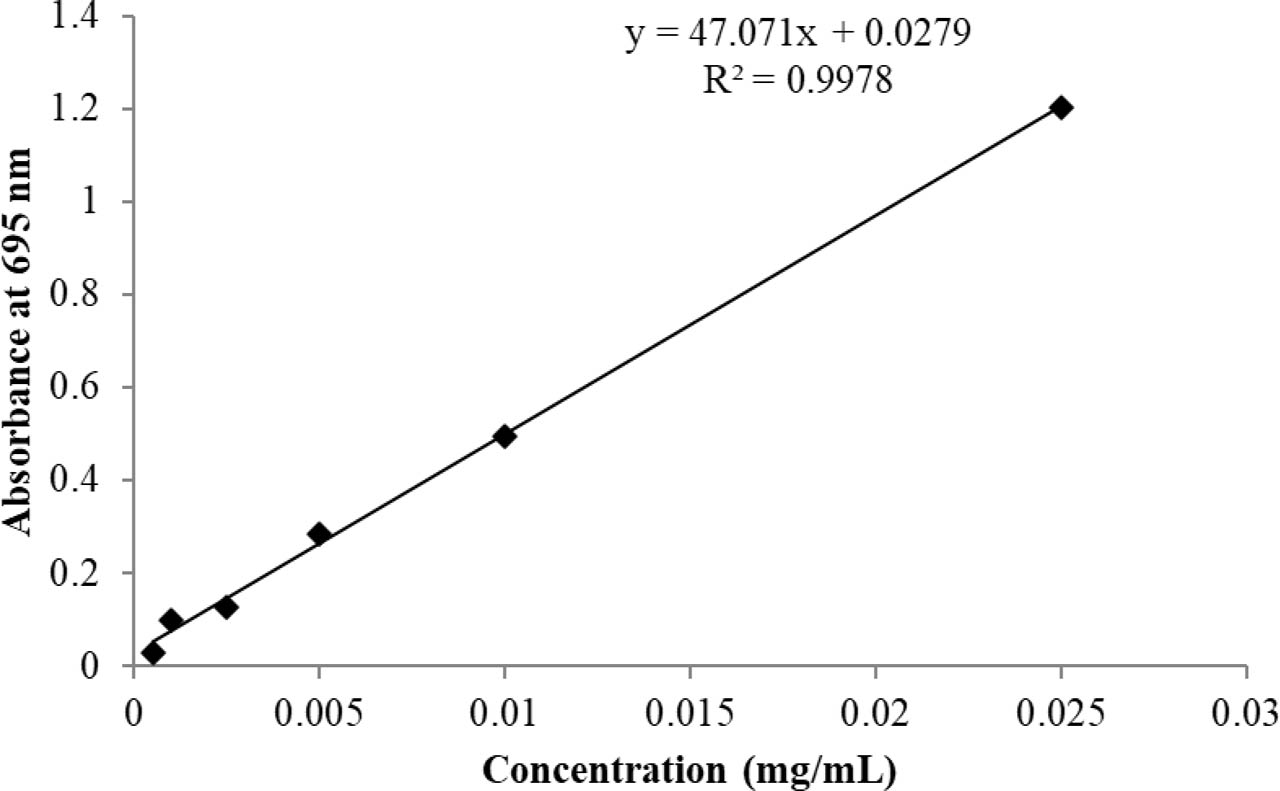
Figure S5.
Straight calibration curve of ascorbic acid for TAC assay
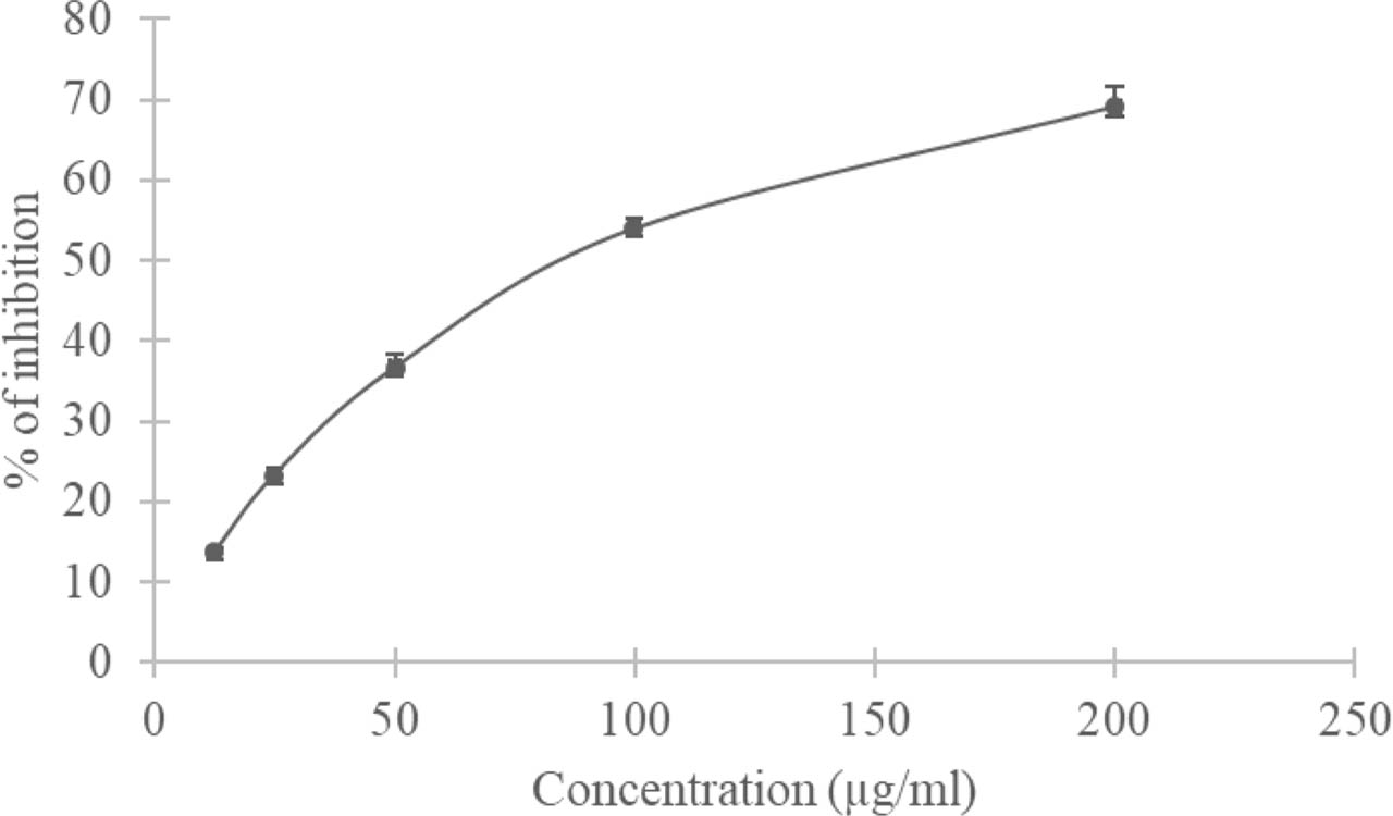
Figure S6.
DPPH radical scavenging activity Linaria scariosa herbal cream