
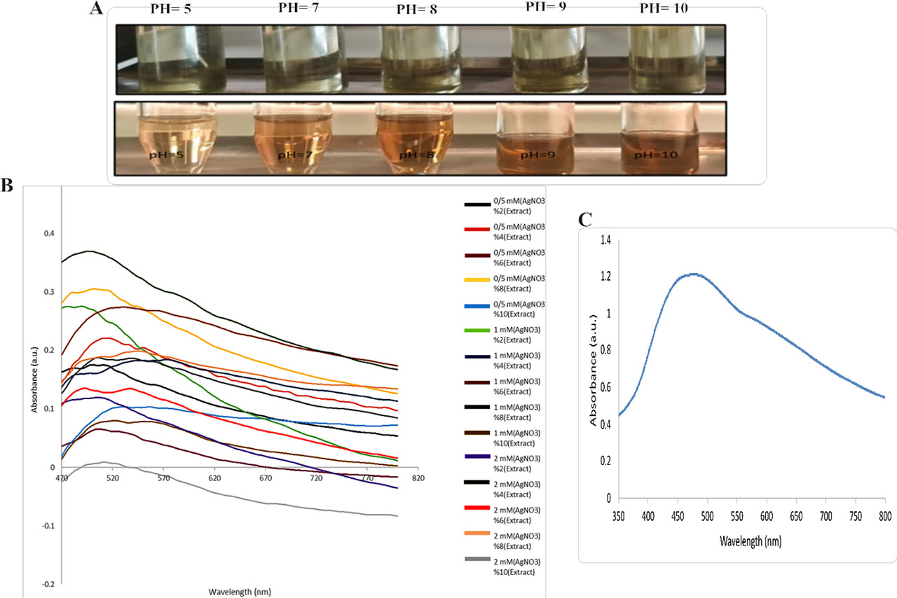
Figure 1.
Silver nanoparticle synthesis and characterization. (A) Visual observation of color changes during synthesis: top row shows reaction mixtures before pH adjustment; bottom row shows mixtures after pH adjustment and 24 h incubation. (B) UV-Vis spectra from the screening matrix illustrating the combined effects of AgNO3 concentration (0.5, 1.0, 2.0 mM) and extract fraction (2–10% v/v); each line is identified by AgNO3 concentration and extract percentage. (C) Representative UV-Vis spectrum under optimized conditions (2.0 mM AgNO3, 4% v/v extract, pH 8.0, 24 h), showing the characteristic surface plasmon resonance (SPR) peak near 400–450 nm. Panel A illustrates the pH-dependent effects, panel B summarizes the multiparameter screening of AgNO3 and the extract fraction, and panel C shows the outcome of the optimization.
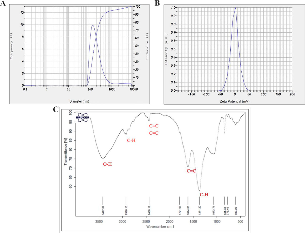
Figure 2.
(A) Size distribution analysis, (B) Zeta potential spectra measurements, and (C) FTIR spectra of biosynthesized AgNP
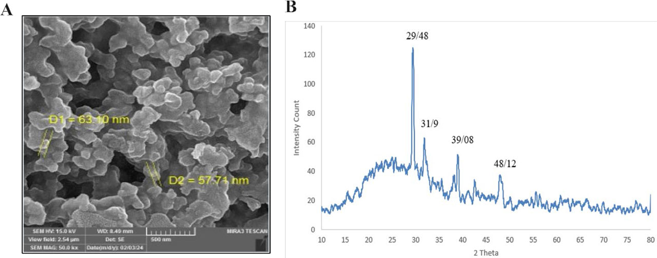
Figure 3.
(A) FESEM and (B) XRD spectrum analysis of biosynthesized AgNPs
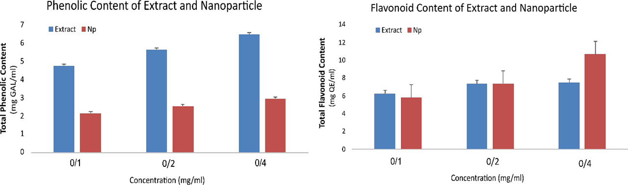
Figure 4.
Quantitative comparison of (A) total phenolic and (B) flavonoid content between different concentrations (0.1, 0.2, 0.4 mg/ml) of Melissa officinalis extract (Extract) and silver nanoparticles (NPs)
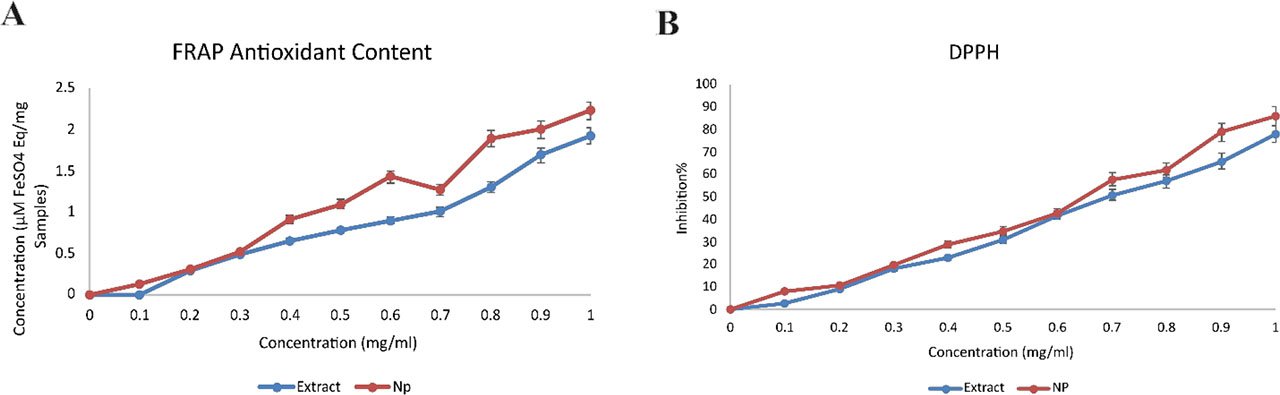
Figure 5.
Antioxidant potency of Melissa officinalis extract (Extract) and silver nanoparticles (NPs). (A) FRAP and (B) DPPH
Table 1.
Comparison of antibacterial efficacy expressed as MIC and MBC values for Melissa officinalis extract and its biosynthesized silver nanoparticles (AgNPs) against three bacterial strains. AgNPs exhibited significantly stronger inhibition toward Staphylococcus aureus, whereas both samples showed limited activity against E. coli and P. mirabilis
| Bacterial strain | Sample | MIC (µg/ml) | MBC (µg/ml) | Observation summary |
|---|---|---|---|---|
| Staphylococcus aureus | Extract | 32 | 64 | Moderate activity; extract inhibited growth only at high concentrations |
| AgNPs | 8 | 16 | Strong activity; AgNPs four-fold lower MIC than extract | |
| Escherichia coli | Extract | >128 | >128 | No measurable inhibition or bactericidal effect |
| AgNPs | 64 | 128 | Weak activity; partial inhibition at the highest dose | |
| Proteus mirabilis | Extract | >128 | >128 | No activity detected |
| AgNPs | 64 | 256 | Low activity; growth inhibition only at high concentrations |
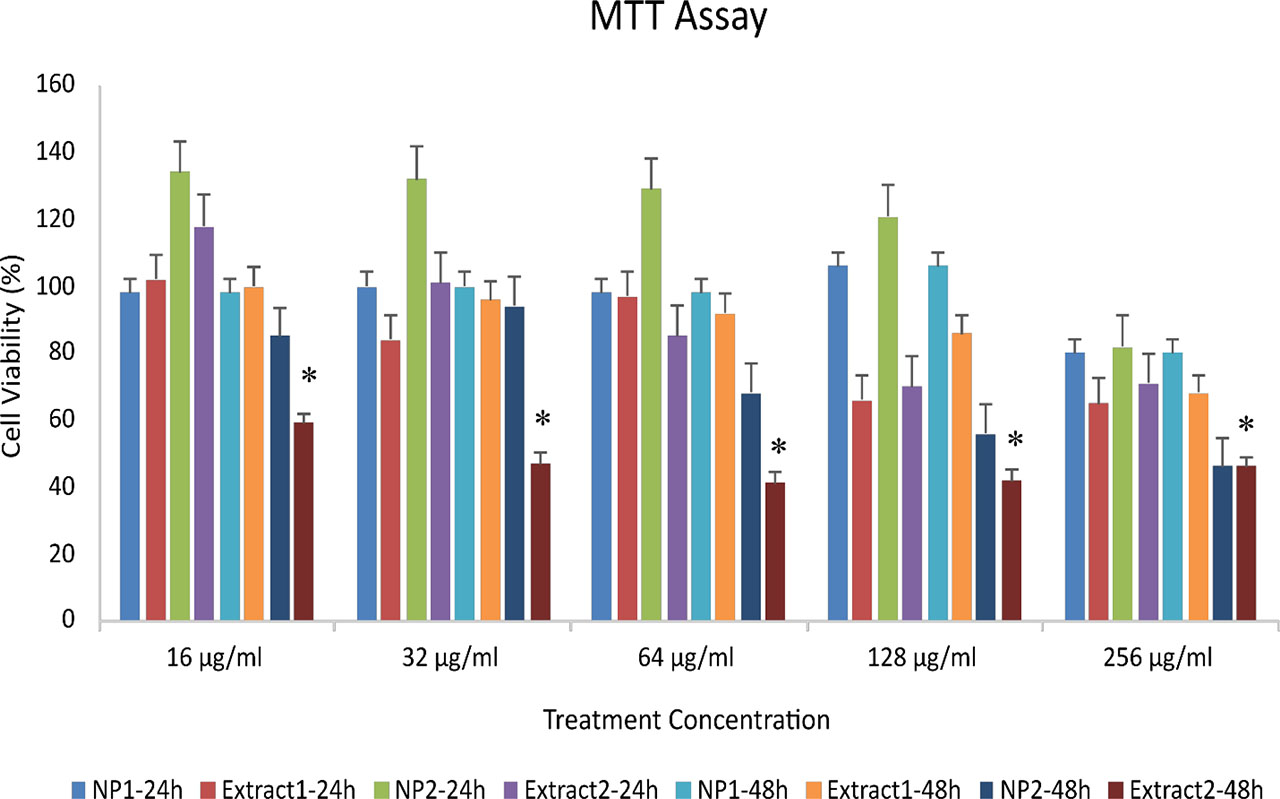
Figure 6.
Cytotoxic effects of green-synthesized silver nanoparticles (AgNPs) and aqueous Melissa officinalis extract on MCF-7 breast cancer cells after 24 and 48 hours of exposure. NP1–24 h: AgNPs synthesized by method 1 (24 h treatment); Extract 1–24 h: plant extract prepared by method 1 (24 h); NP2–24 h: AgNPs synthesized by method 2 (24 h); Extract 2–24 h: plant extract prepared by method 2 (24 h); NP1–48 h, Extract 1–48 h, NP2–48 h, and Extract 2–48 h correspond to the same treatments after 48 hours. Data are presented as mean ± SD (p ≤ 0.05).