
INTRODUCTION
Silver nanoparticles (AgNPs) have garnered significant interest due to their remarkable antimicrobial, antioxidant, and anticancer properties (1, 2). Nevertheless, traditional AgNPs synthesis methods, such as chemical reduction and physical methods, are typically conducted using hazardous chemicals, organic solvents, and a significant amount of energy, raising environmental and biocompatibility concerns (3, 4). Consequently, this has led to a growing desire to develop environmentally friendly, sustainable, and biocompatible methods for biomedical applications (5, 6). Green synthesis utilizes biological agents, such as plant extracts, bacteria, fungi, and algae, to produce nanoparticles under mild conditions (7). Among these methods, the use of plants for mediating the synthesis process stands out due to its simplicity of application, scalability, cost efficiency, and utilization of natural phytochemicals, such as polyphenols, flavonoids, terpenoids, and alkaloids, as both reducing and stabilizing agents (8, 9). The biomolecules promote the reduction of metal salts and stabilize the resultant nanoparticles (7, 10). Melissa officinalis (lemon balm), a member of the Lamiaceae family and native to the Mediterranean region, is particularly suitable for green synthesis due to its diverse phytochemical profile (11,12,13). Traditionally used for its antiviral, antimicrobial, antiviral, and sedative attributes, M. officinalis contains prominent quantities of rosmarinic acid, caffeic acid, flavonoids, and essential oils such as citronellal and geraniol (12, 14). Its broad therapeutic profile and high antioxidant content make it a promising candidate for the synthesis of biologically active nanomaterials (15). AgNP biosynthesis using plant extracts is typically indicated by a visual color change in the reaction mixture, as observed through surface plasmon resonance (SPR), confirming the presence of the nanoparticles (16,17,18). Several analytical techniques, such as ultraviolet-visible (UV-Vis) spectroscopy, Fourier transform infrared (FTIR) spectroscopy, dynamic light scattering (DLS), zeta potential determination, and X-ray diffraction (XRD), are utilized to determine the size of the particles, stability, morphology, and crystal form (19,20,21). AgNPs exhibit strong antimicrobial efficacy against a wide variety of pathogens. In this study, particular focus is placed on three clinically significant bacteria: the Gram-positive Staphylococcus aureus, associated with a range of infections including skin conditions and pneumonia; the Gram-negative Escherichia coli, a common causative agent of urinary tract infections and foodborne illnesses; and the Gram-negative opportunistic pathogen Proteus mirabilis, known for its role in complicated urinary tract infections and its capacity for biofilm formation (22,23,24,25). These pathogens were selected due to their medical relevance and increasing resistance to conventional antibiotics. Besides acting as antimicrobials, AgNPs have shown potential anticancer activity by causing oxidative stress, mitochondrial damage, and apoptosis of cancer cells (26,27,28,29). In the present study, MCF-7 human breast cancer cells have been selected as a model for estrogen receptor-positive breast cancer to evaluate the cytotoxicity of the synthesized AgNPs. Understanding the differential cytotoxicity between cancer and normal cells is essential for the development of safe drugs.
In this study, AgNPs were synthesized using a green synthesis approach using M. officinalis extract, with detailed physicochemical characterization and evaluation of their antioxidant, antimicrobial, and cytotoxic activity reconstructed. Characterization of the AgNPs was performed by UV-Vis spectroscopy, FTIR, DLS, zeta potential, and XRD studies. The phytochemical composition and antioxidant potential of both the extract and AgNPs were determined, and the antimicrobial activity was evaluated against selected bacterial strains. Cytotoxicity was assessed using MCF-7 human breast cancer cells. Through an investigation of the green synthesis of AgNPs using Melissa officinalis, this study aims to provide a basis for generating biocompatible nanomaterials with potential applications in biomedical and pharmaceutical fields.
MATERIAL AND METHODS
Optimization of Green Synthesis of Silver Nanoparticles Using Melissa officinalis Leaves
Melissa officinalis leaves were collected from the Iran Botanical Garden along the Tehran–Karaj Highway (Tehran Province, Iran) and taxonomically authenticated. The leaves were thoroughly rinsed with tap water followed by distilled water, then air-dried at room temperature, and protected from direct sunlight. Dried leaves were ground into a fine powder. For aqueous extraction, 2 g of powdered leaves was boiled in 100 mL of distilled water at 60 °C for 30 min. After cooling, the mixture was filtered through a Whatman No. 1 filter paper and centrifuged at 6,000 rpm for 10 min. The resulting supernatant was stored at 4 °C and used within 4 weeks. The stock extract concentration was 20 mg/mL (2 g/100 mL), and working dilutions were freshly prepared for each experiment. A 2-mM silver nitrate (AgNO3) stock solution was prepared by dissolving 34 mg of AgNO3 in 100 mL of deionized water. For standard nanoparticle synthesis, 40 µL of plant extract (20 mg/mL stock) was added to 1,960 µL of 2 mM AgNO3 solution (final extract content: 4% v/v), and the reaction mixture was incubated at 37 °C in the dark for 24 h.
To optimize synthesis parameters, reactions were performed using varying AgNO3 concentrations (0.5, 1.0, and 2.0 mM), extract concentrations (2%, 4%, 6%, 8%, and 10% v/v), and pH values ranging from 5 to 10, adjusted using 0.1 M HCl or NaOH. Reaction temperatures of room temperature and 60 °C were tested under two processing conditions: sonication (30 min × 3) and magnetic stirring (6 h). Reaction progress was monitored at 24, 48, 72, and 96 h. Characterization and biological assays were conducted using nanoparticles synthesized under optimized standard conditions (2 mM AgNO3, 4% extract, 37°C, 24 h, pH 8). For comparative biological assays, a synthesis-matched extract (4% v/v, corresponding to the same dilution used during AgNP synthesis) was prepared from the stock solution and tested alongside the AgNPs.
Purification of Synthesized Nanoparticles
Nanoparticles were separated by centrifugation at 9,000 rpm for 3 min at 27 °C. The supernatant was discarded, and the pellet was washed three times with deionized water. The washed nanoparticles were vortexed and sonicated for 30 minutes to achieve uniform resuspension, then adjusted to a 2.5% aqueous suspension, and stored at 4 °C in sterile microtubes for subsequent experiments. A single optimized batch of AgNPs was synthesized under optimized standard conditions (2 mM AgNO3, 4% extract, 37 °C, 24 h, pH 8) and then purified, resuspended, and used for all characterization and biological assays to ensure consistency across experiments.
Characterization of Silver Nanoparticles
The optical properties of AgNPs were analyzed by UV-Visible spectrophotometry (Shimadzu UV-1800, Shimadzu Corp., Japan)over the range of 200–800 nm to confirm surface plasmon resonance (SPR) at 400–450 nm. Fourier transform infrared spectroscopy (FTIR, KBr pellet method) (Bruker Tensor 27, Bruker, Germany) was used to identify functional groups involved in reduction and stabilization. Particle size and surface charge were measured using dynamic light scattering (DLS) and zeta potential analysis (Malvern Instruments, UK), respectively. Crystallinity was assessed by X-ray diffraction (XRD) (Rigaku, Japan), and morphology and size distribution were examined by scanning electron microscopy (SEM) (TESCAN, Czech Republic).
Phytochemical and Antioxidant Assay
The total phenolic content (TPC) and total flavonoid content (TFC) of the Melissa officinalis extract and its biosynthesized silver nanoparticles (AgNPs) were determined following standard spectrophotometric procedures. TPC was quantified using the Folin–Ciocalteu reagent method and expressed as milligrams of gallic acid equivalents per gram of sample (mg GAE/g). TFC was determined using the aluminum chloride colorimetric method and expressed as milligrams of quercetin equivalents per gram of sample (mg QE/g). Absorbances were recorded at 765 nm for TPC and 415 nm for TFC using an ELISA microplate reader (SpectraMax Plus, Molecular Devices, USA) (28). For both assays, working sample concentrations of 0.1, 0.2, and 0.4 mg/mL were prepared for the crude extract and the AgNP suspensions. These values represent the test concentrations of the samples used in the colorimetric assays, not the extract concentrations employed during nanoparticle synthesis. The highest concentration (0.4 mg/mL) corresponds to the effective extract concentration present in the standard AgNP synthesis mixture (40 µL extract in 1960 µL of 2 mM AgNO3). All measurements were performed in triplicate, and results were expressed as mean ± SD. Radical scavenging activity was assessed by the ferric-reducing antioxidant power (FRAP) and 2,2-diphenyl-1-picrylhydrazyl (DPPH) assays. Ferrous sulfate and ascorbic acid (1 mg/mL) were used as standards to construct calibration curves, and absorbances were recorded at 593 nm (FRAP) and 517 nm (DPPH), respectively (30).
Antibacterial Assay
Both AgNPs and plant extract were screened for their antibacterial potential against Staphylococcus aureus, Escherichia coli, and Proteus mirabilis by minimum inhibitory concentration (MIC) and minimum bactericidal concentration (MBC), which were determined by a broth micro-dilution method in 96-well plates according to the standards of the CLSI (2007) (28).
Cytotoxicity Assay
Cytotoxicity was evaluated on MCF-7 (breast cancer cells) by MTT assay (31). Cells were maintained in DMEM supplemented with 10% FBS and 1% penicillin-streptomycin. Subsequently, the cells were seeded into 96-well plates and exposed to various concentrations (16–256 μg/mL as final concentrations) of AgNPs and extracts for 24 and 48 hours. Untreated and DMSO-treated cells served as controls. After rinsing with PBS, cells were incubated in serum-free medium containing 10 μL MTT for 4 h at 37°C in the dark. The supernatant was removed, and formazan crystals were dissolved in 100 μL DMSO with gentle shaking at 37°C for 5 min. The absorbance was then measured at 570 nm. Untreated controls (media only) and vehicle controls (DMSO where applicable) were also included on each plate. Cell viability was then calculated using the following formula: (1) Cytotoxicity percentage ((control O.D.−sample O.D.)/control O.D.) ×100%.
Statistical Analysis
All experiments were performed in at least three independent replicates, and data are presented as mean ± standard deviation (SD). For cell assays, untreated and vehicle controls were included as negative controls; for antioxidant assays, ascorbic acid (DPPH) and ferrous sulfate (FRAP) were used as standards. Statistical comparisons were performed by one-way ANOVA followed by Duncan’s multiple range test (p < 0.05) using IBM SPSS Statistics v.23. Post hoc comparisons and the number of biological/technical replicates for each assay are indicated in the corresponding figure legends.
RESULTS
Visual Observation and UV-Vis Confirmation of AgNP Synthesis
Formation of silver nanoparticles was indicated visually by a color change of the reaction mixture from light yellow to dark brown following incubation (24 h, 37 °C) under the standard conditions (2.0 mM AgNO3, 4% v/v extract, pH 8.0). UV–Visible spectroscopy confirmed nanoparticle formation by the appearance of a characteristic surface plasmon resonance (SPR) band in the 350–500 nm region. During the optimization process, the effects of AgNO3 concentration (0.5–2.0 mM), extract fraction (2–10% v/v), and pH (5–10) were systematically evaluated. Under certain non-optimized screening conditions, broader and red-shifted SPR peaks approaching ∼500–520 nm were observed (Fig. 1B), which are attributed to increased nanoparticle size, aggregation, and polydispersity caused by excess extract concentration or non-ideal pH conditions. In contrast, optimized synthesis conditions (pH 8.0, 2.0 mM AgNO3, 4% v/v extract) produced a sharper and more intense SPR peak in the ∼400–450 nm range (Fig. 1C), indicating the formation of smaller and more uniformly dispersed silver nanoparticles. These optimized conditions were therefore selected for all subsequent characterization and bioactivity assays (Fig. 1A–C).
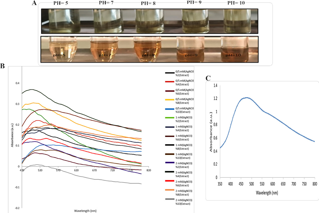
Figure 1.
Silver nanoparticle synthesis and characterization. (A) Visual observation of color changes during synthesis: top row shows reaction mixtures before pH adjustment; bottom row shows mixtures after pH adjustment and 24 h incubation. (B) UV-Vis spectra from the screening matrix illustrating the combined effects of AgNO3 concentration (0.5, 1.0, 2.0 mM) and extract fraction (2–10% v/v); each line is identified by AgNO3 concentration and extract percentage. (C) Representative UV-Vis spectrum under optimized conditions (2.0 mM AgNO3, 4% v/v extract, pH 8.0, 24 h), showing the characteristic surface plasmon resonance (SPR) peak near 400–450 nm. Panel A illustrates the pH-dependent effects, panel B summarizes the multiparameter screening of AgNO3 and the extract fraction, and panel C shows the outcome of the optimization.
FTIR Analysis of Functional Groups
FTIR analysis was used to identify the functional groups involved in the reduction of silver salts to silver nanoparticles using a Melissa officinalis (lemon balm) plant extract (Fig. 2C). The wide absorption band found between 3350 and 3450 cm−1 is ascribed to the O-H stretching vibrations of hydroxyl groups from phenolic and alcoholic chemicals in the plant extract. These groups serve as strong reducing agents, facilitating the conversion of Ag+ to Ag0. The absorption peaks between 2850 and 2950 cm−1 correspond to aliphatic C-H stretching vibrations (from CH2 and CH3 groups), whereas peaks in the 2100–2500 cm−1 range are related with triple-bonded functional groups such as C≡C (alkynes) and conjugated C=C systems.
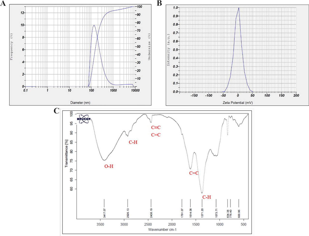
Figure 2.
(A) Size distribution analysis, (B) Zeta potential spectra measurements, and (C) FTIR spectra of biosynthesized AgNP
Carbonyl stretching vibrations at 1610–1620 cm−1 may be caused by carboxylic acids or other unsaturated chemicals in the extract. Additional peaks between 1370 and 1450 cm−1 reveal aliphatic bending vibrations (CH bending), whereas smaller peaks between 600 and 1073 cm−1 indicate the existence of aromatic ring vibrations and secondary metabolites. The FTIR data confirm the presence of hydroxyl, carbonyl, and amine functional groups, which act as reducing and capping agents in the green production of silver nanoparticles. These biomolecules not only help to reduce silver ions but also contribute to the nanoparticles’ long-term stability via surface interactions.
DLS and Zeta Potential Measurements
DLS analysis indicated a broad hydrodynamic size distribution with an intensity-weighted mean diameter of ∼182.8 nm, consistent with aggregation (see Fig. 2A). Zeta potential measurements showed a near-neutral charge (∼+0.2 mV), consistent with limited electrostatic stabilization and partial aggregation (Fig. 2B). SEM micrographs were used to determine primary particle sizes; measurements from SEM images gave an average primary particle diameter of approximately 60 nm (see Fig. 3A and associated legend). We added a note clarifying that DLS measures hydrodynamic clusters, while SEM shows dried primary particles; both measurements are reported for completeness and compared in Discussion.
XRD and SEM Characterization of Nanoparticles
The crystallinity of the nanoparticles was validated using X-ray diffraction (XRD) analysis (Fig. 3B). The detected diffraction peaks at 2θ values of 29.48°, 31.9°, 39.08°, and 48.12° matched the typical planes of face-centered cubic silver structures, showing that the synthesized nanoparticles have a crystalline nature. SEM imaging confirmed these findings by showing that the nanoparticles were primarily spherical in shape, although minor aggregation occurred, likely due to the near-neutral surface charge (Fig. 3A).
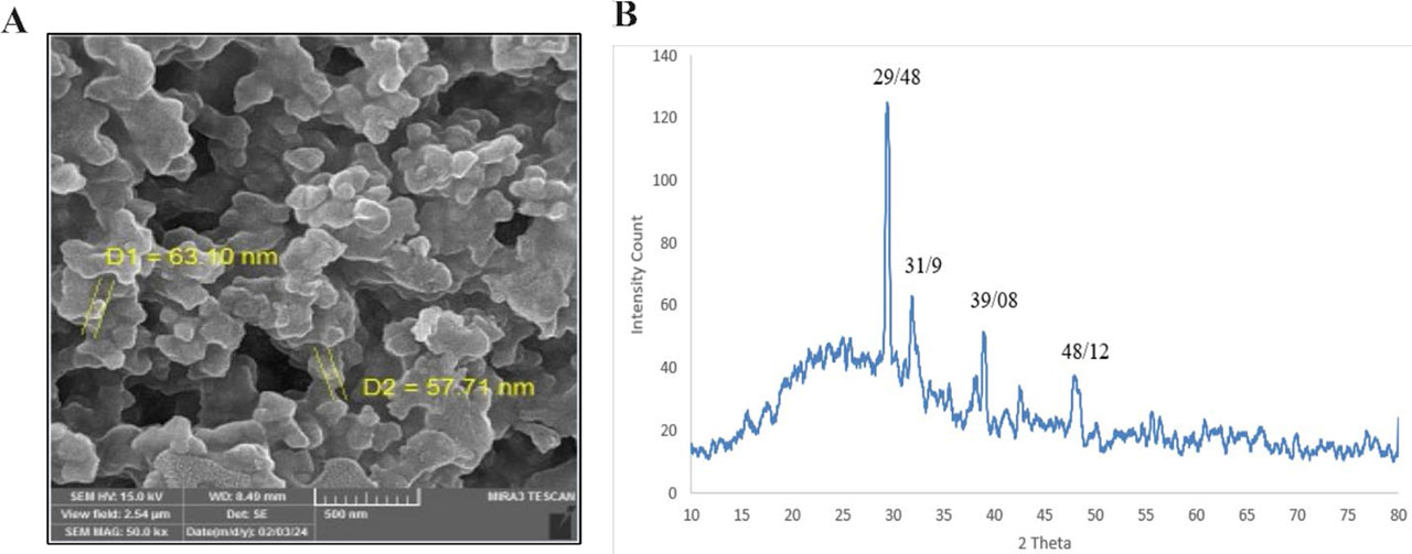
Figure 3.
(A) FESEM and (B) XRD spectrum analysis of biosynthesized AgNPs
Evaluation of Total Phenolic and Flavonoid Content
Phytochemical study demonstrated a significant decrease in both total phenolic content (TPC) and total flavonoid content (TFC) following the production of silver nanoparticles (AgNPs), indicating that these beneficial compounds play a role in the reduction and capping processes. TPC values for Melissa officinalis extract at concentrations of 0.1, 0.2, and 0.4 mg/mL were 4.76, 5.65, and 6.47 (GAE/g), respectively. AgNP samples had substantially lower TPC values of 2.16, 2.56, and 2.96 mg GAE/g (p < 0.05), suggesting that phenolic compounds were consumed during nanoparticle synthesis. TFC in the extract was determined as 6.28, 7.37, and 7.52 ± 0.01 (QE/g) at the same concentrations, while AgNP samples revealed TFC values of 5.82, 7.40, and 10.70 ± 0.05 mg QE/g. Although the AgNPs had a high TFC concentration at 0.4 mg/mL, statistical analysis showed a significant overall decrease relative to the extract (p < 0.05). These results reveal that both phenolic and flavonoid chemicals contributed to nanoparticle production, with phenolics exhibiting a more significant decrease, indicating that phenolics played a more prominent role in Ag+ reduction than flavonoids.
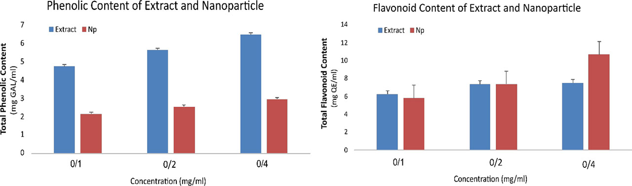
Figure 4.
Quantitative comparison of (A) total phenolic and (B) flavonoid content between different concentrations (0.1, 0.2, 0.4 mg/ml) of Melissa officinalis extract (Extract) and silver nanoparticles (NPs)
Antioxidant Capacity Assessed by FRAP and DPPH Assays
The antioxidant activities of the extract and AgNPs were assessed using FRAP and DPPH tests. In the FRAP experiment, the antioxidant capacities of both samples increased in a concentration-dependent manner. At a concentration of 1, AgNPs exhibited a higher FRAP value (2.23 Eq/mg) than the extract (1.93 Eq/mg) (Fig. 5A), showing superior ferric-reducing activity. DPPH radical scavenging activity followed a similar pattern, with AgNPs inhibiting DPPH radicals by 86.72% compared to 78.31% for the extract at the highest tested concentration of 1 mg/mL (Fig. 5B), indicating that phytochemicals on the nanoparticle surface preserved or slightly enhanced antioxidant activity.
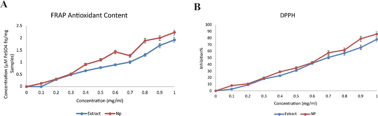
Figure 5.
Antioxidant potency of Melissa officinalis extract (Extract) and silver nanoparticles (NPs). (A) FRAP and (B) DPPH
Antibacterial Activity of Melissa officinalis Using Minimum Inhibitory Concentration (MIC) and Minimum Bactericidal Concentration (MBC)
The antibacterial properties of the aqueous Melissa officinalis extract and its green-synthesized silver nanoparticles (AgNPs) were evaluated against Staphylococcus aureus, Escherichia coli, and Proteus mirabilis using the broth microdilution method to determine MIC and MBC values (Table 1). The AgNPs exhibited substantially greater antibacterial activity than the crude extract, particularly against S. aureus, where MIC and MBC values were 8 µg/mL and 16 µg/mL, respectively, compared to 32 µg/mL and 64 µg/mL for the extract. Against the Gram-negative strains E. coli and P. mirabilis, AgNPs displayed only weak to low activity (MIC = 64 µg/mL; MBC = 128–256 µg/mL), while the extract showed no detectable inhibition. The stronger response of S. aureus is attributed to its thick peptidoglycan layer, which facilitates AgNP attachment and silver ion penetration, whereas the outer lipopolysaccharide barrier in Gram-negative bacteria limits nanoparticle access.
Table 1.
Comparison of antibacterial efficacy expressed as MIC and MBC values for Melissa officinalis extract and its biosynthesized silver nanoparticles (AgNPs) against three bacterial strains. AgNPs exhibited significantly stronger inhibition toward Staphylococcus aureus, whereas both samples showed limited activity against E. coli and P. mirabilis
| Bacterial strain | Sample | MIC (µg/ml) | MBC (µg/ml) | Observation summary |
|---|---|---|---|---|
| Staphylococcus aureus | Extract | 32 | 64 | Moderate activity; extract inhibited growth only at high concentrations |
| AgNPs | 8 | 16 | Strong activity; AgNPs four-fold lower MIC than extract | |
| Escherichia coli | Extract | >128 | >128 | No measurable inhibition or bactericidal effect |
| AgNPs | 64 | 128 | Weak activity; partial inhibition at the highest dose | |
| Proteus mirabilis | Extract | >128 | >128 | No activity detected |
| AgNPs | 64 | 256 | Low activity; growth inhibition only at high concentrations |
Overall, biosynthesized AgNPs enhanced antibacterial potency, reducing the inhibitory and bactericidal thresholds approximately fourfold against S. aureus and demonstrating selective efficacy toward Gram-positive bacteria.
Cytotoxicity Against MCF-7 Breast Cancer Cells
The MTT test results show that Melissa officinalis extract had higher cytotoxicity properties against MCF-7 breast cancer cells than its green-synthesized AgNPs (NP1 and NP2). Cell viability declined in a dose- and time-dependent manner, with the extract having the greatest effect at 48 hours, particularly at 64 µg/mL, where cell viability was reduced to approximately 35%. For the 48-hour extract treatment, the IC50 was 32 µg/mL, corresponding to approximately 50% cell survival. In contrast, nanoparticles had a lesser impact across all doses and time periods.
DISCUSSION
In this study, the green synthesis of silver nanoparticles (AgNPs) using Melissa officinalis aqueous extract as a reducing and stabilizing agent was successfully achieved. After increasing the pH to 8, the reaction mixture changed color from pale yellow to dark brown, and a surface plasmon resonance (SPR) peak in the 350–500 nm area developed after 24 hours, indicating nanoparticle formation. Baltazar et al. (2017) observed a noticeable color shift and an SPR peak about 450 nm without requiring pH adjustment, suggesting minor changes in nanoparticle synthesis (11).
Further analysis using Fourier transform infrared spectroscopy (FTIR) revealed the presence of hydroxyl, carbonyl, and amine groups—evidence that phenolic and flavonoid compounds in Melissa officinalis were key to both reducing the silver ions and stabilizing the nanoparticles. Compared to a study by Hamady et al. (2019), the FTIR spectrum in this research also showed new peaks in the 600–1073 cm−1 range, likely due to variations in the phytochemical composition of the plant extract used (32). These findings are consistent with those of Pirtarighat et al. (2017), who identified similar functional groups in other plant-based silver nanoparticle syntheses (33).
The synthesized AgNPs had a mean hydrodynamic diameter of 182.8 nm, with a zeta potential of roughly +0.2 mV, indicating low electrostatic stability and a tendency toward aggregation. In contrast, Hamady et al. (2019) created smaller AgNPs (12–38 nm) with a higher negative zeta potential (−15.11 mV), resulting in increased nanoparticle stability. Jebril et al. (2020) found negative zeta potentials (∼−13.1 mV) in AgNPs, indicating the critical role of surface charge in nanoparticle dispersion and aggregation (34).
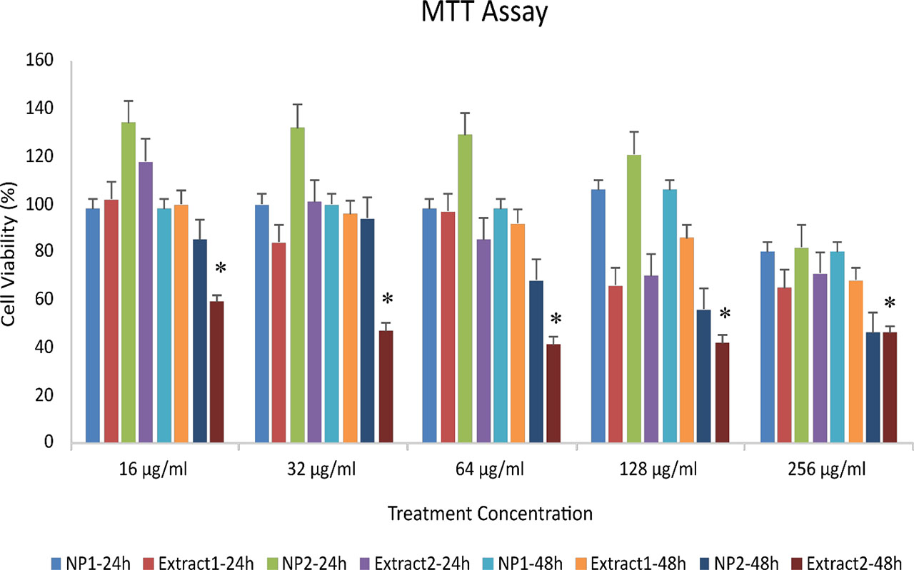
Figure 6.
Cytotoxic effects of green-synthesized silver nanoparticles (AgNPs) and aqueous Melissa officinalis extract on MCF-7 breast cancer cells after 24 and 48 hours of exposure. NP1–24 h: AgNPs synthesized by method 1 (24 h treatment); Extract 1–24 h: plant extract prepared by method 1 (24 h); NP2–24 h: AgNPs synthesized by method 2 (24 h); Extract 2–24 h: plant extract prepared by method 2 (24 h); NP1–48 h, Extract 1–48 h, NP2–48 h, and Extract 2–48 h correspond to the same treatments after 48 hours. Data are presented as mean ± SD (p ≤ 0.05).
Scanning electron microscopy (SEM) investigation demonstrated the spherical shape of the synthesized AgNPs. SEM photos also showed lower particle sizes than DLS data, a known effect due to dehydration during SEM sample preparation. These findings are consistent with prior research by Haggag et al. (2019) and Pirtarighat et al. (2017), who also found spherical nanoparticles with similar morphology (33, 35).
Phytochemical examination revealed a substantial reduction in total phenolic and flavonoid concentrations in synthesized AgNPs compared to pure plant extract, indicating that these chemicals actively participate in nanoparticle synthesis as reducing and capping agents. This is consistent with Jalilian et al. (2020), who found a more significant decrease in phenolic content than flavonoid content during green nanoparticle production, emphasizing the importance of phenolic compounds in the reduction and stabilization process (36).
The antioxidant activity of both the extract and the synthesized AgNPs increased with concentration, as shown by the FRAP and DPPH tests, respectively. AgNPs outperformed the plant extract in terms of antioxidant capacity, supporting the findings of Pirtarighat et al. (2017) and de Baltazar et al. (2017). The increased antioxidant activity is most likely due to the bioactive chemicals that coat the nanoparticle surface (11, 33).
The minimum inhibitory concentration (MIC) and minimum bactericidal concentration (MBC) results (Table 1) clearly show that Melissa officinalis-mediated silver nanoparticles (AgNPs) are more effective antibacterial agents than the crude extract. The fourfold reduction in MIC and MBC values against Staphylococcus aureus suggests a synergistic effect between the plant-derived phytochemicals coating the nanoparticles and the released silver ions. Phenolic and flavonoid compounds, such as rosmarinic and caffeic acids, may help stabilize Ag+ ions on the nanoparticle surface and promote gradual ion release, thereby improving interaction with bacterial membranes. In contrast, Escherichia coli and Proteus mirabilis showed weaker responses, consistent with the natural resistance of Gram-negative bacteria. Their outer lipid membrane restricts the entry of Ag+ ions and nanoparticles, reducing antimicrobial efficiency. A similar Gram-selective pattern has been reported in other plant-based AgNP studies (11, 33).
Additionally, the near-neutral zeta potential (+0.2 mV) of the synthesized AgNPs may reduce electrostatic attraction to the negatively charged surfaces of Gram-negative bacteria, while the more permeable peptidoglycan layer of Gram-positive cells favors stronger binding and localized silver ion release. These interactions lead to oxidative stress, protein denaturation, and DNA damage, explaining the strong bactericidal effect observed against S. aureus (33). Overall, using MIC and MBC data instead of inhibition-zone measurements provides a clearer and more quantitative understanding of antibacterial potency. The findings confirm that green-synthesized AgNPs from M. officinalis enhance antimicrobial efficacy, particularly against Gram-positive bacteria, while maintaining an environmentally friendly synthesis approach.
Cytotoxicity investigations using the MTT technique demonstrated that the Melissa officinalis extract exhibited higher anticancer activity against MCF-7 breast cancer cells than the synthesized AgNPs, as shown in Hanachi et al. (2021). This contradicts the findings of Hamady et al. (2019), who observed higher cytotoxic effects of AgNPs (32, 37). The greater cytotoxicity of the crude extract relative to AgNPs likely reflects several factors: (i) the immediate availability of free bioactive compounds such as rosmarinic acid, caffeic acid, and flavonoids in the extract; (ii) partial consumption or surface binding of these molecules during nanoparticle synthesis, reducing their free concentration; and (iii) the aggregation and relatively large hydrodynamic diameter of the AgNPs (DLS ≈182.8 nm) combined with a near-neutral zeta potential (+0.2 mV), which may limit nanoparticle uptake by cells. These mechanisms are supported by our phytochemical analysis, which showed a decrease in total phenolic content following nanoparticle formation. Together, these factors may explain the reduced cytotoxicity of the AgNPs observed in this study. Further experiments assessing nanoparticle dispersion, stability, and cellular uptake under culture conditions would help confirm these interpretations.
Compared to previous research by Wypij et al. (2021), Haggag et al. (2019), and Hamady et al. (2019), the synthesized AgNPs in this study had bigger particle sizes and a lower zeta potential, resulting in reduced antibacterial and cytotoxic activity. Nonetheless, the synthesized Melissa officinalis-based AgNPs demonstrated remarkable antioxidant activity and selective antibacterial activities, indicating their potential for biomedical and pharmacological use (32, 35, 38). Manosalva et al. (2019) and Rodríguez-Félix et al. (2021) have shown that improving the particle size and surface stability of green-synthesized AgNPs can increase their biological potential (11, 39).
CONCLUSION
In this study, AgNPs were successfully synthesized using a green method, where Melissa officinalis aqueous extract served as both a natural reducing and stabilizing agent. The synthesis was validated through a visible color change, the appearance of characteristic SPR peaks in UV-Vis spectra, and FTIR analysis, which identified the functional groups involved in nanoparticle formation. The resulting AgNPs exhibited spherical morphology with a moderate particle size and low zeta potential, indicating some degree of aggregation.
Phytochemical analysis confirmed that phenolic and flavonoid compounds played a critical role in nanoparticle synthesis, as reflected by their significant reduction following AgNP formation. The synthesized AgNPs demonstrated enhanced antioxidant activity compared to the crude extract and showed selective antibacterial effects, particularly against Gram-positive bacteria such as Staphylococcus aureus. However, their cytotoxicity against MCF-7 breast cancer cells was lower than that of the plant extract likely due to their larger particle size and tendency to aggregate.
Although the synthesized AgNPs had larger sizes and lower stability compared to other biogenic nanoparticles reported in previous studies, they still exhibited promising antioxidant and selective antimicrobial activities. These findings highlight the potential of Melissa officinalis-mediated AgNPs for biomedical and pharmaceutical applications. Future work should focus on optimizing synthesis parameters to achieve smaller, more stable nanoparticles with enhanced biological activities.