
TABLE 1
Characterization of cell lines used in this study
| Cell line | Origin | Chemotherapy | Treatment outcome |
|---|---|---|---|
| W1 | tumor tissue, ovarian serous adenocarcinoma | No | N/A |
| A2780 | tumour tissue, ovarian endometrioid adenocarcinoma | No | N/A |
| SKOV-3 | ascites, ovarian serous adenocarcinoma | ThioTEPA | ineffective |
| OVCAR-3 | ascites, ovarian serous adenocarcinoma, high grade | cyclophosphamide, DOX, CIS | ineffective |
| PEA1 | pleural effusion, serous adenocarcinoma, high grade | No | N/A |
| PEA2 | ascites, serous adenocarcinoma, high grade | CIS, Prednimustine | ineffective |
| PEA O23 | ascites, serous adenocarcinoma, low grade | CIS, chlorambucil | ineffective |
[i] N/A- not applicable
TABLE 2
Oligonucleotide sequences used for Q-PCR analysis
| Transcript | Sequence (5′-3′ direction) | ENST number | Product size | |
|---|---|---|---|---|
| MCTP1 | F | AGAACCTCAACCCTGTGTGG | 00000312216 | 123 bp |
| R | AGGCTGAGCCCATAAAGTCA | |||
| IFIH1 | F | GGGGCATGGAGAATAACTCA | 00000263642 | 132 bp |
| R | TGCCCATGTTGCTGTTATGT | |||
| SAMD4 | F | CCAAAGGTGCAAGACACAAA | 00000251091 | 146 bp |
| R | CGGAGTCAGGATCATCTGGT | |||
| HERC5 | F | CTTCCTGCATGTGGTTTCCT | 00000264350 | 128 bp |
| R | AAACAGTGCCAGTGGGAAAG | |||
| SEMA3A | F | TGTTGGAGCAAAGGATCACA | 00000265362 | 109 bp |
| R | AGCCCACTTGCATTCATCTC | |||
| GAPDH | F | GAAGGTGAAGGTCGGAGTCA | 00000229239 | 199 bp |
| R | GACAAGCTTCCCGTTCTCAG | |||
| β-actin | F | TCTGGCACCACACCTTCTAC | 00000331789 | 169 bp |
| R | GATAGCACAGCCTGGATAGC | |||
| HPRT1 | F | CTGAGGATTTGGAAAGGGTG | 00000298556 | 156 bp |
| R | AATCCAGCAGGTCAGCAAAG | |||
| B2M | F | CGCTACTCTCTCTTTCTGGC | 00000558401 | 133 bp |
| R | ATGTCGGATGGATGAAACCC | |||
TABLE 3
Summary of cell lines resistance to cisplatin, paclitaxel, doxorubicin and topotecan treatment
| Cell line | IC50 (ng/ml) | |||
|---|---|---|---|---|
| cisplatin | paclitaxel | doxorubicin | topotecan | |
| W1 | 151 (96–216) 1 | 3,37 (3,18–3,62) 1 | 13,6 (9,1–18,0) 1 | 2,03 (1,94–2,11) 1 |
| A2780 | 439 (283–565) 2,9 ↑* | 3,43 (1,91–4,73) 1,02 ↑ | 19,79 (12,52–24,44) 1,46 ↑ | 3,13 (1,48–4,71) 1,54 ↑ |
| SKOV-3 | 890 (595–1222) 5,8 ↑** | 0,784 (0,73–0,84) 4,30 ↑** | 17,76 (13,7–22,84) 1,31 ↑ | 51,9 (40,34–60,71) 25,6 ↑*** |
| OVCAR-3 | 2613 (2125–2884) 17,3 ↑*** | 458 (372–594) 136 ↑*** | 354 (203–571) 26 ↑** | 398 (283–505) 196↑*** |
| PEA1 | 2737 (2031–3763) 18,1 ↑** | 5,66 (3,25–9,07) 1,68 ↑ | 81 (72–90) 5,96 ↑** | 29 (19,2–46,9) 14,3 ↑** |
| PEA2 | 2469 (2426–2557) 16,4 ↑*** | 6,88 (4,31–8,38) 2,04 ↑* | 173 (148–195) 12,7 ↑** | 132 (79–188) 65 ↑** |
| PEO23 | 2171 (1839–2438) 14,4 ↑*** | 1404 (1000–1600) 417 ↑*** | 275 (198–375) 20,2 ↑*** | 498 (300–750) 245 ↑** |
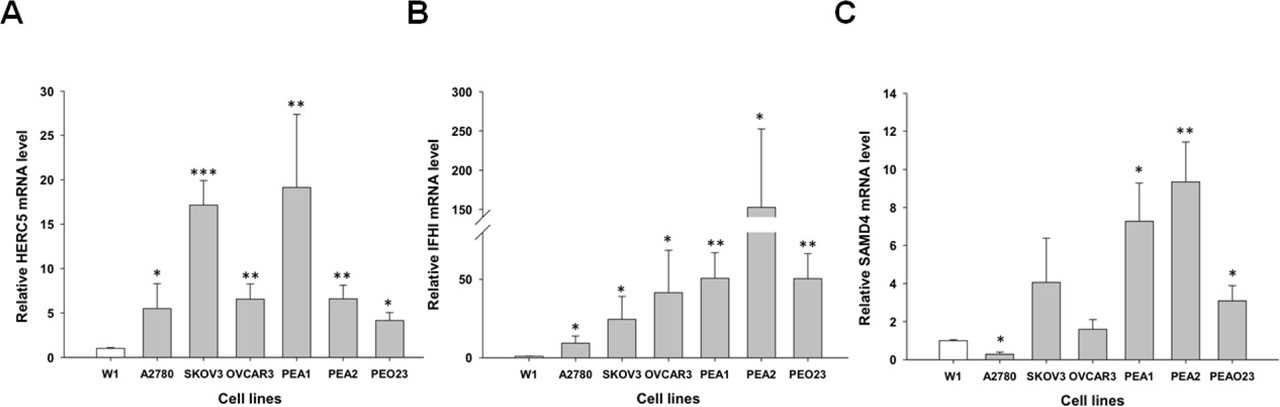
FIGURE 1
Expression analysis (Q-PCR) of the HERC5 (A), IFIH1 (B), and SAMD4 (C) transcripts in different ovarian cancer cell lines. The figure presents the relative gene expression in cell lines (grey bars) with respect to the W1 cell line (white bar), which was assigned a value of 1. The values were considered significant at *p<0.05, **p<0. 0 and ***p<0.001
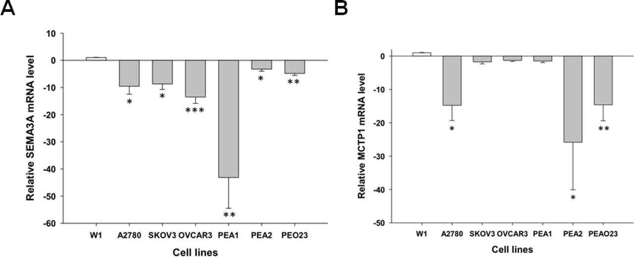
FIGURE 2
Expression analysis (Q-PCR) of the SEMA3A (A) and MCTP1 (B) transcripts in different ovarian cancer cell lines. The figure presents the relative gene expression in cell lines (grey bars) with respect to the W1 cell line (white bar), which was assigned a value of 1. The values were considered significant at *p<0.05, **p<0.01 and ***p<0.001