
Figure 1
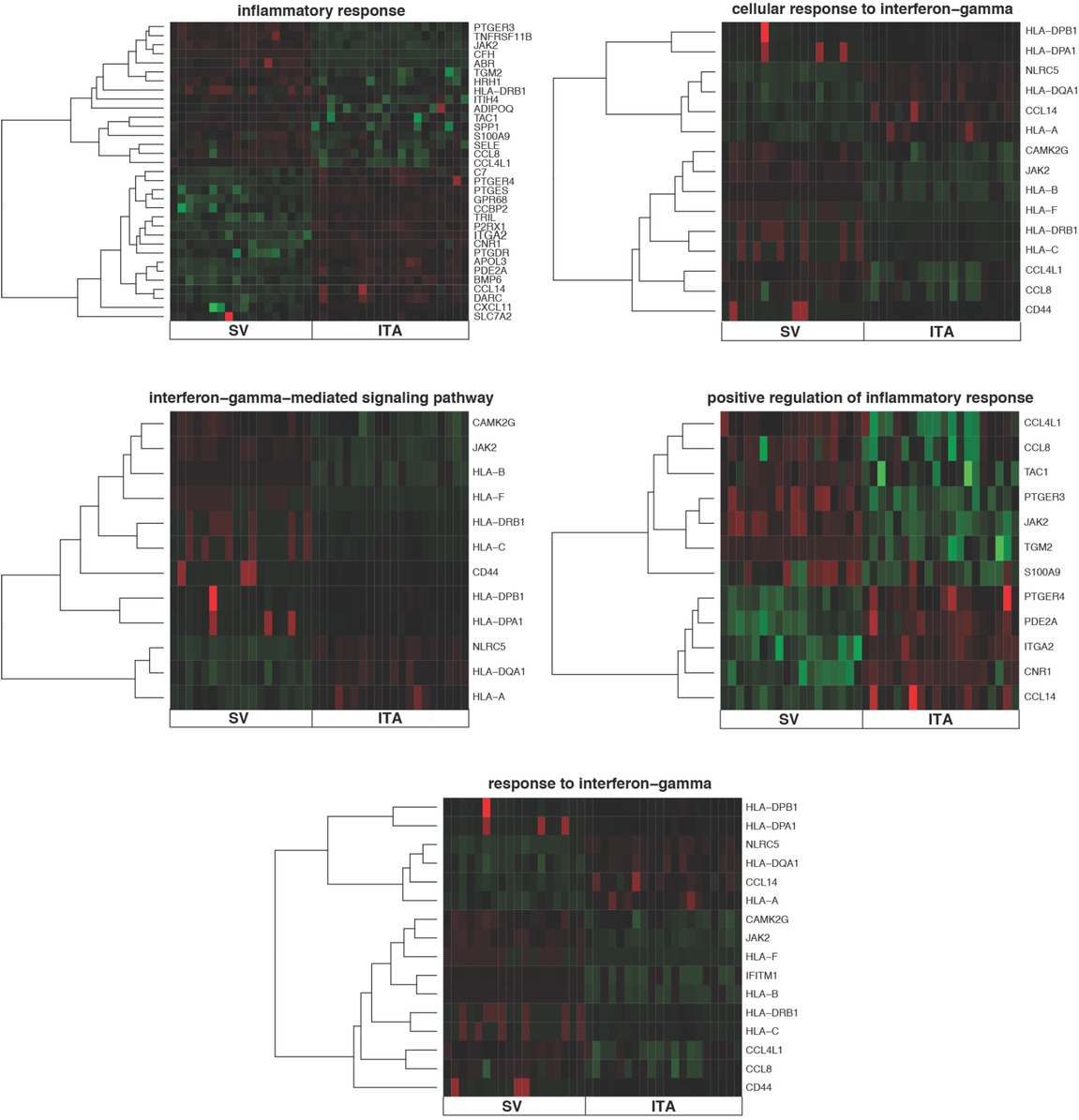
Heatmaps presenting differentially expressed genes involved in “cellular response to interferon-gamma”, “inflammatory response”, “interferon-gamma-mediated signaling pathway”, “response to interferon-gamma” and “positive regulation of inflammatory response” based on GO BP terms. Each row on the Y axis represents a single transcript. The red color indicates downregulated genes while the green are upregulated
Table 1
The 10 most significantly upregulated and 10 most significantly downregulated genes involved in inflammatory response
| Gene symbol | Gene name | Fold change | Adj. p.val |
|---|---|---|---|
| TNFRSF11B | tumor necrosis factor receptor superfamily, member 11b | 4.74 | <0.01 |
| CCL4L1 | chemokine (C-C motif) ligand 4—like 1 | 4.53 | <0.01 |
| SELE | selectin E | 3.76 | 0.01 |
| PTGER3 | prostaglandin E receptor 3 (subtype EP3) | 3.53 | <0.01 |
| CCL8 | chemokine (C-C motif) ligand 8 | 3.23 | <0.01 |
| JAK2 | Janus kinase 2 | 3.18 | <0.01 |
| TAC1 | tachykinin, precursor 1 | 3.17 | <0.01 |
| S100A9 | S100 calcium binding protein A9 | 3.15 | 0.02 |
| SPP1 | secreted phosphoprotein 1 | 2.97 | <0.01 |
| TGM2 | transglutaminase 2 (C polypeptide, protein-glutamine-gamma-glutamyltransferase) | 2.58 | <0.01 |
| CCL14 | chemokine (C-C motif) ligand 14 | -2.43 | <0.01 |
| BMP6 | bone morphogenetic protein 6 | -2.45 | <0.01 |
| CFH | complement factor H | -2.63 | <0.01 |
| CD44 | CD44 molecule (Indian blood group) | -2.73 | <0.01 |
| C7 | complement component 7 | -2.78 | <0.01 |
| TRIL | TLR4 interactor with leucine-rich repeats | -2.95 | <0.01 |
| PDE2A | phosphodiesterase 2A, cGMP-stimulated | -2.98 | <0.01 |
| NLRC5 | NLR family, CARD domain containing 5 | -3.16 | <0.01 |
| CNR1 | cannabinoid receptor 1 (brain) | -3.27 | <0.01 |
| P2RX1 | purinergic receptor P2X, ligand-gated ion channel, 1 | -6.48 | <0.01 |
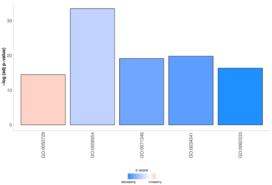
Figure 2
The bar plot presenting z-scores of processes involved in inflammatory response
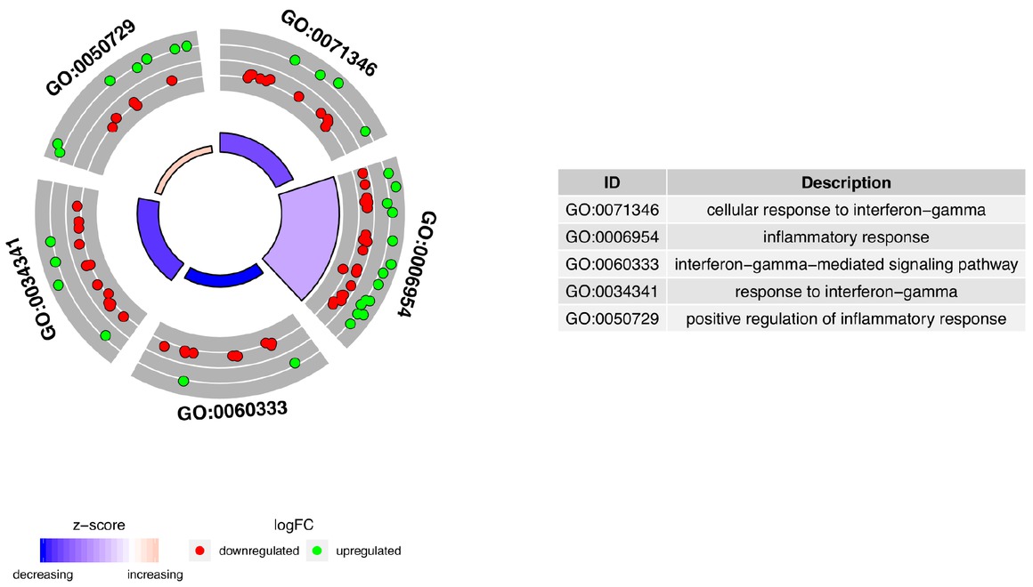
Figure 3
The circular scatter plots of differentially expressed genes involved in “cellular response to interferon-gamma”, “inflammatory response”, “interferon-gamma-mediated signaling pathway”, “response to interferon-gamma” and “positive regulation of inflammatory response” GO BP terms. Each dot represents a single gene. The z-scores were presented as segments of inner circles
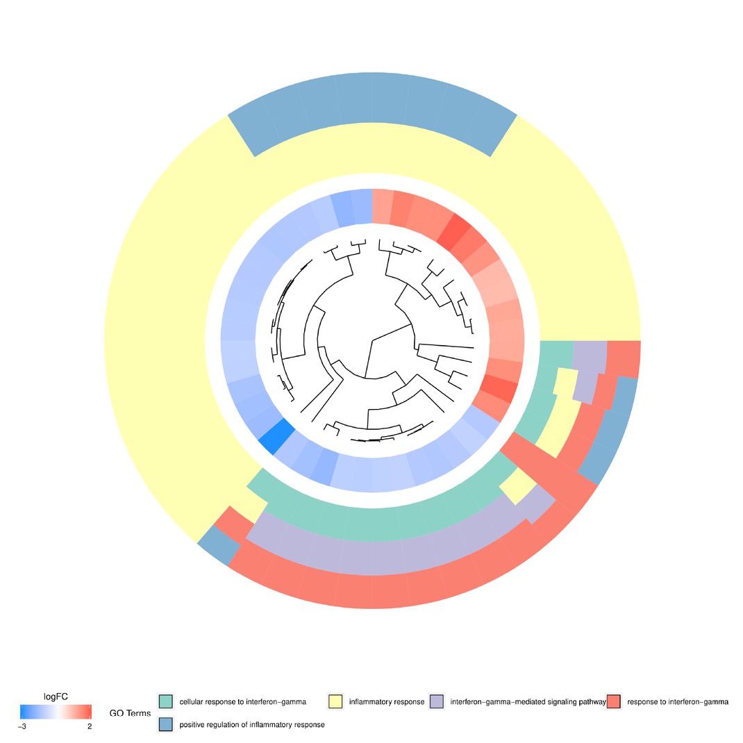
Figure 4
The dendrogram of differentially expressed genes involved in “cellular response to interferon-gamma”, “inflammatory response”, “interferon-gamma-mediated signaling pathway”, “response to interferon-gamma” and “positive regulation of inflammatory response” GO BP terms. The DEGs were clustered based on their logFC values
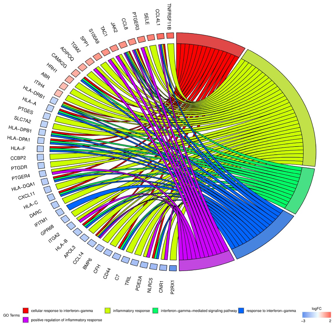
Figure 5
Analysis of enriched gene ontological groups involved in inflammatory response. The network plot presenting the linkages of genes and GO BP terms

Figure 6
Heatmap presenting the relationship between genes and selected GO BP terms. The yellow color of tiles indicates the absence of logFC values
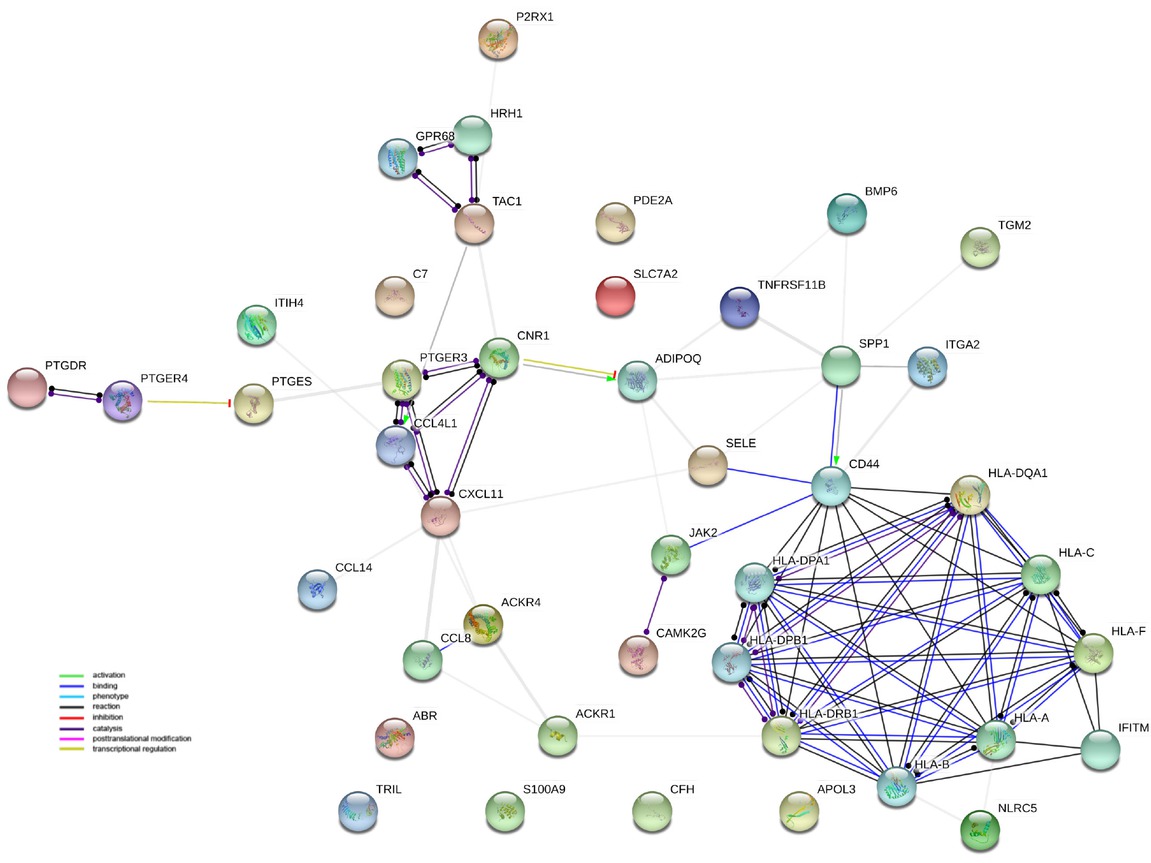
Figure 7
Interaction network of proteins encoded by DEGs belonging to “cellular response to interferon-gamma”, “inflammatory response”, “interferon-gamma-mediated signaling pathway”, “response to interferon-gamma” and “positive regulation of inflammatory response” GO BP terms. The network was generated by STRING software. Network nodes represent proteins. Empty nodes indicate proteins of unknown 3D structure
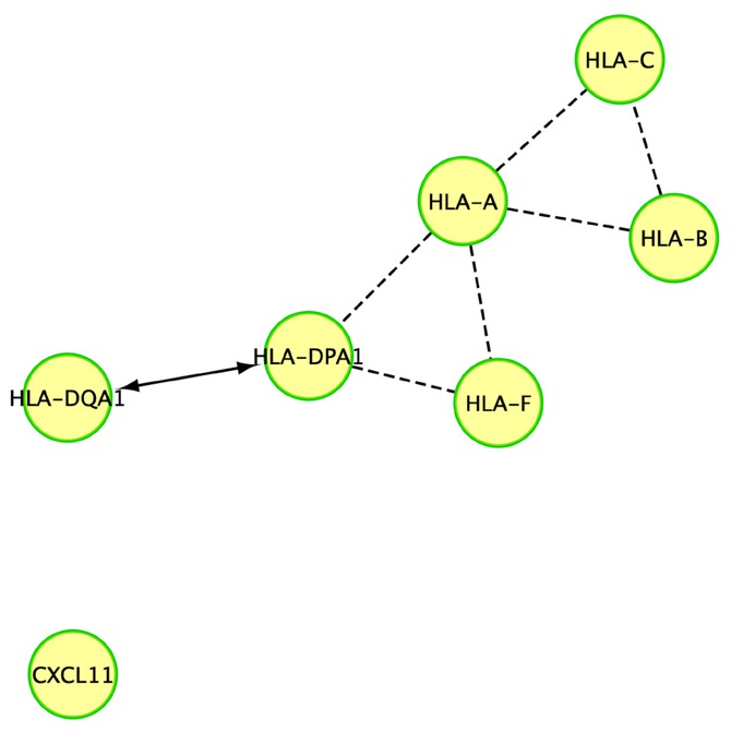
Figure 8
Reactome FI network for “Immune response”. “--->” indicates activating/catalyzing, “-“ FIs extracted from complexes or inputs and “---” predicted FIs
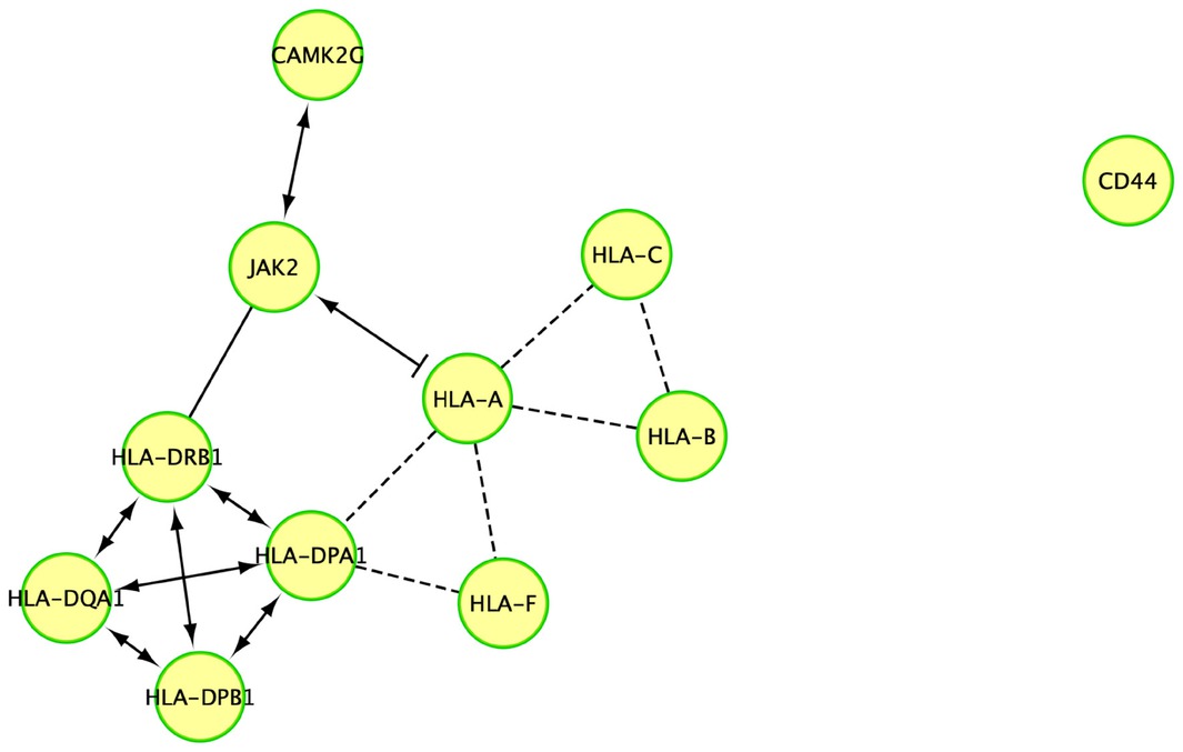
Figure 9
Reactome FI network for “Interferon-gamma-mediated signaling pathway”. “--->” indicates activating/catalyzing, “-|” for inhibition, “-“ FIs extracted from complexes or inputs and “---” predicted FIs