
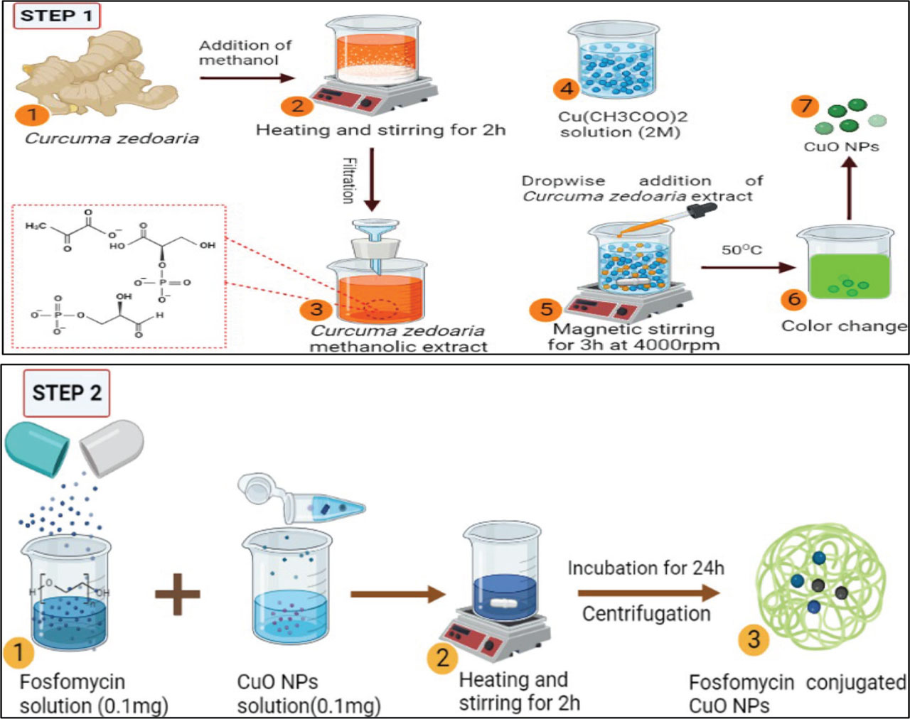
Figure 1.
Schematic representation of C. zedoaria mediated synthesis of CuO NPs (Step-1); Fos-CuO NPs (Step-2). CuO NPs, copper oxide nanoparticles; Fos-CuO NPs, Fosfomycin conjugated copper oxide nanoparticles.
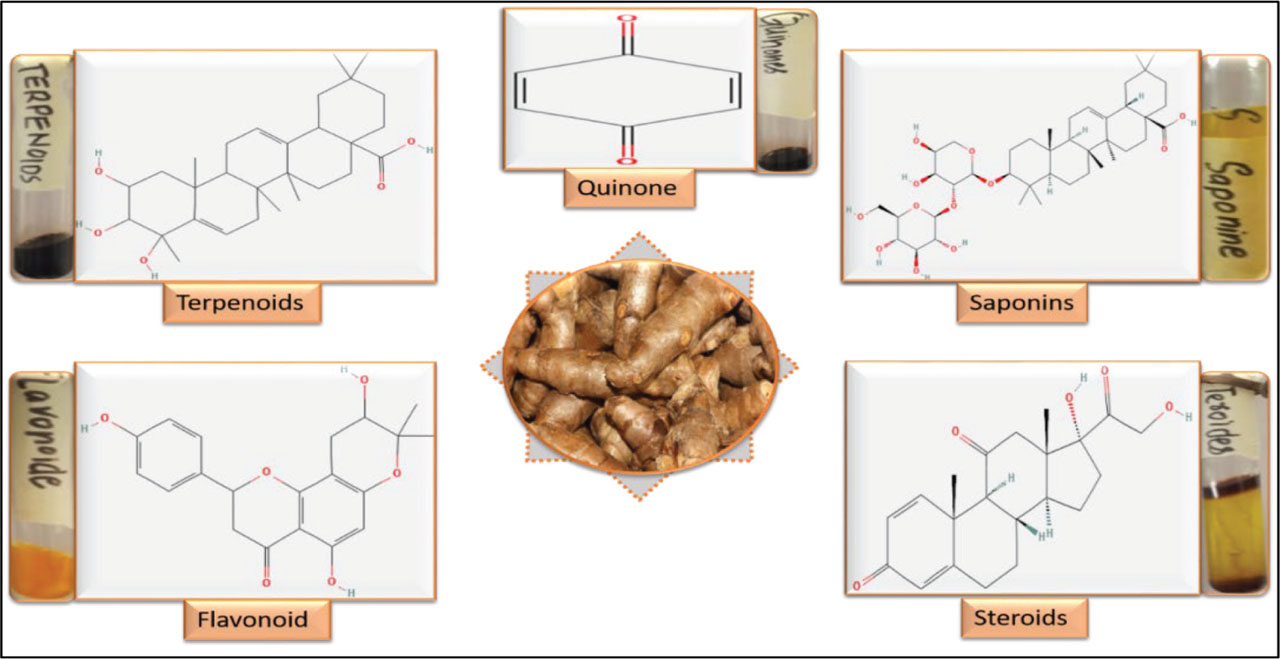
Figure 2.
Phytochemical analysis of methanolic extract of C. zedoaria powder.
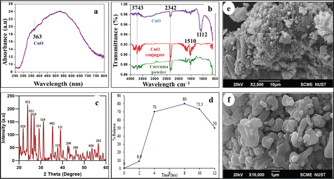
Figure 3.
(A–C) UV visible spectroscopic and XRD analysis of CuO NPs. (B) FTIR analysis of CuO NPs, Fos-CuO conjugate, and C. zedoaria powder. (D) In vitro release profile of fosfomycin from CuO NPs and (E,F) SEM images of CuO NPs at 10 μm and 1 μm. CuO NPs, copper oxide nanoparticles; Fos-CuO, Fosfomycin conjugated copper oxide; SEM, scanning electron microscopy.
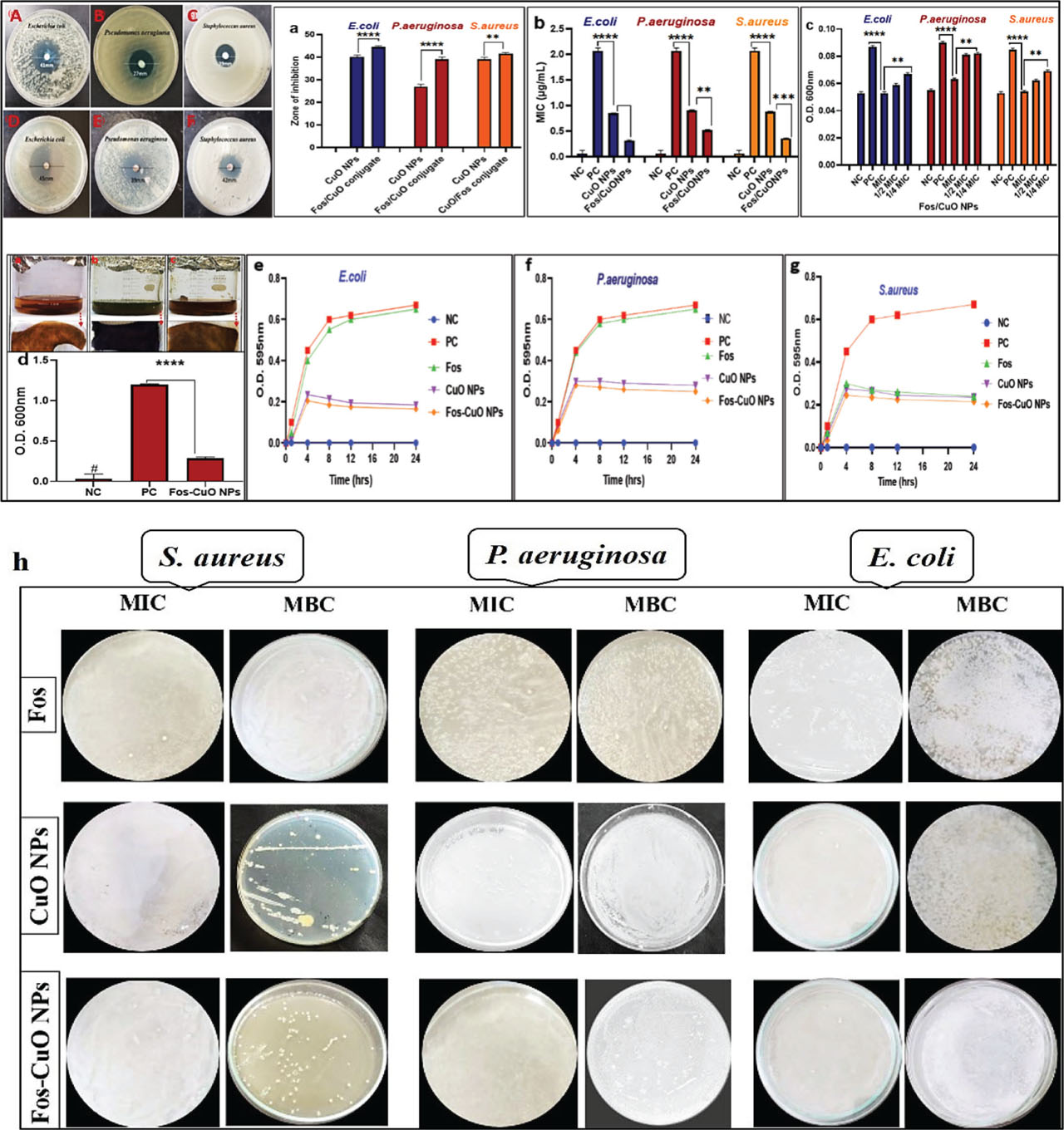
Figure 4.
Antibacterial activity of biogenic CuO NPs (A–C) and Fos-CuO NPs (D–F). Graphical representation of antibacterial potential (A); MIC assay of CuO NPs and Fos/CuO NPs (B); biofilm inhibition assay of the most effective Fos/CuO conjugate at MIC, ½ MIC and ¼ MIC on microtiter plate (C) and teeth sample (D) time kill assay graph (E–G) and bactericidal activity (MIC: MBC), based on CFU enumeration (H) of Fos, CuO NPs and Fos-CuO NPs against E. coli, S. aureus, and P. auroginosa. CuO NPs, copper oxide nanoparticles; Fos-CuO NPs, fosfomycin conjugated copper oxide nanoparticles; MBC, minimum bactericidal concentrations; MIC, minimum inhibition concentration; NC, negative control; PC, positive control.
Table 1.
Minimum inhibition concentration and MBC of Fosfomycin, CuO NPs, and Fos-CuO NPs
| Test microorganism | Fos | CuO NPs | Fos-CuO NPs | |||
|---|---|---|---|---|---|---|
| MIC (mg/mL) | MBC (mg/mL) | MIC (μg/mL) | MBC (μg/mL) | MIC (μg/mL) | MBC (μg/mL) | |
| E. coli | 50 ± 0.5 | 65 ± 0.5 | 0.86 ± 0.5 | 1.72 ± 0.5 | 0.32 ± 0.5 | 0.64 ± 0.5 |
| P. aeruginosa | 56 ± 0.5 | 70 ± 0.5 | 0.91 ± 0.5 | 1.82 ± 0.5 | 0.53 ± 0.5 | 1.06 ± 0.5 |
| S. aureus | 32 ± 0.5 | 46 ± 0.5 | 0.89 ± 0.5 | 1.78 ± 0.5 | 0.37 ± 0.5 | 0.74 ± 0.5 |
Table 2.
Fractional inhibition concentration of Fos-CuO NPs
| Test microorganism | Fos MIC (μg/mL) | CuO MIC (μg/mL) | Fos-CuO MIC (μg/mL) | FICI | Interpretation |
|---|---|---|---|---|---|
| E. coli | 50,000 ± 0.5 | 0.86 ± 0.5 | 0.32 ± 0.5 | 0.37 | Synergy |
| P. aeruginosa | 56,000 ± 0.5 | 0.91 ± 0.5 | 0.53 ± 0.5 | 0.58 | Additive (borderline synergy) |
| S. aureus | 32,000 ± 0.5 | 0.89 ± 0.5 | 0.37 ± 0.5 | 0.42 | Synergy |
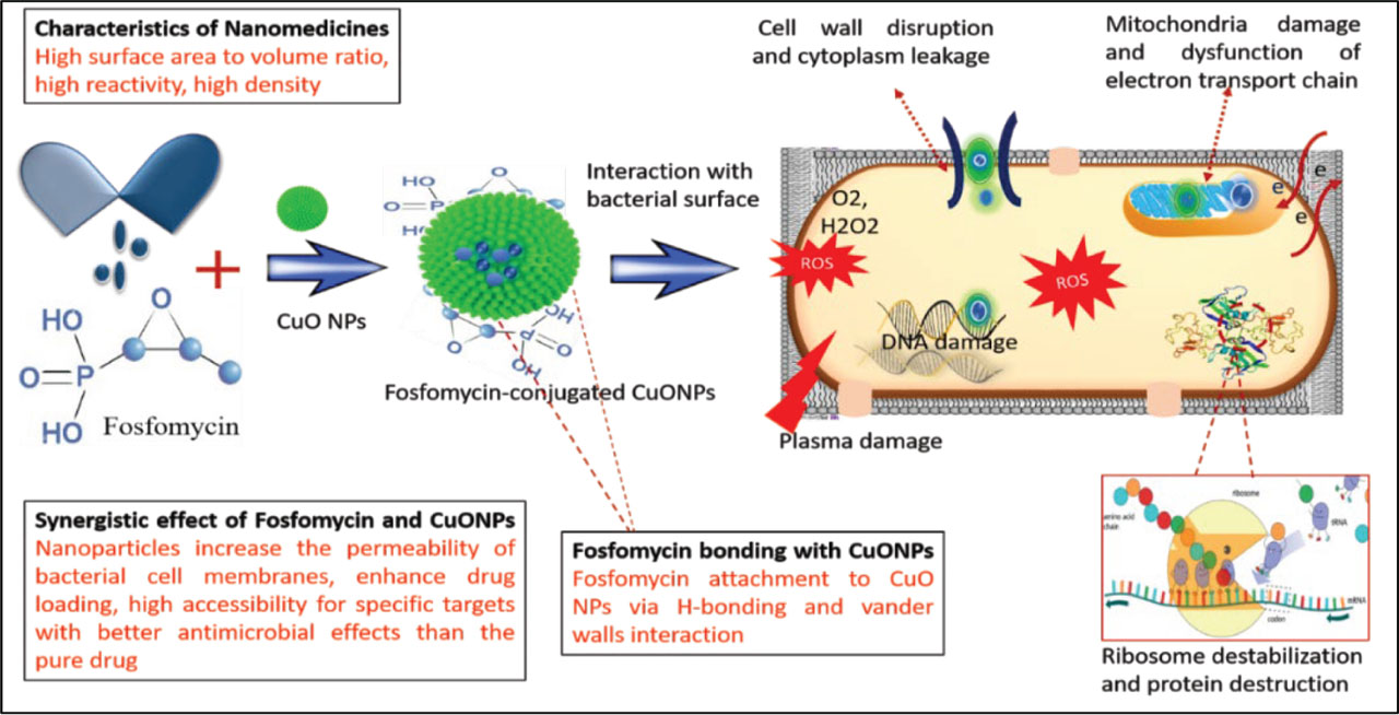
Figure 5.
Hypothetical antibacterial mechanism of Fos-CuO NPs. Possible mechanisms include ROS generation, leading to dysfunction of the electron transport chain, and disruption of cell membrane, protein, and DNA. Fos-CuO NPs, fosfomycin conjugated copper oxide nanoparticles.
Table 3.
The molecular docking scores between ligands and biofilm-associated proteins in P. aeruginosa
| Protein | Ligands | Binding affinity (kcal/mol) |
|---|---|---|
| Lec A | CuO | −2.6 |
| Fosfomycin | −4.1 | |
| Fosfomycin-conjugated | −4.4 | |
| Pel A | CuO | −2.5 |
| Fosfomycin | −4 | |
| Fosfomycin-conjugated | −4.9 | |
| Pel B | CuO | −2.5 |
| Fosfomycin | −3.7 | |
| Fosfomycin-conjugated | −3.7 | |
| gacA | CuO | −2.6 |
| Fosfomycin | −4 | |
| Fosfomycin-conjugated | −3.9 | |
| pslA | CuO | −2.7 |
| Fosfomycin | −4.7 | |
| Fosfomycin-conjugated | −3.8 |
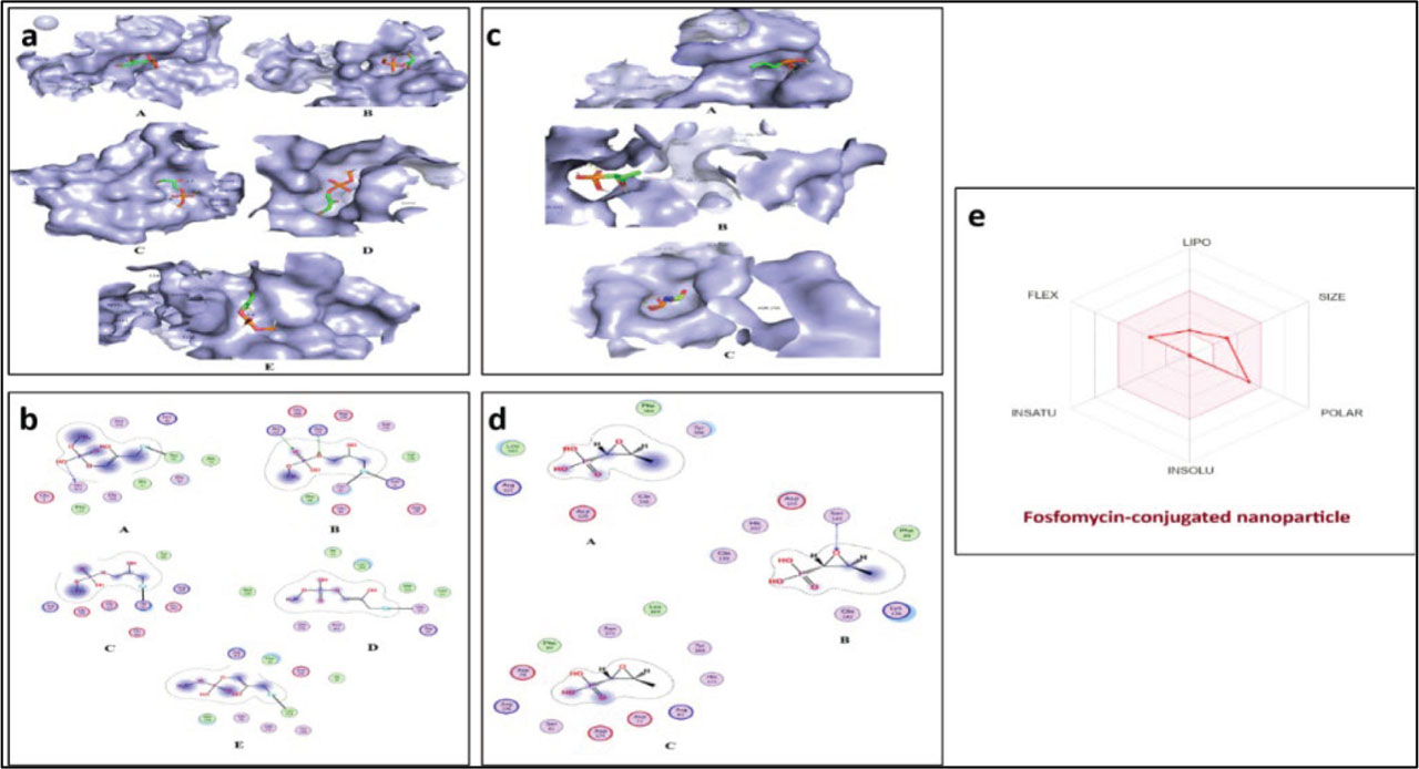
Figure 6.
The 3D interaction (A) and 2D interaction (B) between biofilm-associated proteins and Fos-CuO NPs. (A), Lec A (PDB: 3ZYH); (B), Pel A (PDB: 5TCB); (C) Pel B (PDB: 5WFT); (D), gacA (Accession: Q51373); (E), pslA (Accession: Q9I1N). The 3D interaction (C) and 2D interaction (D) between biofilm-associated proteins and Fosfomycin. (A), Pel B (PDB: 5WFT); (B), gacA (Accession: Q51373); (C), pslA (Accession: Q9I1N) and 3D ADME Radar (E) for the graphical representation of Fosfomycin-conjugated nanoparticle for lipophilicity, flexibility, instauration, insolubility, size, and polarity. The pink region of the radar indicates an ideal range of bioavailability to a drug. ADME, absorption, distribution, metabolism, and excretion; CuO NPs, copper oxide nanoparticles; Fos-CuO NPs, fosfomycin conjugated copper oxide nanoparticles.