
Table 1.
Results of 8th, 15th, and 30th days tail flick tests of control and sevoflurane groups
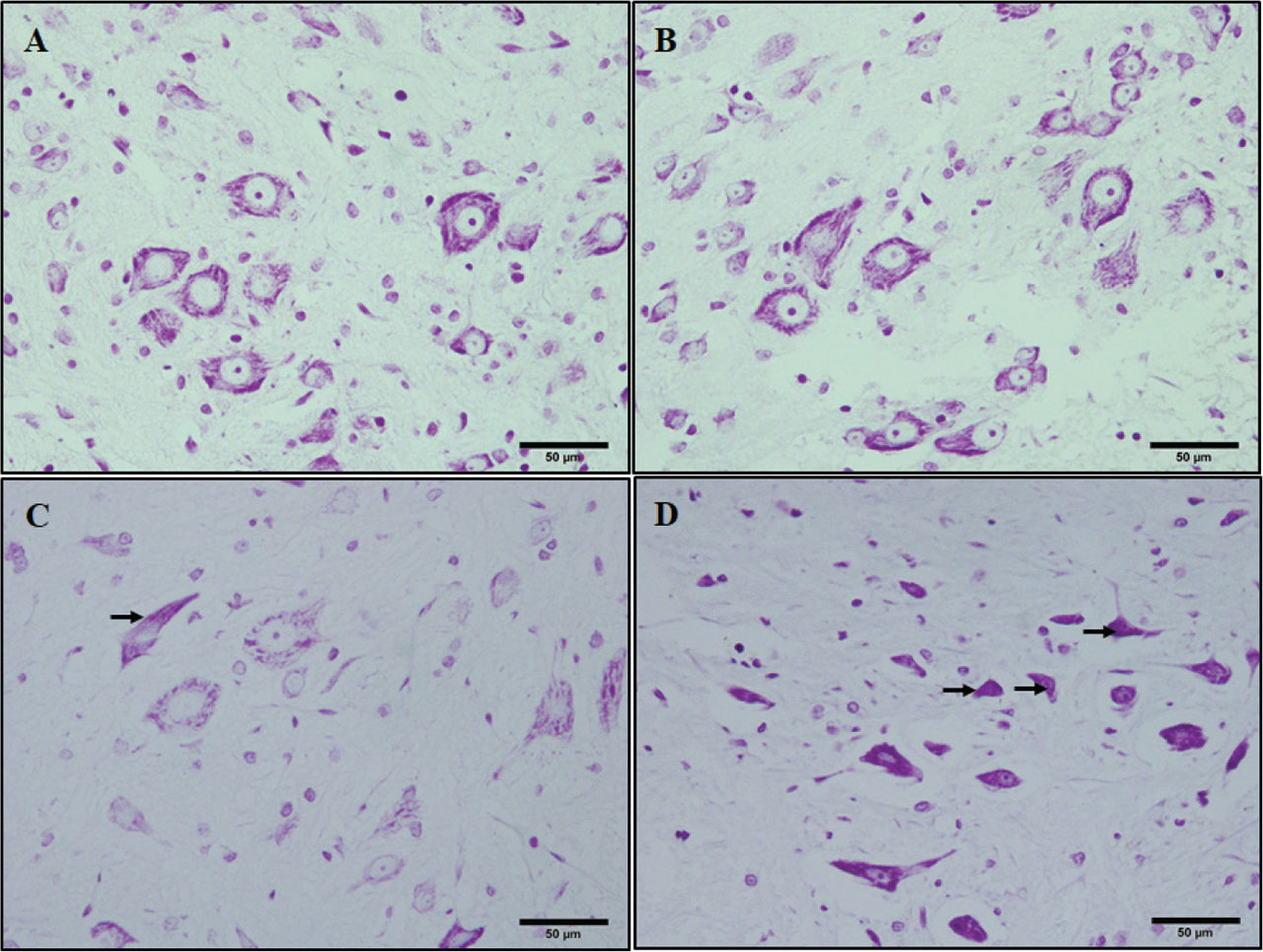
Figure 1.
Crystal violet-stained sections of the rat spinal cords. (A) acute control, (B) chronic control, (C) acute sevoflurane, and (D) chronic sevoflurane groups. Arrows showing pyknotic cells. Bars = 50 μm.
Table 2.
The percentage of TUNEL-positive cells of ACont, ASevo, CCont, and CSevo groups
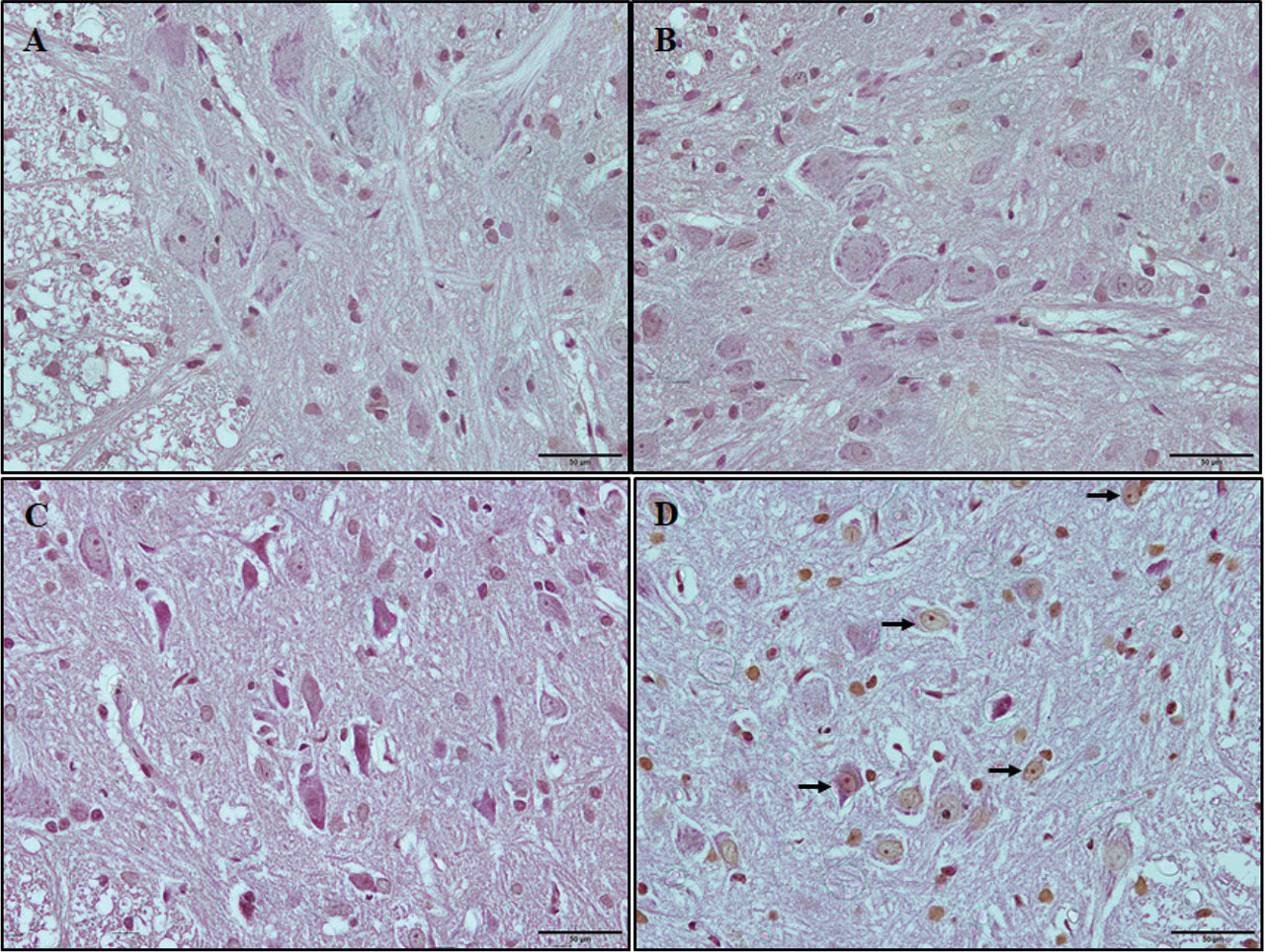
Figure 2.
TUNEL staining. Representative photomicrographs of TUNEL-positive cells (arrows). (A) acute control, (B) chronic control, (C) acute sevoflurane, and (D) chronic sevoflurane groups. Bars = 50 μm.