
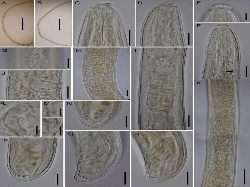
Figure 1:
Xiphinemella esseri (Chitwood, 1957) (light photomicrographs). A: Male, entire body; B: Female, entire body; C: Anterior region in dorso-ventral median view; D, F: Anterior region in lateral median view (F, arrow pointing on odontophore basal flanges); E: Lip region in surface lateral view; G: Lateral chord; H: Uterine egg; I: Pharyngeal expansion; J: Vagina; K: Anterior genital branch in part (showing the oviduct-uterus junction and uterus morphology); L: Spicule; M, O: Lateral guiding piece; N, P: Female caudal region; Q, R: Male caudal region. (Scale bars: A, B = 500 μm; C, D, F, G, I-K, N, P-R = 20 μm; E, L, M, O = 10 μm; H = 25 μm).
Table 1.
Morphometrics of Xiphinemella esseri Chitwood, 1957.
| Population | Florida, USA | ||||||
|---|---|---|---|---|---|---|---|
| Reference | Present paper | Chitwood (1957) | De Bruin and Heyns (1991) | ||||
| Character | n | 9♀♀ | 7♂♂ | ♀♀ | ♂♂ | 5♀♀ | 4♂♂ |
| L | 2.82 ± 0.23 (2.47-3.32) | 2.55 ± 0.26 (2.30-3.07) | 2.4–3.5 | 2.2-3.0 | 3.10 (2.62-3.74) | 3.17 (3.06-3.25) | |
| a | 53.2 ± 3.1 (50-59) | 52.1 ± 6.4 (45-62) | 40-43 | 35-45 | 47.1 (39.7-58.4) | 53.2 (51.6-54.2) | |
| b | 8.6 (n = 1) | 8.2 (n = 2) | 11.4-14.0 | 7.6-11.0 | 10.3 (8.7-11.6) | 10.8 (9.9-12.2) | |
| c | 117 ± 21 (100-158) | 101 ± 5.5 (94-110) | 93-130 | 77-120 | 137 (95-161) | 124 (105-152) | |
| c′ | 0.7 ± 0.1 (0.6-0.8) | 0.8 ± 0.1 (0.7-0.9) | 0.8a | 0.9a | 0.6 (0.5-0.7) | 0.85 (0.7-1.0) | |
| V | 47.3 ± 1.5 (45.4-49.7) | – | 42-47 | – | 46 (45-47) | – | |
| Lip region diam. | 16.8 ± 0.4 (16.5-17.5) | 16.6 ± 0.2 (16.5-17.0) | 17a | – | 17.2 (17-18) | 17 (16-18) | |
| Odontostyle length | 47.0 ± 1.0 (46-49) | 46.3 ± 0.4 (46-47) | 72-75b | – | 46 (42-50) | 46 (45-48) | |
| Odontophore length | 35.0 ± 0.8 (34-36) | 34.9 ± 1.0 (33-36) | – | – | 33 (31-35) | 34 (31-36) | |
| Guiding ring from ant. end | 36.3 ± 1.5 (34-39) | 36.5 ± 1.0 (35-38) | 35-38 | – | 36 (30-42) | 37 (32-42) | |
| Neck length | 288 (n = 1) | 288, 296 (n = 2) | – | – | 300 (276-333) | 296 (267-322) | |
| Pharyngeal expansion length | 84.0 ± 2.8 (81-87) | 82.2 ± 1.9 (80-85) | 75-85 | – | 84 (80-89) | 80 (76-85) | |
| Diam. at neck base | 45.1 ± 2.8 (42-50) | 44.6 ± 2.5 (42-48) | – | – | – | – | |
| at midbody | 53.4 ± 4.8 (49-64) | 50.0 ± 3.4 (46-55) | – | – | – | – | |
| at anus | 34.0 ± 2.6 (32-39) | 30.4 ± 1.2 (29-32) | – | – | 39 (30-42) | 31 (30-32) | |
| Prerectum length | 611, 683 (n = 2) | 594 ± 142,3 (444-727) (n = 3) | – | – | 557 (427-695) (n = 3) | – | |
| Rectum length | 36.9 ± 6.0 (27-43) | 45.1 ± 4.4 (40-52) | – | – | 39 (36-42) | – | |
| Tail length | 24.0 ± 4.0 (18-28) | 25.2 ± 2.0 (23-28) | – | – | 23 (20-30) | 26 (21-31) | |
| Spicule length | – | 42.6 ± 1.4 (41-45) | – | 40-45 | – | 34 (31-36) | |
| Ventromedian supplements | – | 9 ± 0.6 (8-10) | – | – | – | 8-9 | |
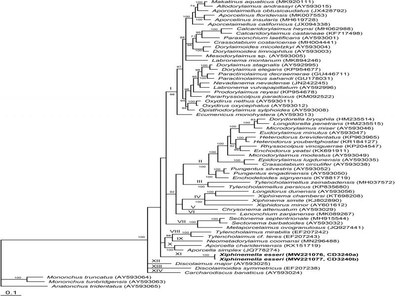
Figure 2:
Bayesian 50% majority rule consensus tree as inferred from the D2-D3 expansion segments of 28S rRNA gene sequence alignment under the GTR + I + G model. Posterior probability values more than 70% are given on appropriate clades. New sequences are indicated by bold letters.
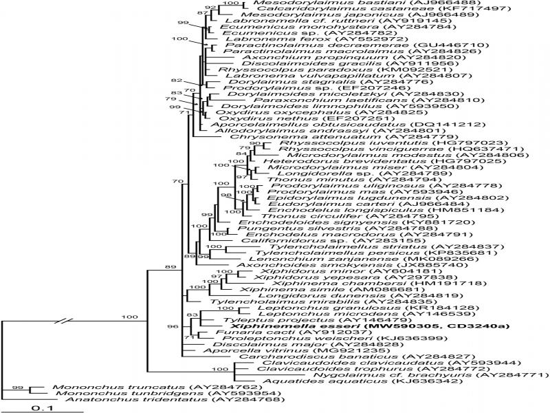
Figure 3:
Bayesian 50% majority rule consensus tree as inferred from the 18S rRNA gene sequence alignment under the GTR + I + G model. Posterior probability values more than 70% are given on appropriate clades. A new sequence is indicated by bold letters.