
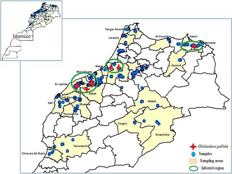
Figure 1:
Sampled areas and regions infested with Globodera pallida in Morocco.
Table 1.
Morphological and morphometric characteristics of Moroccan population of cysts and second-stage juveniles compared to the standard Globodera pallida (EPPO, 2004).
| Species | Shape of knob | J2 stylet length (µm) | Number of cuticula ridges | Granek’s ratio |
|---|---|---|---|---|
| G. rostochiensisa | Rounded | (21.8) | 16-31 (>14) | 1.3-9.5 (>3) |
| G. pallida a | Pointed | 22-24 (23.8) | 8-20 (<14) | 1.2-3.5 (<3) |
| Moroccan population G. pallida | Pointed | 22.6 | 9 | 2.2 |
1 Notes: aStandard measurement OEPP/EPPO (2004) OEPP/EPPO Bulletin 34: 155-16.
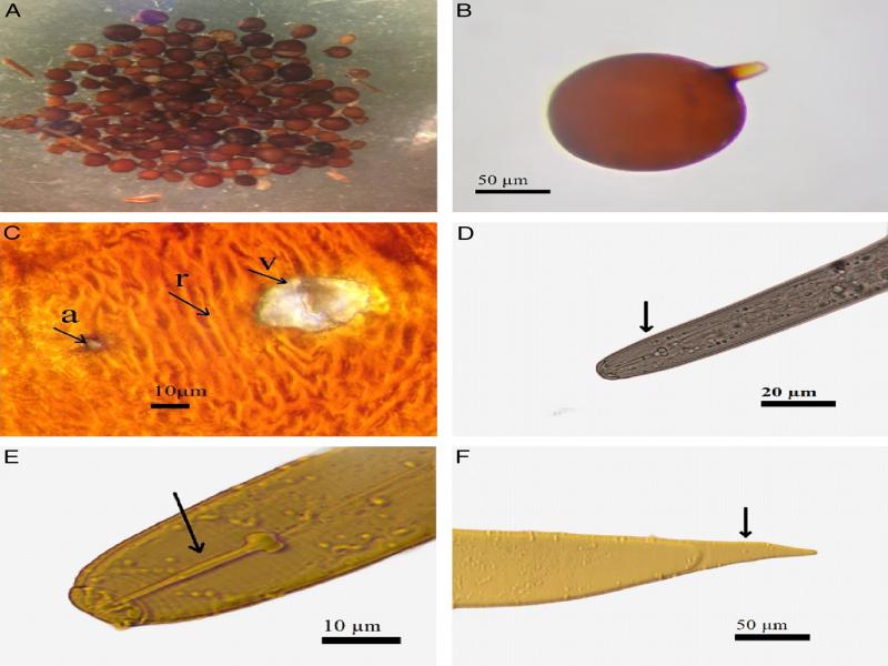
Figure 2:
Photomicrographs of morphological characterization of cyst, egg, and second-stage juvenile. A, B: Cyst of Globodera pallida, C: Vulva (v), anus (a), and cuticular ridges (r) of cyst, D, E: Stylet knob shape of J2, F: Juvenile tail.
Table 2.
Primers used in the present study.
| Primer name | Nematode | 5′-3′ Sequence | Band size | References |
|---|---|---|---|---|
| ITS5 | Forward | GGAAGTAAAAGTCGTAACAAGG | – | White et al. (1990) |
| PITSp4 | Globodera pallida | ACAACAGCAATCGTCGAG | 265 bp | Bulman and Marshall (1997); Skantar et al. (2007) |
| PITSr3 | Globodera rostochiensis | AGCGCAGACATGCCGCAA | 434 bp |
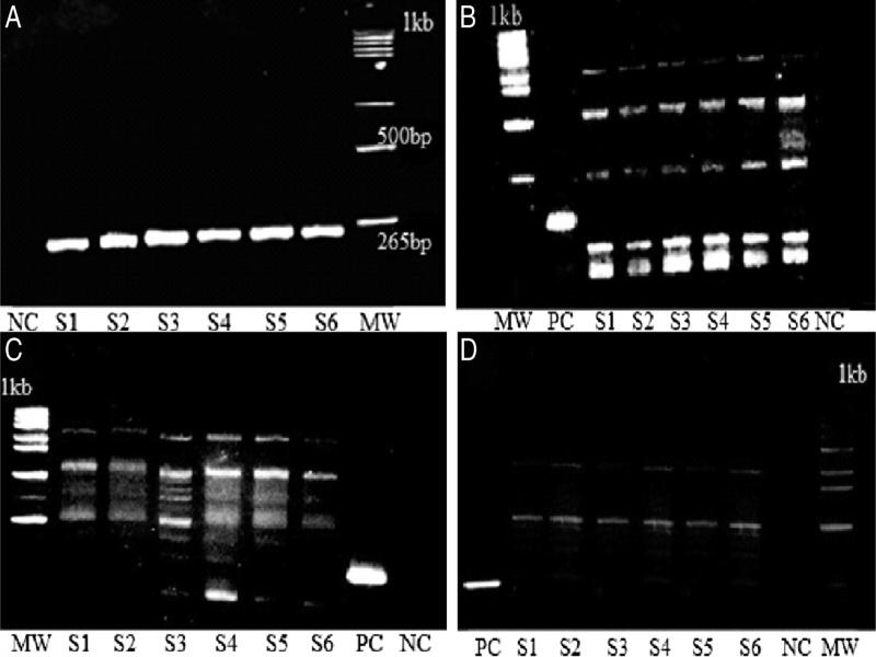
Figure 3:
Amplified PCR products from Globodera spp. digested by three enzymes AluI, MboI, and RsaI. A: Amplified PCR products, B: AluI, C: RsaI, D: MboI, MW: molecular weight markers (1 kb), NC: negative control, PC: undigested DNA, S1-S2: Eastern region samples, S3-S4: Western region samples (Gharb), S5-S6: Doukkala region samples.