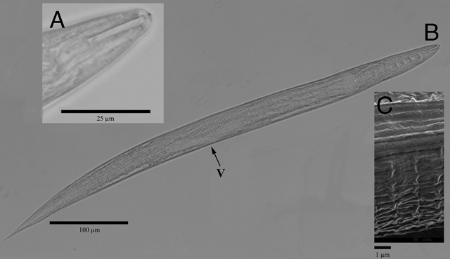
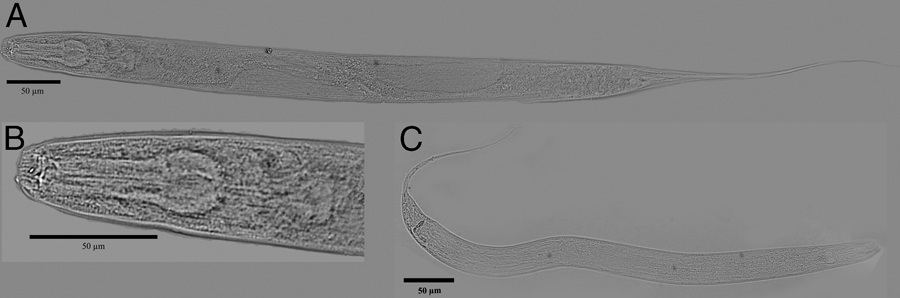
The invasive phytophagous millipede Chamberlinius hualienensis Wang, 1956 was originally described from Taiwan, where it is extremely common (Chen et al., 2011). It was discovered in Okinawa, Japan in 1986 (Higa and Kishimoto, 1986) and outlying islands of the Ryukyu Archipelago (Nakamura and Korsós, 2010). Fourteen years ago it was detected on Hachijo Island, Japan (Fujiyama et al., 2012). Remarkably these millipedes are not known to swarm in Taiwan (Chen et al., 2011), but in Japan mass occurrences of the species have even led to disruptions of railway traffic (Niijima and Arimura, 2002). While most millipedes in the world are harmless to humans, they can vector plant and animal diseases caused by bacteria such as Citrobacter, Enterobacter, Salmonella, and Raoultella species (Kania and Klapeć, 2012). Millipedes may cause unexpected damage when they are introduced to new locations, becoming invaders that leave behind their old parasites and predators. A taxonomic inventory of their invertebrate associates may shed light on their biology and ecology. Therefore, it was interesting that nematodes of the Rhabditida were recently discovered within the gut of the invasive species C. hualienensis (Meyer-Rochow, 2015). Their morphological and molecular characterization, phylogenetic relationships and ecological associations are detailed in this report.
Materials and methods
Nematodes were discovered to be associated with the phytophagous millipede C. hualienensis, specimens of which were collected in November on Hachijojima (33o05'N; 139o47'E) from mass aggregations on the concrete walls of a freeway (Meyer-Rochow, 2015). Nematodes were rinsed under tap water strictly from the internal cavity (hemocoel and intestine were not specified) of millipedes after dissection and placed in 65% ethanol before sending to Beltsville in December, 2014 and January 2015 to process for slides and PCR. Culture was not possible due to inadequate laboratory facilities in Japan.
Microscopy
Nematodes were imaged at ×40‒60 on an Olympus BX51 microscope with a DP71 camera (Olympus America Inc., Center Valley, PA) equipped with polarization optics. Measurements in micrometers were made with an ocular micrometer on a Zeiss Ultraphot II compound microscope with Nomarski optics on alcohol-distorted specimens before formalin fixation, and images were also directly measured with CellSens ver 1.6 imaging software integrated with the camera (Olympus America LLC, Center Valley, PA). Fixed specimens of Rhabditidae juveniles were processed for permanent slides according to the formalin-glycerine method (Golden, 1990). Imaged specimens of Diplogastridae were subsequently processed for PCR so no vouchers are available. For scanning electron microscopy (SEM) Oscheius nematodes were fixed according to a method recently described by Takaku et al. (2013). Specimens were observed in a JEM7100F JEOL high-pressure environmental scanning electron microscope at 1 kV.
DNA analysis
Specimens of each nematode were mechanically disrupted in 20 µl of extraction buffer (Baldwin et al., 1997) then stored in PCR tubes at –80°C until needed. Extracts were prepared from thawed pools by incubating the tubes at 60°C for 60 min, followed by 95°C for 15 min. to deactivate proteinase K. Two microliters of the extract was used for each 25 µl PCR reaction.
The ribosomal LSU D2-D3 expansion segment was amplified with primers D2A 5′-ACAAGTACCGTGAGGGAAAGTTG-3′ and D3B 5′TCGGAAGGAACCAGCTACTA-3′ (Nunn et al., 1996) using previously published amplification procedures (Baldwin et al., 1997 for O. necromenus; Ye et al., 2007 for others).
The 18S sequences reaction components included, per 25 µL reaction: 17.55 µL H2O, 2.5 µL 10X PCR buffer, 0.5 µL dNTP mix (10 mM each dNTP), 0.75 µL MgCl2, 50 mM, 0.75 µL 18S-G18S4 primer, 10 µM, 0.75 µL 18S-18P primer, 10 µM, 0.2 µL Taq (Invitrogen platinum, 1 unit), 23 µL of the above mix + 2 µL template DNA; cycling conditions were 94 C – 2 min, 94 C – 30 sec, 50 C – 30 sec, 68 C – 2 min, repeat 40 times: steps 2 through 4, 68 C – 10 min, 4 C – Hold, with primers of Thomas et al. (1997) used for PCR and sequencing.
PCR products were visualized and purified within the Lonza FlashGelTM DNA system (VWR International, Radnor, PA), and sequence was generated with an ABI BigDye Terminator v3.1 kit with sample sequence data analyzed on an ABI 3130XLAutomated DNA sequencer (Applied Biosystems, Foster City, CA, USA). The 28S sequence was determined on both strands using D2A and D3B primers.
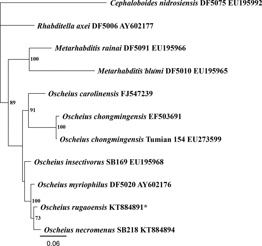
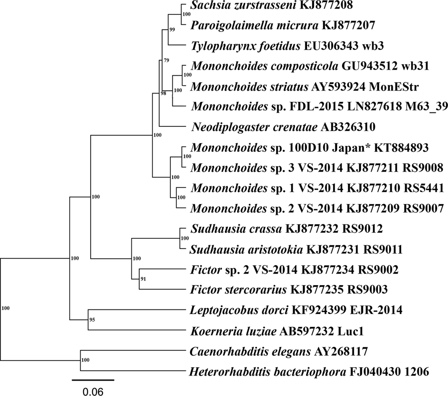
The 28S rDNA sequences related to Oscheius (Table 1) and 18S rDNA sequences of Mononchoides Rahm, 1928 and relatives (Table 2) were aligned with MAFFT ver 7.017 (Katoh, et al., 2005). Bayesian likelihood trees were made with the MrBayes (Huelsenbeck and Ronquist, 2001) plugin within Geneious 7.1.7 (Biomatters, Auckland, New Zealand) using ModelTest ver. 3.7 (Posada and Crandall, 1998) AIC parameters generated within PAUP* (Sinauer Associates, Sunderland, MA).
Table 1
Nematode 28S rDNA sequences for selected taxa in Figure 3.
| Species | Isolate | Locality | Accession |
|---|---|---|---|
| Cephaloboides nidrosiensis | DF5075 | The UK | EU195992 |
| Metarhabditis blumi | DF5010 | Spain | EU195965 |
| Metarhabditis rainai | DF5091 | Fiji | EU195966 |
| Oscheius carolinensis | USA | FJ547239 | |
| Oscheius chongmingensis | China | EF503691 | |
| Oscheius chongmingensis | Tumian154 | China | EU273599 |
| Oscheius insectivorus | SB169 | Germany | EU195968 |
| Oscheius myriophilus | DF5020 | The USA | AY602176 |
| Oscheius necromenus | SB218 | Australia | This paper |
| Oscheius sp. | YNb59 | China | AY177182 |
| Oscheius sp. | Japan | This paper | |
| Rhabditella axei | DF5006 | France | AY602177 |
| Species | Isolate | Locality | Accession |
|---|---|---|---|
| Caenorhabditis elegans | N2 | The UK | AY268117 |
| Heterorhabditis bacteriophora | 1206 | – | FJ040430 |
| Fictor stercorarius | RS9003 | Germany | KJ877235 |
| Fictor sp. 2 | RS9002 | The USA | KJ877234 |
| Koerneria luziae | Luc1 | Japan | AB597232 |
| Leptojacobus dorci | EJR2014 | Japan | KF924399 |
| Mononchoides cf. americanus | 100D10 | Japan | This paper |
| Mononchoides composticola | wb31 | Belgium | GU943512 |
| Mononchoides striatus | MonEStr | – | AY593924 |
| Mononchoides sp. | FDL-2015 M63_39 | Italy | LN827618 |
| Mononchoides sp 1 | VS-2014 RS5441 | France | KJ877210 |
| Mononchoides sp 2 | VS-2014 RS9007 | Mexico | KJ877209 |
| Mononchoides sp 3 | VS-2014 RS9008 | New Caledonia | KJ877211 |
| Neodiplogaster crenatae | NK126 | Japan | AB326310 |
| Paroigolaimella micrura | VS-TU-2014-2 | Germany | KJ877207 |
| Sachsia zurstrasseni | VS-TU-2014-3 | Germany | KJ877208 |
| Sudhausia aristotokia | RS9011 | Ghana | KJ877231 |
| Sudhausia crassa | RS9012 | South Africa | KJ877232 |
| Tylopharynx foetidus | wb3 | Belgium | EU306343 |