
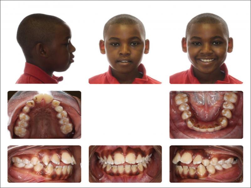
Figure 1.
Facial and intraoral photographs taken at the initial visit (age 12 years, 0 months).
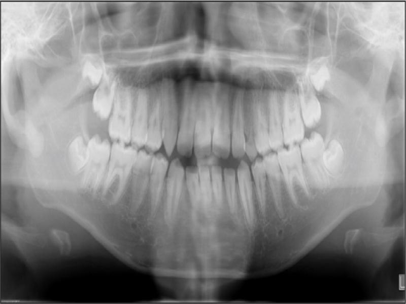
Figure 2.
Panoramic radiograph taken at the initial visit (age 12 years, 0 months).
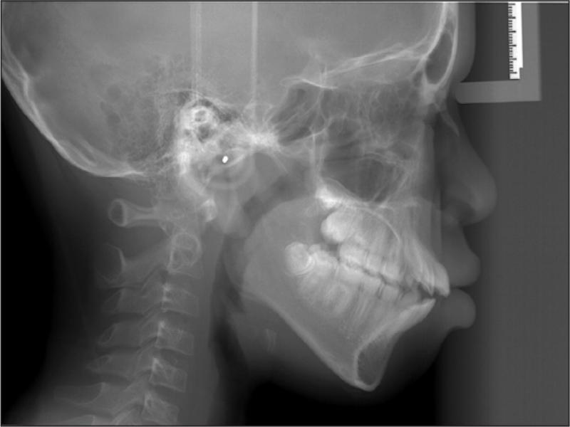
Figure 3.
Lateral cephalogram taken at the initial visit (age 12 years, 0 months).
Table I.
Cephalometric measurements before and after treatment.
| Measurement | Initial taken at initial visit (12 y 0 mo) | Final taken at debond visit (15 y 3 mo) |
|---|---|---|
| Facial Angle (FH-NPo) (°) | 84.2 | 83.9 |
| Convexity (NA-APo) (°) | 6.8 | 2.3 |
| SNA (°) | 79.5 | 78.1 |
| SNB (°) | 76.7 | 77 |
| ANB (°) | 2.8 | 1.1 |
| AB – NPo (°) | 2.8 | 1.3 |
| FMA (MP-FH) (°) | 32.8 | 34.3 |
| Y-Axis -- Downs (SGn-FH) (°) | 64.5 | 66.6 |
| Occ Plane to FH (°) | 12.7 | 14.7 |
| Interincisal Angle (U1-L1) (°) | 96.3 | 127 |
| L1 to Mand Plane -90 (°) | 13.8 | -3.9 |
| U1 – SN (°) | 119 | 105.7 |
| U-Incisor Protrusion (U1-APo) (mm) | 15.8 | 9.5 |
| L1 Protrusion (L1-APo) (mm) | 11.3 | 6.2 |
| FH – SN (°) | 8.1 | 6.9 |
| Wits Appraisal (mm) | -3.2 | -5.4 |
| Lower Face Height (ANS-Me) (mm) | 72 | 80.1 |
| LFH/TFH (ANS-Me:N-Me) (%) | 58.1 | 61.8 |
| Upper Lip to E-Plane (mm) | 2.4 | 2.1 |
| Lower Lip to E-Plane (mm) | 10.2 | 7.7 |
| Nasolabial Angle (Col-Sn-UL) (°) | 83 | 82.8 |
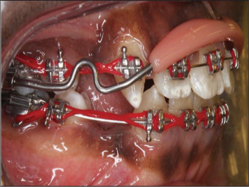
Figure 4.
A prefabricated upper lip bumper inserted to reinforce the upper anchorage during upper right canine retraction due to its long root.
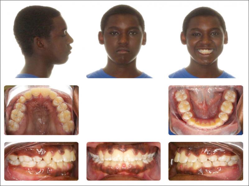
Figure 5.
Facial and intraoral photographs taken at the debond visit (age 15 years, 3 months).
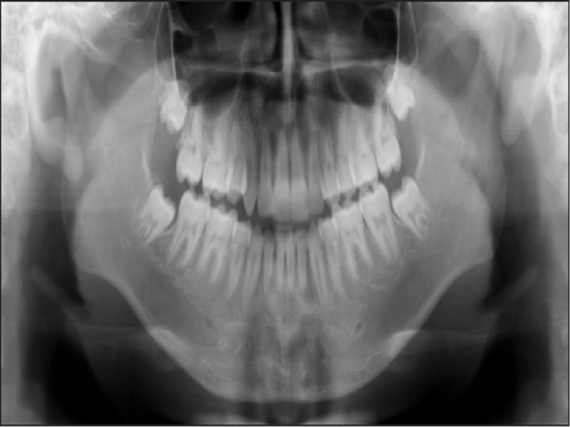
Figure 6.
Panoramic radiograph taken at the debond visit (age 15 years, 3 months).
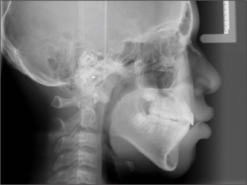
Figure 7.
Lateral cephalogram taken at the debond visit (age 15 years, 3 months).
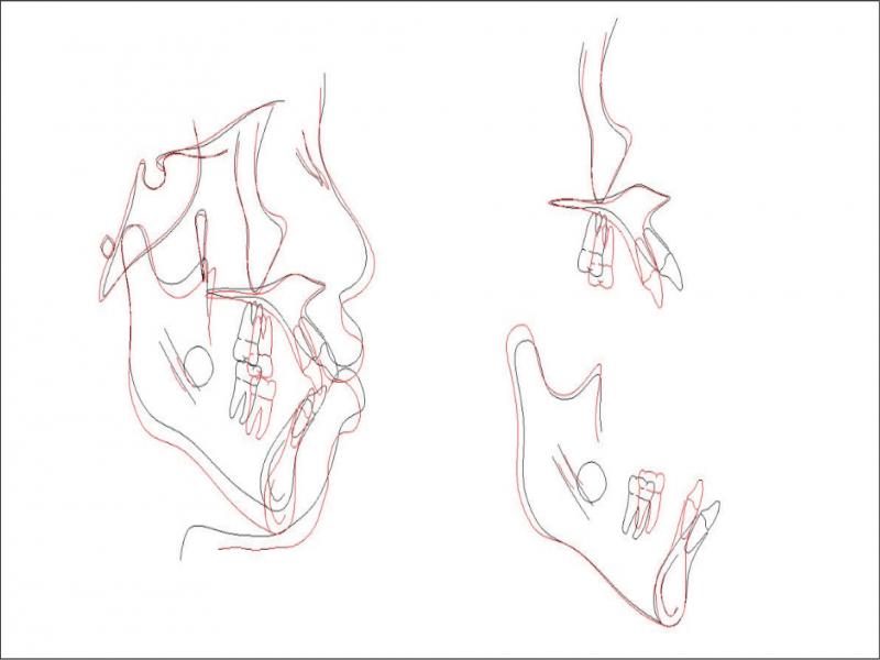
Figure 8.
Cephalometric superimposition of before and after treatment
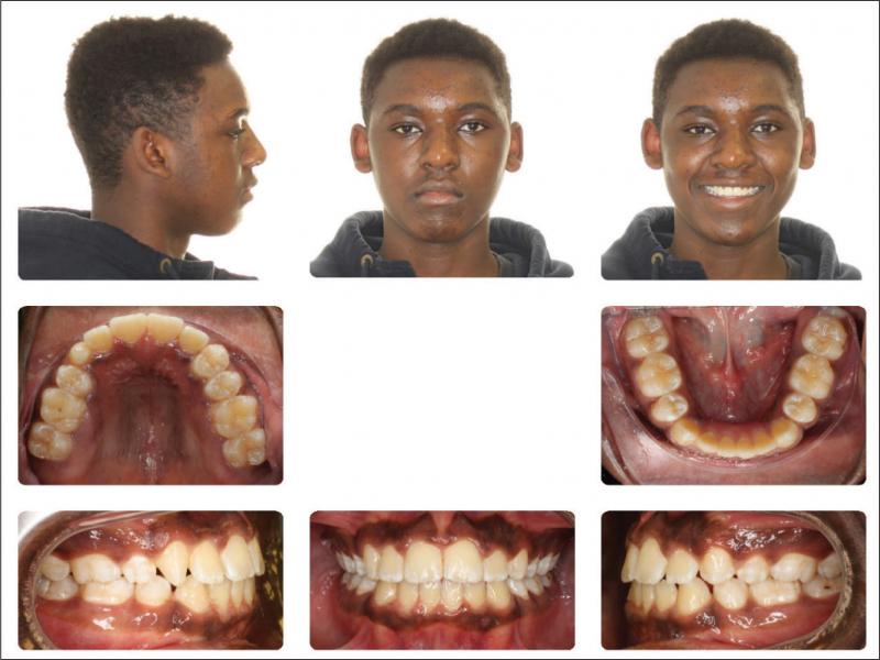
Figure 9.
Facial and intraoral photographs taken at the 19 month follow-up visit (age 16 years, 10 months).