
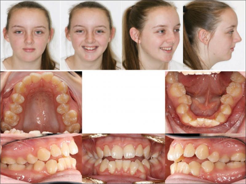
Figure 1.
Pretreatment photographs.
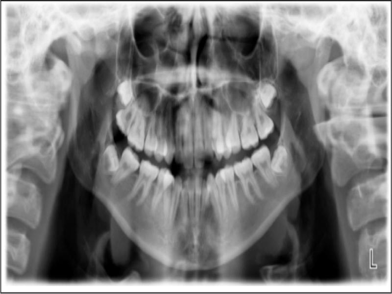
Figure 2.
Pretreatment OPG.
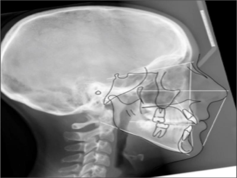
Figure 3.
Pretreatment lateral cephalogram.
Table I.
Cephalometric measurements – initial and prefinishing.
| Measurement | Standard | 18/8/2017 | 30/10/2019 | Change | ||
|---|---|---|---|---|---|---|
| SKELETAL | Maxilla | SNA | 82 ± 2° | 72.6 | 72.8 | 0.2 |
| A Point Convexity | 3 ± 2 mm | -2 | -2.1 | -0.1 | ||
| NaVert – A point | 1 mm | -1.3 | -1.2 | 0.1 | ||
| Mandible | SNB | 80 ± 2° | 72.6 | 73.0 | 0.4 | |
| Facial Angle | 87 ± 3° | 90.7 | 90.9 | 0.2 | ||
| NaVert – Po | -8 to -6 mm -2 to +4 mm | 1.3 | 1.8 | 0.5 | ||
| Basal Arch Relationship | ANB | 2 ± 2° | 0 | -0.2 | -0.2 | |
| WITS | F: 0mm | -2.1 | -1.9 | 0.2 | ||
| Vertical | Md Plane Angle | 26 ± 4° | 28.6 | 27.7 | -0.9 | |
| LFH Angle | 47 ± 4° | 49.8 | 48.3 | -1.5 | ||
| Md Arc | 26 ± 4° | 35.2 | 37.6 | 2.4 | ||
| Facial Axis Angle | 90 ± 3° | 83.2 | 83.7 | 0.5 | ||
| Growth | CVMI | 1-6 | 6 | 6 | 0 | |
| DENTAL | Upper Incisors | UI to FH | 110° | 120.9 | 118.9 | -2 |
| UI to Pal Plane | 110° | 112 | 110.9 | -1.1 | ||
| UI to Na Vert | 5 mm | 6.8 | 7.6 | 0.8 | ||
| Lower Incisors | LI to Md Plane | 92.5° | 80.7 | 86.8 | 6.1 | |
| LI to Apo | 1 ± 2 mm | 0.2 | 2.4 | 2.2 | ||
| LI to Apo | 22 ± 4° | 22.1 | 27.7 | 5.6 | ||
| Interincisal | UI to LI | 130 ± 2° | 129.7 | 126.6 | -3.1 | |
| FACE DEPTH | U Face G’-Sn | 50% | 46.6 | 45.8 | -0.8 | |
| L Face Sn-Me’ | 50% | 53.4 | 54.2 | 0.8 | ||
| U Lip Sn-Stom | 33% | 30.8 | 27.7 | -3.1 | ||
| L Lip Stom-Me’ | 67% | 69.2 | 72.3 | 3.1 | ||
| PFH:AFH | 59 to 63% | 56.5 | 57.3 | 0.8 | ||
| SOFT ISSUE | L Lip – E Line | -2 ± 2 mm | -5.1 | -4.0 | 1.1 | |
| SnVert – U Lip | 1 mm | -1.7 | -0.9 | 0.8 | ||
| SnVert – L Lip | -1 mm | -4.1 | -2.0 | 2.1 | ||
| SnVert – Po’ | -3 mm | -7.6 | -8.0 | -0.4 | ||
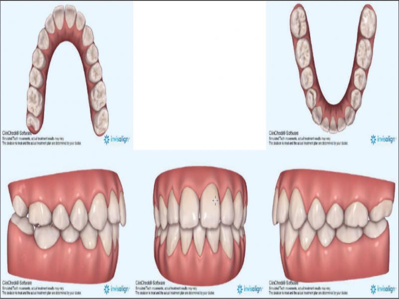
Figure 4.
Invisalign® predicted treatment outcome.
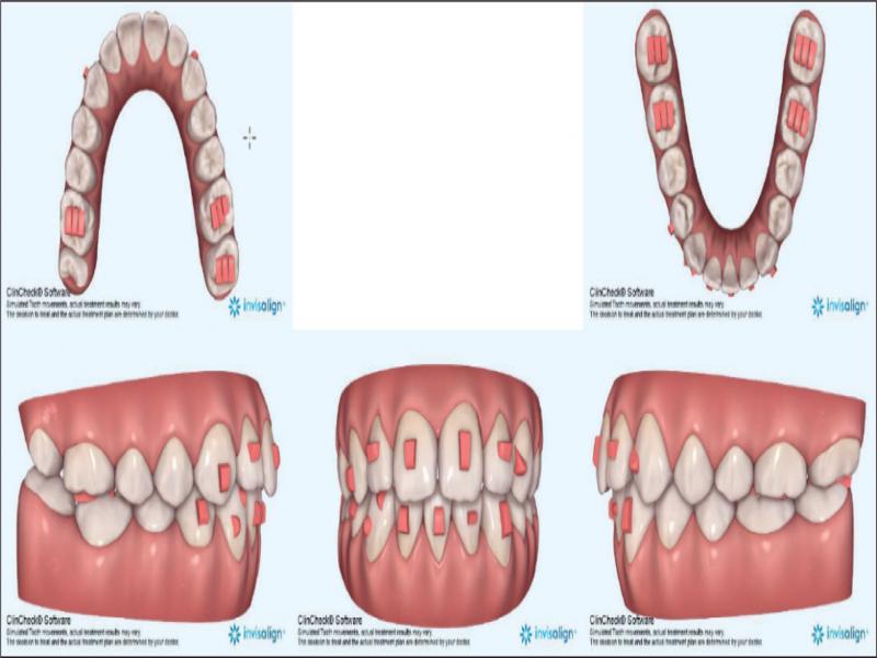
Figure 5.
Invisalign® predicted outcome showing attachments.
| Date | Stage |
|---|---|
| 16/5/2018 | Aligners inserted – 29 aligners in initial treatment |
| 17/4/2019 | 32, 12, 22 not tracking at aligner 29 Attachments removed and scan for Additional Aligners |
| 15/5/2019 | Bonded new attachments. 26 aligners in Additional Aligner order Commence cross-elastics on left side at Aligner 1 |
| 18/7/2019 | Aligner 6 12, 22 not tracking. Commence bootstrap elastics |
| 16/10/2019 | Aligner 12 31, 42 not tracking vertically so attachments removed to allow passive vertical settling, Commence powerarm mechanics for 32 |
| 30/10/2019 | Prefinishing OPG and Lateral Ceph ordered |
| 26/2/2020 | Completed aligners; removed bonded attachments, bonded retainers |
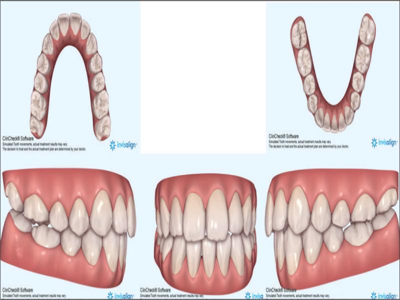
Figure 6.
Actual treatment outcome.
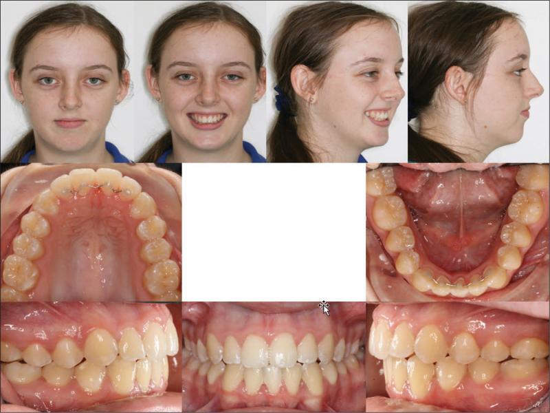
Figure 7.
Final photographs 26.02.2020.
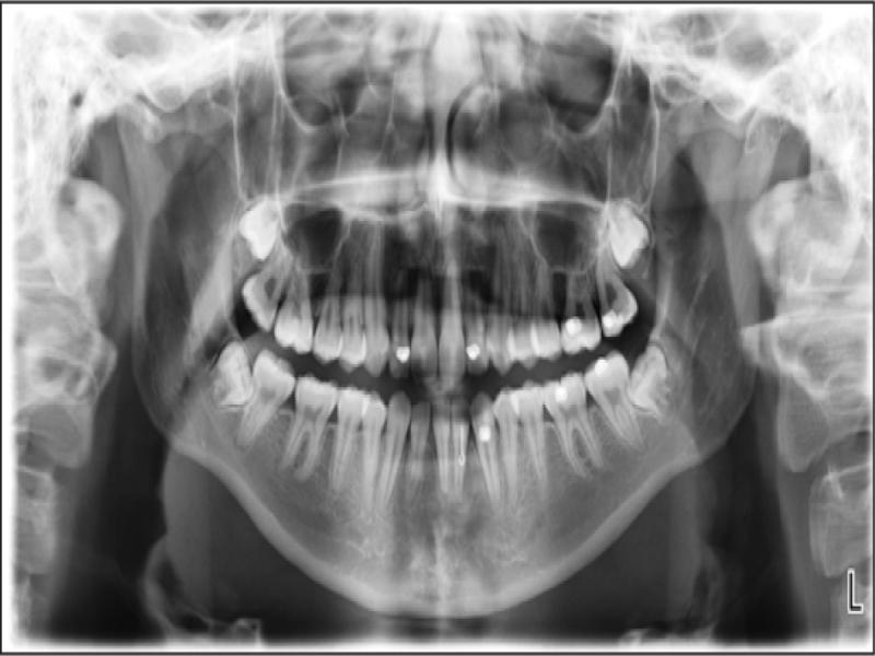
Figure 8.
Prefinishing OPG 30.10.19.
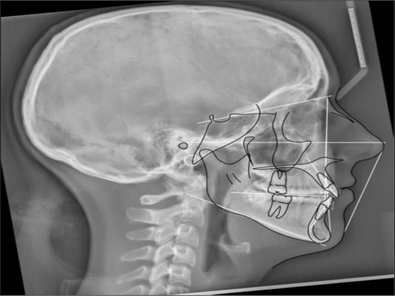
Figure 9.
Prefinishing lat cephalogram 30.10.19.
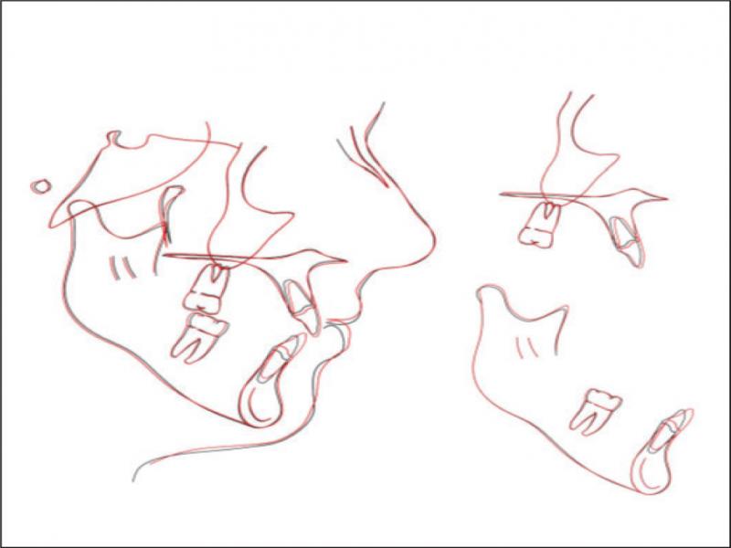
Figure 10.
Cephalometric superimposition (Bjork) pretreatment vs prefinish.
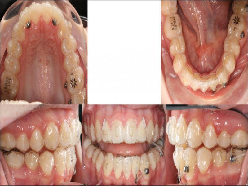
Figure 11.
Auxiliary treatment during aligner treatment.
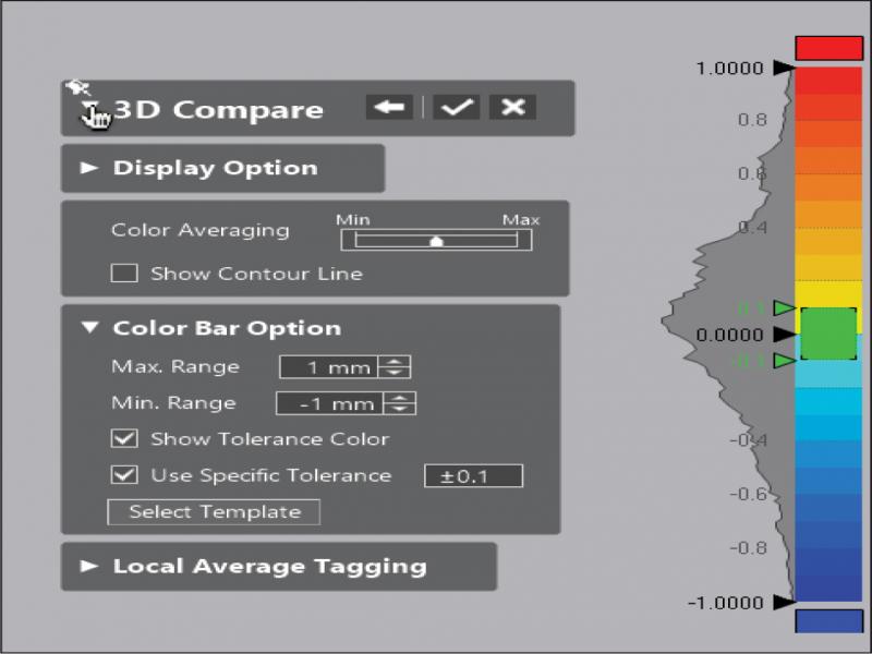
Figure 12.
Heat map definition: Scale is 1 mm either side of ideal, in 0.1 mm gradations. Deviations that are moving out of or away from the reference model are towards the red end of the spectrum. Deviations that are moving into the reference model are towards the blue end of the spectrum. All deviations greater than 1 mm are recorded as either solid red or solid blue.
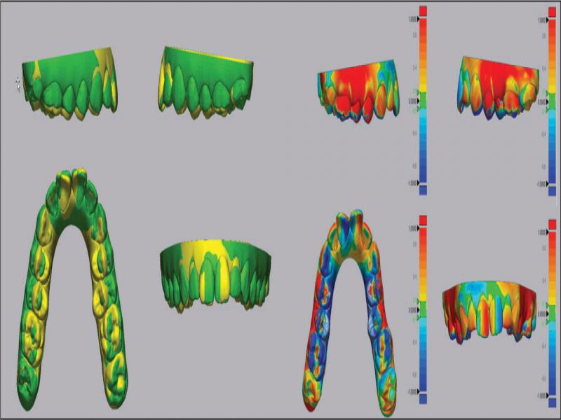
Figure 13a.
Maxillary arch .stl superimpositions. Yellow – Initial .stl, Green – Final Outcome .stl and Heat Maps (Deviation is 1 mm either side of Nil, reference model is the Initial .stl).
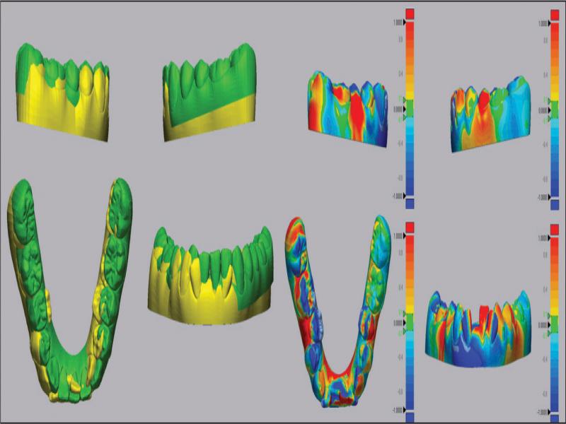
Figure 13b.
Mandibular arch .stl superimpositions. Yellow – Initial .stl, Green – Final Outcome .stl and Heat Maps (scale is 1 mm, reference model is the Initial .stl).
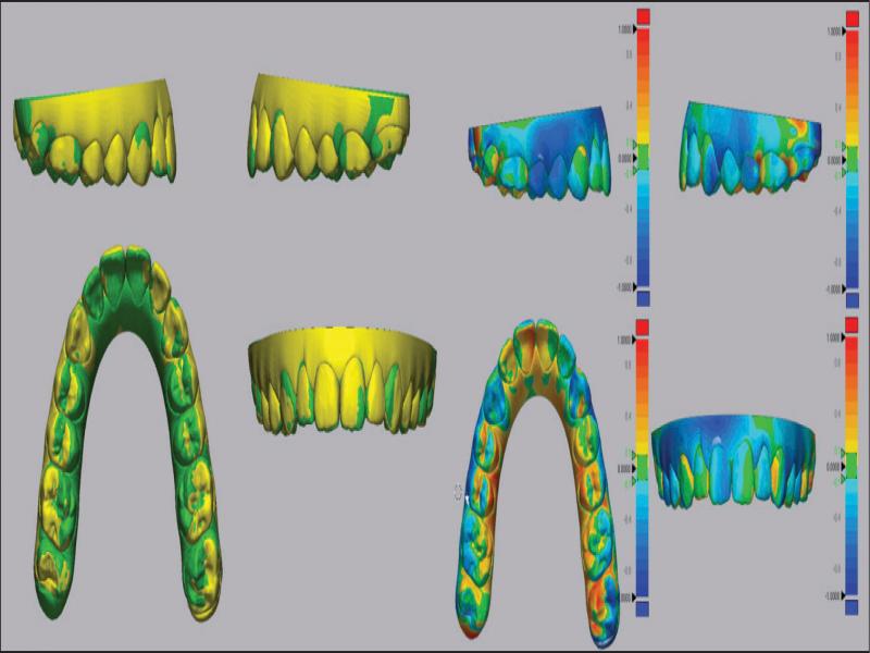
Figure 14a.
Maxillary arch .stl superimpositions. Yellow – Predicted Outcome .stl, Green – Final Outcome .stl and Heat Maps (scale is 1 mm, reference model is the Predicted Outcome .stl).
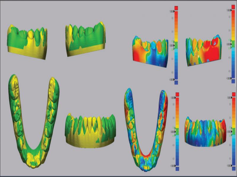
Figure 14b.
Mandibular arch .stl superimpositions. Yellow – Predicted Outcome .stl, Green – Final Outcome .stl and Heat Maps (scale is 1 mm, reference model is the Predicted Outcome .stl).