Introduction
Following the first report of treatment of a lower incisor extraction case using Invisalign® appliances in 2002, 1 a 12 consecutively-treated patient case series 2 and a three-patient case report 3 were published by 2016. From these publications, it appears that the Invisalign® appliance is able to produce satisfactory clinical outcomes in the management of lower incisor extraction cases.
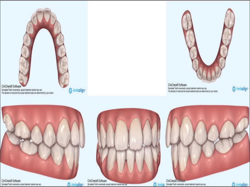
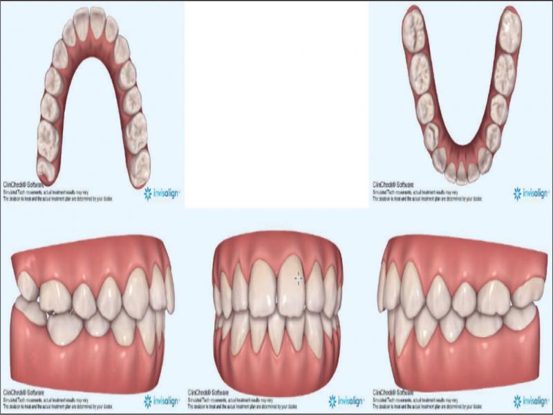
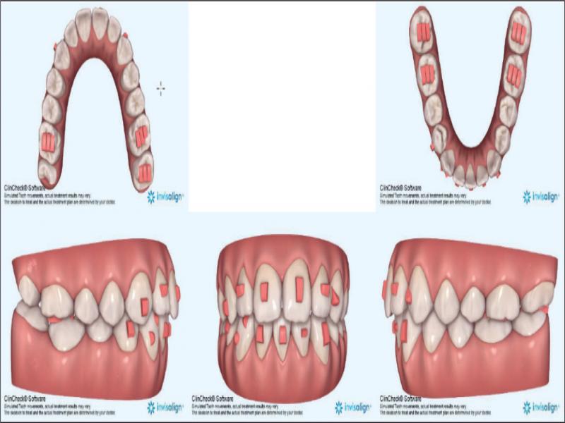
It is essential for the successful treatment of any lower incisor extraction case to have an appropriate diagnosis accompanied by a well-designed digital treatment plan (ClinCheck®) if Invisalign® aligners are to be used. The treating clinician may use ClinCheck® software to perform a virtual diagnostic wax-up of a case to facilitate the treatment planning process. Once an extraction decision is confirmed, the software may be used to determine the appropriate attachments, treatment sequence, movement speed of the teeth and the three-dimensional visualisation of the case at any treatment stage. Once the plan is accepted and the aligners manufactured, the clinician needs to closely monitor the case, employ auxiliary mechanics when necessary, and order additional aligners if required.
A decision to extract a lower incisor in orthodontic treatment is based on factors including:
Minimal upper incisor and moderate to severe lower incisor crowding.
Bilateral Class I or mild Class III molar relationships.
An acceptable soft tissue profile.
Minimal overbite and overjet.
Minimal growth potential.
Mandibular tooth-size excess (Bolton discrepancy) present.
Poor prognosis of a lower incisor due to pathology such as gingival recession, severe wear or fracture.
When potential relapse is deemed significant (severe rotations or displacements).
Patient preference for lower arch only treatment may require lower incisor extraction to provide a positive overjet. 3-7
In relation to post-treatment stability, lower incisor extraction cases have been considered to be significantly more successful than alternative treatment options. 8 However, caution must be taken to minimise potential negative outcomes, including increases in overjet and overbite and gingival problems related to recession and open interproximal embrasures. 7
There is limited evidence to support claims that clear aligners can effectively treat open bite problems in high mandibular plane angle cases without extruding posterior teeth. 9,10 It is theorised that intrusive forces from the occlusion and pressing on the clear aligners that are interposed occlusally may be the mechanism for vertical control during treatment.
Transverse expansion using aligners has been demonstrated to fall short of the predicted outcome, implying either the need to overprescribe the expansion in order to achieve the desired clinical result or employ auxiliaries such as intermaxillary elastics to augment the aligner forces. 11-13
Diagnosis
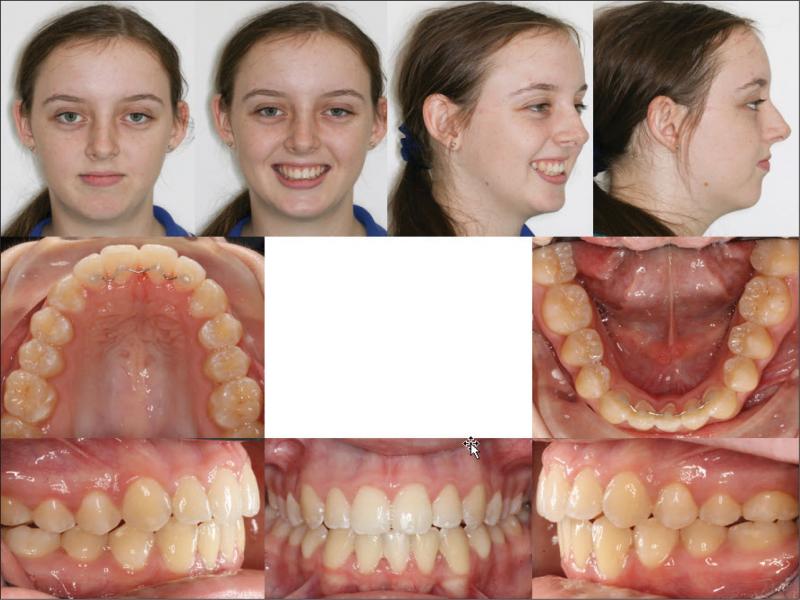
A 14-year 7-month-old Caucasian female presented to the University of Queensland postgraduate orthodontic clinic concerned about the crowding of her teeth (Figure 1). The facial appearance was asymmetric, and the profile straight but with an increased lower anterior facial height. The lips were competent, thin, and retrusive. An examination of the smile revealed consonance of the incisal edges with the lower lip, acceptable upper incisor display, coincidence of the upper dental and facial midlines, and an increased buccal corridor width and reduced posterior tooth display. Her dental malocclusion was characterised by a mild Class III buccal segment relationship with moderate to severe upper and lower crowding, an anterior open bite of 2 mm, an overjet of 6.8 mm, and tapered, lingually inclined buccal segments. All teeth were present, and a Bolton’s mandibular excess of 1.4 mm (3-3) and 2.9 mm (6-6) was noted.
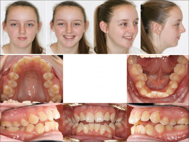
Figure 1.
Pretreatment photographs.
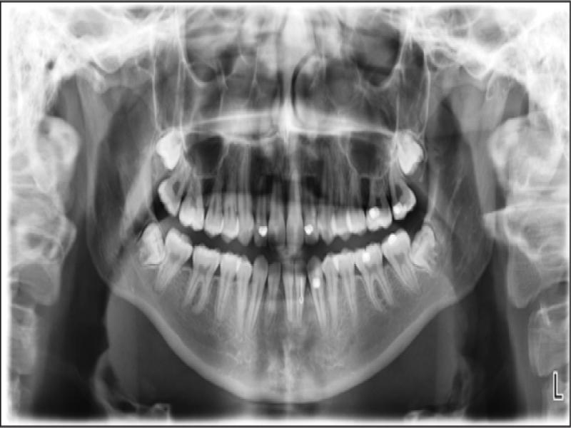
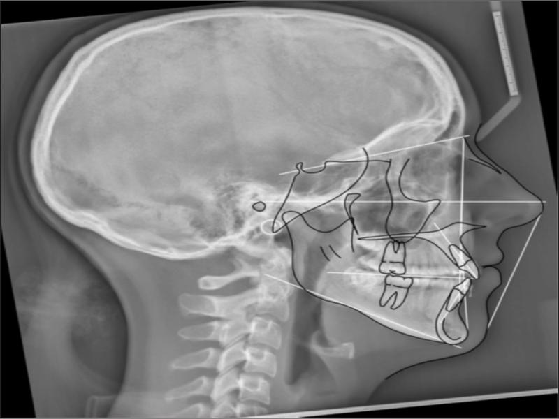
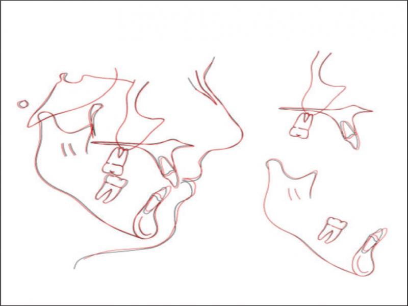
A radiographic examination was unremarkable on the OPG (Figure 2). Cephalometrically (Figure 3, Table I) a Class III skeletal relationship was evident, with a Wits discrepancy of -2.1 mm (due primarily to maxillary retrusion – SNA 72.6°). However, the Class III was camouflaged by the significant vertical disproportion, highlighted by an increased mandibular plane angle (28.6°), reduced facial axis angle (83.2°), reduced Jarabak’s ratio (56.5%), and increased lower anterior facial height (53.4%), all of which indicated a dolichofacial pattern. The maxillary retrusion, combined with a mandibular downward and backward rotation, resulted in a bimaxillary retrusive skeletal relationship (SNB 72.6°). The upper incisors were at an acceptable angulation relative to the palatal plane (112°) and the anteroposterior position relative to Na-Vert. The lower incisors were at an acceptable position relative to APo but were retroclined relative to the mandibular plane (80.7°) according to mesofacial norms.
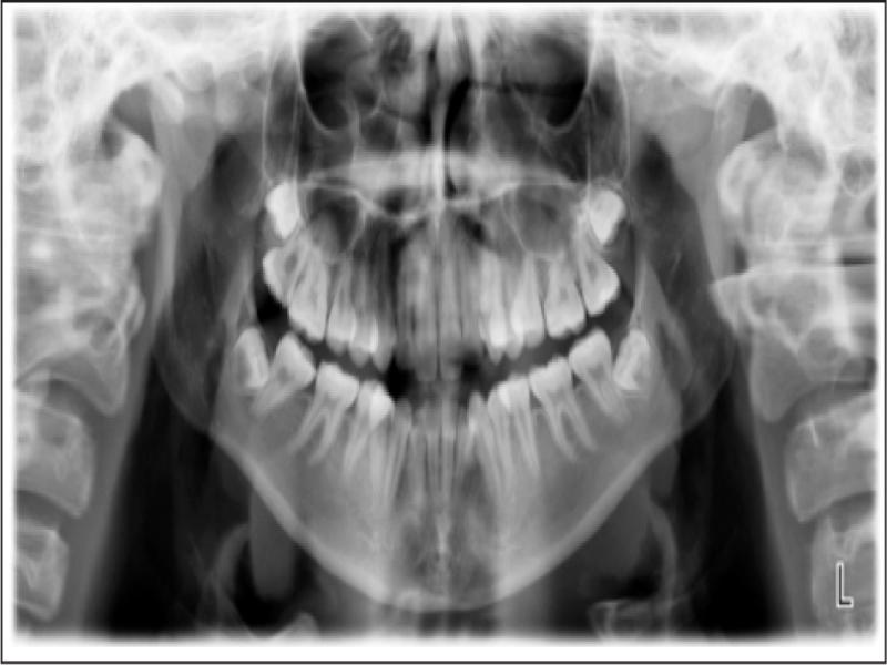
Figure 2.
Pretreatment OPG.
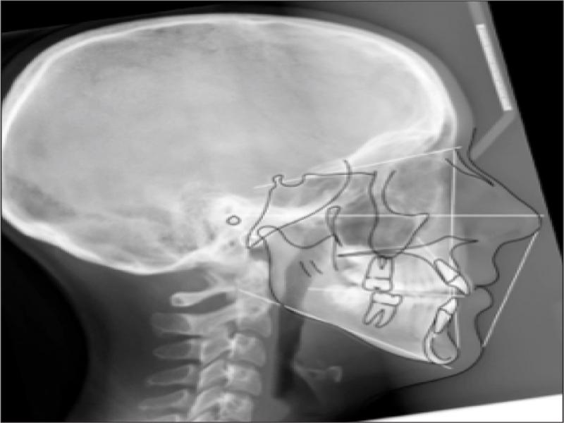
Figure 3.
Pretreatment lateral cephalogram.
Table I.
Cephalometric measurements – initial and prefinishing.
| Measurement | Standard | 18/8/2017 | 30/10/2019 | Change | ||
|---|---|---|---|---|---|---|
| SKELETAL | Maxilla | SNA | 82 ± 2° | 72.6 | 72.8 | 0.2 |
| A Point Convexity | 3 ± 2 mm | -2 | -2.1 | -0.1 | ||
| NaVert – A point | 1 mm | -1.3 | -1.2 | 0.1 | ||
| Mandible | SNB | 80 ± 2° | 72.6 | 73.0 | 0.4 | |
| Facial Angle | 87 ± 3° | 90.7 | 90.9 | 0.2 | ||
| NaVert – Po | -8 to -6 mm -2 to +4 mm | 1.3 | 1.8 | 0.5 | ||
| Basal Arch Relationship | ANB | 2 ± 2° | 0 | -0.2 | -0.2 | |
| WITS | F: 0mm | -2.1 | -1.9 | 0.2 | ||
| Vertical | Md Plane Angle | 26 ± 4° | 28.6 | 27.7 | -0.9 | |
| LFH Angle | 47 ± 4° | 49.8 | 48.3 | -1.5 | ||
| Md Arc | 26 ± 4° | 35.2 | 37.6 | 2.4 | ||
| Facial Axis Angle | 90 ± 3° | 83.2 | 83.7 | 0.5 | ||
| Growth | CVMI | 1-6 | 6 | 6 | 0 | |
| DENTAL | Upper Incisors | UI to FH | 110° | 120.9 | 118.9 | -2 |
| UI to Pal Plane | 110° | 112 | 110.9 | -1.1 | ||
| UI to Na Vert | 5 mm | 6.8 | 7.6 | 0.8 | ||
| Lower Incisors | LI to Md Plane | 92.5° | 80.7 | 86.8 | 6.1 | |
| LI to Apo | 1 ± 2 mm | 0.2 | 2.4 | 2.2 | ||
| LI to Apo | 22 ± 4° | 22.1 | 27.7 | 5.6 | ||
| Interincisal | UI to LI | 130 ± 2° | 129.7 | 126.6 | -3.1 | |
| FACE DEPTH | U Face G’-Sn | 50% | 46.6 | 45.8 | -0.8 | |
| L Face Sn-Me’ | 50% | 53.4 | 54.2 | 0.8 | ||
| U Lip Sn-Stom | 33% | 30.8 | 27.7 | -3.1 | ||
| L Lip Stom-Me’ | 67% | 69.2 | 72.3 | 3.1 | ||
| PFH:AFH | 59 to 63% | 56.5 | 57.3 | 0.8 | ||
| SOFT ISSUE | L Lip – E Line | -2 ± 2 mm | -5.1 | -4.0 | 1.1 | |
| SnVert – U Lip | 1 mm | -1.7 | -0.9 | 0.8 | ||
| SnVert – L Lip | -1 mm | -4.1 | -2.0 | 2.1 | ||
| SnVert – Po’ | -3 mm | -7.6 | -8.0 | -0.4 | ||
| Date | Stage |
|---|---|
| 16/5/2018 | Aligners inserted – 29 aligners in initial treatment |
| 17/4/2019 | 32, 12, 22 not tracking at aligner 29 Attachments removed and scan for Additional Aligners |
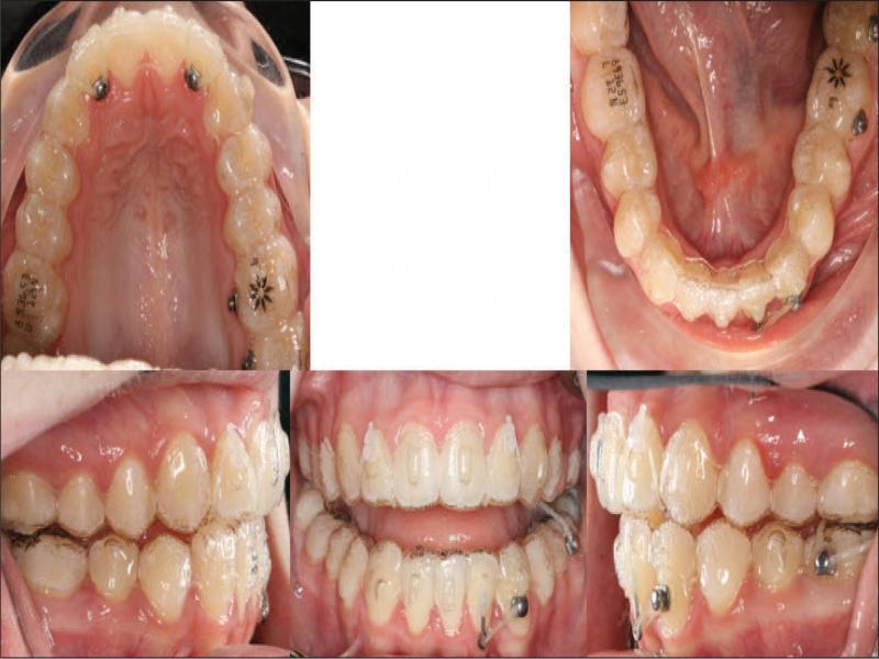
| 15/5/2019 | Bonded new attachments. 26 aligners in Additional Aligner order Commence cross-elastics on left side at Aligner 1 |
| 18/7/2019 | Aligner 6 12, 22 not tracking. Commence bootstrap elastics |
| 16/10/2019 | Aligner 12 31, 42 not tracking vertically so attachments removed to allow passive vertical settling, Commence powerarm mechanics for 32 |
| 30/10/2019 | Prefinishing OPG and Lateral Ceph ordered |
| 26/2/2020 | Completed aligners; removed bonded attachments, bonded retainers |