
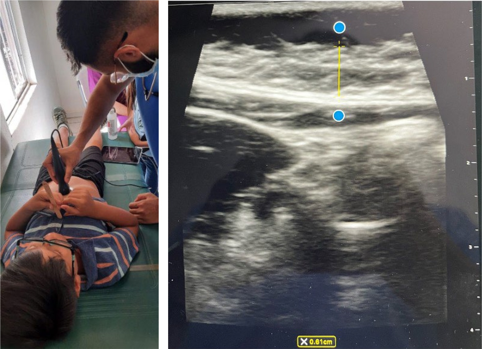
Figure 1
Obtaining ultrasound images
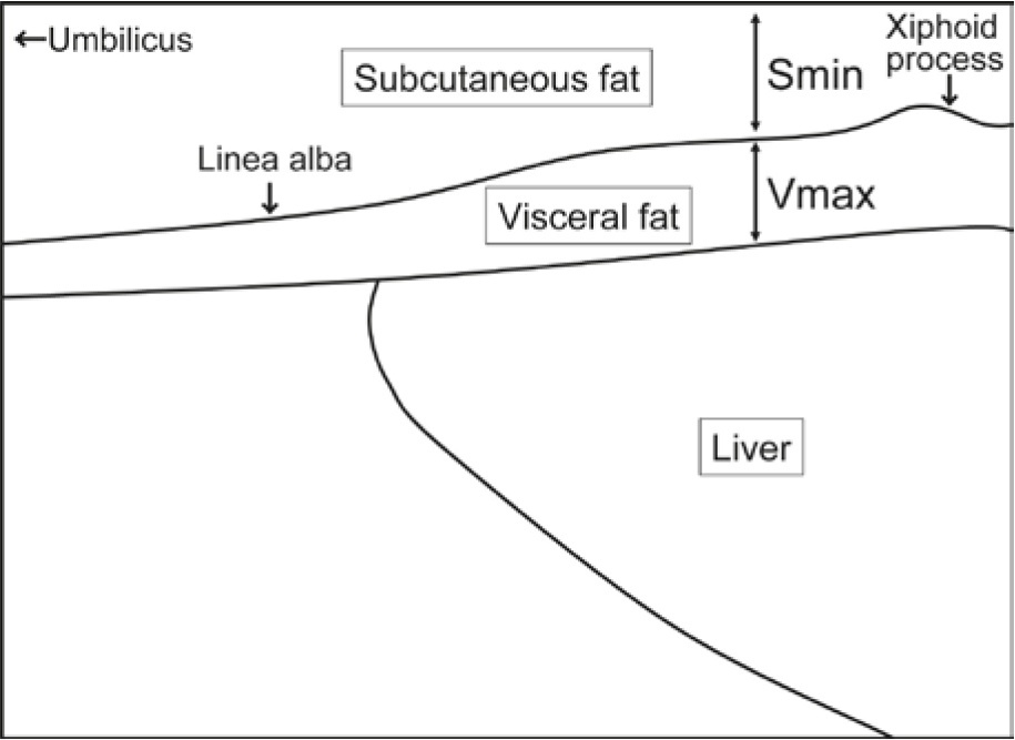
Figure 2
Diagram shows landmarks used for the measurement of abdominal fat thickness
Table 1
Participant characteristics (N = 16)
| Characteristic | Value |
|---|---|
| N | 16 |
| Age (years), mean (SD) | 9.8 (2.7) |
| Sex: Female, n (%) | 7 (44%) |
| Sex: Male, n (%) | 9 (56%) |
| BMI-for-age percentile, mean (SD) | 81.2 (20.1) |
| BMI-for-age ≥85th percentile, n (%) | 10 (63%) |
| Waist circumference (cm), mean (SD) | 68.1 (13.1) |
| Visceral fat thickness (mm), mean (SD) | 8.3 (4.9) |
| Visceral fat thickness (mm), range | 3.1–18.8 |
Table 2
Correlations of visceral fat thickness with anthropometrics (N = 16)
| Variable | r | 95% CI | p-value |
|---|---|---|---|
| BMI-for-age percentile | 0.61 | 0.16–0.85 | 0.012 |
| Waist circumference (cm) | 0.81 | 0.51–0.93 | <0.001 |
| Age (years) | 0.23 | –0.30–0.65 | 0.383 |
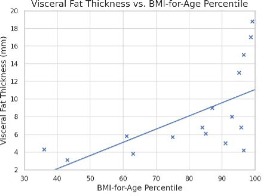
Figure 3
VFT vs. BMI-for-Age Percentile
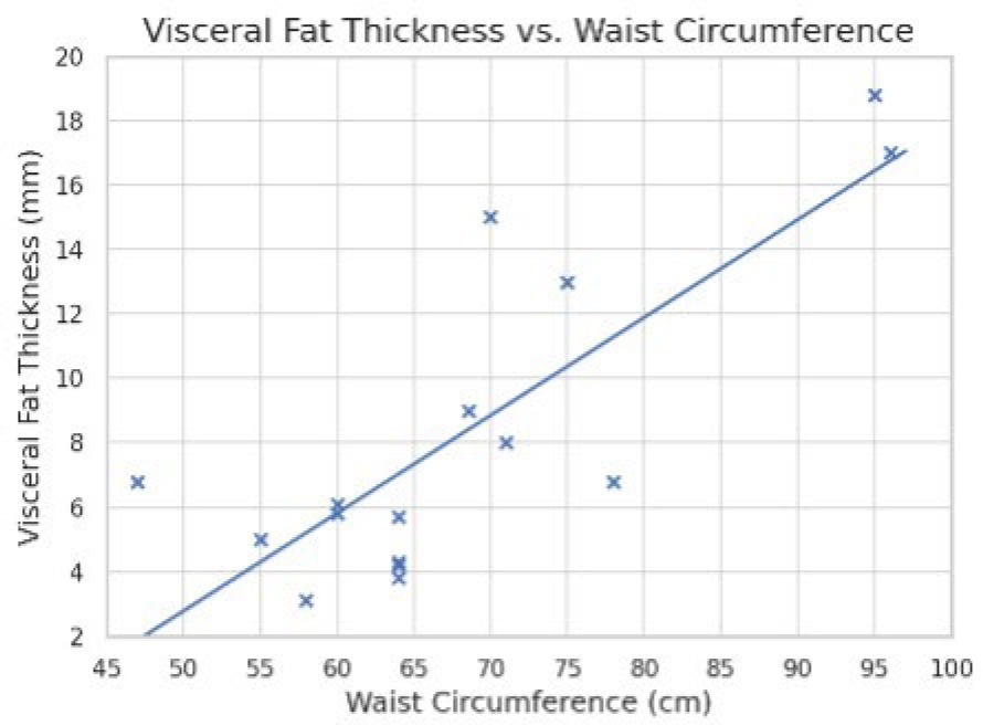
Figure 4
VFT vs. Waist Circumference
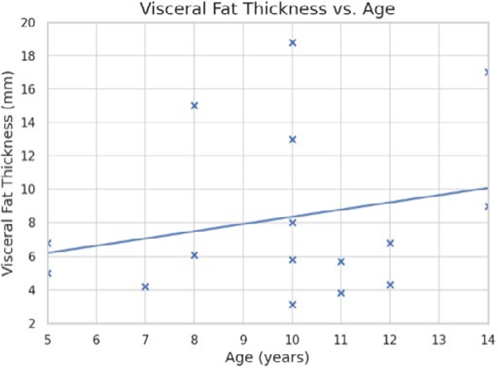
Figure 5
VFT vs. Age
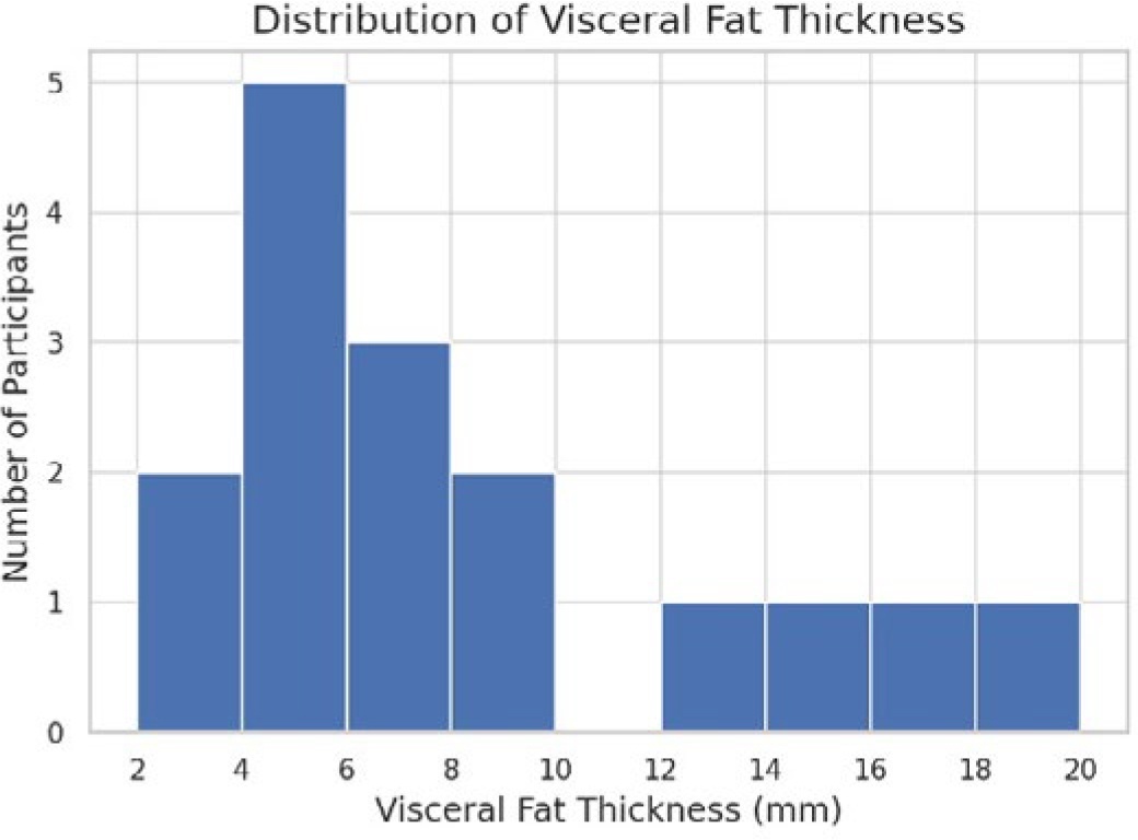
Figure 6
Range of Visceral Fat Thickness in study participants