
Introduction
Childhood obesity has become a critical global health concern, with early manifestations of metabolic and cardiovascular diseases increasingly observed in younger populations. In Mexico, where our study took place, recent data from the 2020-2022 ENSANUT survey revealed that among school-age children (5-11 years), 18.1% were obese and 19.2% overweight. Among adolescents (12–19 years), the prevalence was 17.2% for obesity and 23.9% for overweight, with notably higher rates documented in urban areas and among males. These figures indicate a significant rise in obesity, especially among boys and adolescents, compared to previous years.1 This places them at elevated risk for metabolic syndrome, insulin resistance, dyslipidemia, hypertension, cardiovascular disease, and nonalcoholic fatty liver disease (NAFLD).2-5
Traditional screening tools, such as body mass index (BMI) and waist circumference (WC), remain widely utilized, but inadequately capture variations in visceral fat, a compartment more strongly associated with cardiometabolic risk.6,7 Bedside ultrasound provides a practical, accurate, and low-cost method for directly measuring visceral fat thickness (VFT). This measure also correlates strongly with gold-standard, imaging techniques such as MRI and CT.7,8 This study investigates the potential role of ultrasound-measured VFT in improving metabolic risk screening among underserved pediatric populations, overcoming limitations associated with conventional anthropometric assessments2,7.
Limitations of BMI in Pediatric Metabolic Risk Assessment
Although BMI is a widely utilized pediatric obesity screening tool, it only indirectly reflects adiposity by failing to differentiate between lean and fat mass. Gallagher, et al. demonstrated significant variability in percent body fat at identical BMI values, notably influenced by age and sex, with females and older individuals having substantially higher fat percentages.7 Additionally, BMI measurements may be confounded during pubertal growth, as variations in the timing and progression of puberty can distort BMI interpretations, particularly affecting children with delayed or accelerated maturation.9 These limitations highlight the need for direct adiposity assessment techniques, such as ultrasound-based VFT measurements, to accurately evaluate metabolic risk in pediatric populations.
Bedside Ultrasound-Measured VFT as an Alternative
In contrast to traditional anthropometric methods such as BMI and WC, ultrasound provides a direct measurement of visceral adiposity via VFT. VFT has demonstrated stronger associations with metabolic dysfunction and cardiovascular risk factors in pediatric populations, making it potentially superior for assessing cardiometabolic risk related to fat distribution compared to overall body size alone.2,3,5
Accuracy, Cost, and Feasibility
MRI remains the gold standard for visceral adipose tissue assessment; however, its high cost and limited accessibility restrict widespread use, especially in pediatric populations and low-resource settings. Ultrasound-based VFT measurements provide a practical, cost-effective alternative with demonstrated accuracy. In pediatric studies, ultrasound-measured VFT correlated moderately well with MRI-derived visceral fat volume (r = 0.57), validating its reliability as a non-invasive proxy for visceral adiposity.7 Similarly, in adult populations with type 2 diabetes, ultrasound-derived VFT has shown a strong correlation with CT-derived visceral fat area (r = 0.858), underscoring its cross-modality reliability.8
Potential for Scaled Implementation
Given its portability and ease of use, ultrasound-based VFT measurement holds significant potential for routine integration into clinical care or school-based health screening programs in Mexico. Task-shifting this skill to trained community health workers could further enhance accessibility and enable early identification and monitoring of visceral adiposity, potentially preventing future metabolic complications. For example, in rural Uganda, implementation of a structured, two-year, ultrasound training program for non-physician emergency providers resulted in more than 2,100 ultrasound exams, surpassing the utilization of traditional radiology services.1 This successful example highlights the feasibility and effectiveness of expanding ultrasound skills to community-level providers in resource-limited settings.
Materials and Methods
Study Design and Ethical Compliance
A retrospective analysis was conducted using de-identified data from 16 pediatric patients (ages 5–14 years) seen at community health centers in Puerto Peñasco and Sonoyta, Mexico. Children were scanned during routine primary-care or urgent-care visits while awaiting the clinician; each scan added ≈2-3 minutes and, therefore, minimized disruptions to routine care, an important consideration in resource-limited settings.10 The study was classified as a non-research, quality improvement activity under 45 CFR 46.102(l) and was approved by the University of Arizona College of Medicine – Phoenix Institutional Review Board (STUDY #2023-123). Written parent/guardian consent and child assent (≥7 y) were obtained, and all data were fully de-identified prior to analysis as part of this quality-improvement ultrasound-screening initiative.
Ultrasound Protocol and Operator Training
VFT was assessed using a Butterfly iQ+ ultrasound with a 7.5 MHz linear probe. Ultrasound scans were performed longitudinally along the linea alba from the xiphoid process to the umbilicus, with the transducer lightly touching the skin to avoid compression of fat layers, as described by Hamagawa, et al.3 This approach specifically measures the maximum preperitoneal visceral fat thickness (Vmax), representing preperitoneal visceral fat, distinct from the overlying subcutaneous fat (Figure 2). The preperitoneal region was selected due to its superficial location, clear visibility on ultrasound, and superior reproducibility compared to other ultrasound methods measuring intra-abdominal visceral fat thickness.4 Additionally, Vmax has been independently associated with cardiometabolic risks such as coronary artery disease (CAD), further validating its clinical utility.3
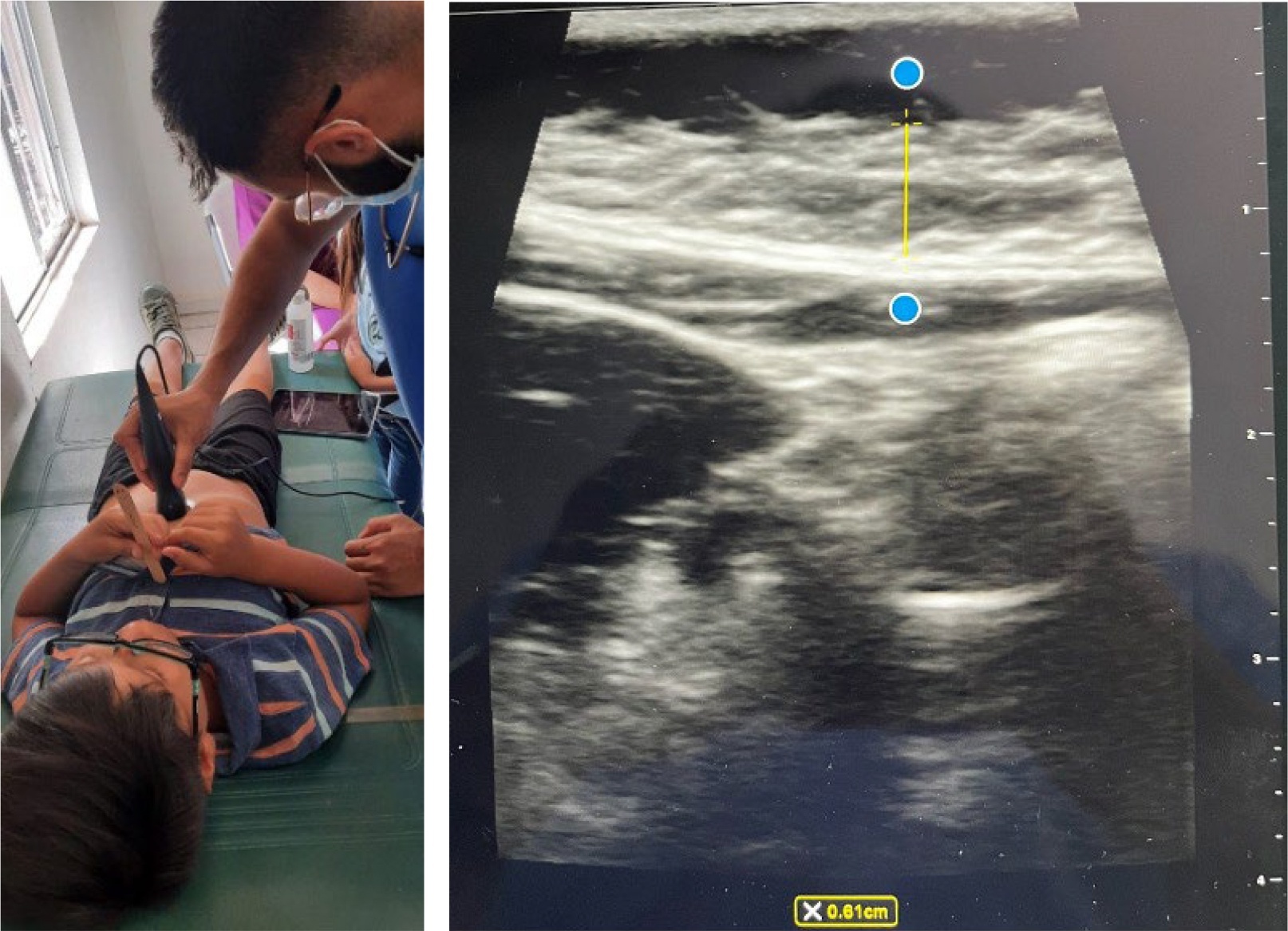
Figure 1
Obtaining ultrasound images
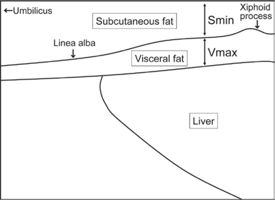
Figure 2
Diagram shows landmarks used for the measurement of abdominal fat thickness
Scanning was standardized with consistent device settings, and images were obtained at end-expiration to ensure uniform measurements. All scans were obtained by a single medical student after a 2-hour, hands-on, training session that included five practice patients. This shows that brief instruction is sufficient for novice operators, and that any medical student, advanced practice provider, or physician can learn the protocol. During training, the operator was taught to freeze the image only after the bright echogenic linea alba (anterior landmark) and the peritoneal line just beneath the preperitoneal fat (posterior landmark) were both clearly visible, ensuring that the calipers captured the preperitoneal, visceral fat layer rather than overlying subcutaneous fat, Figure 1. An informal, repeat-measurement check on the first five participants showed a maximum within-operator difference of <2 mm, providing practical assurance of consistency for this pilot phase. Each scan required approximately 3 minutes from probe placement to final measurement, with thickness recorded directly in the Butterfly iQ app at the bedside. Images could be exported for specialist review, but bedside measurements were adequate for this pilot. The Butterfly iQ+ that was used costs about US $2,699 as a one-time purchase, with an optional US $299 annual software subscription. Comparable handheld ultrasound devices range from US $1,500 to US $5,000.11
Anthropometric Data Collection
Alongside VFT, standard anthropometric data were collected:
BMI-for-age percentile (BMI percentile): Weight (kg)/height² (m²) converted to age- and sex-specific percentiles using CDC growth charts.12
WC: Measured at the midpoint between the iliac crest and the lowest rib, according to standardized methods described by Gallagher, et al.6
Blood Pressure (BP): Obtained using an automated oscillometric sphygmomanometer following established pediatric hypertension guidelines by Flynn, et al.13
Statistical Analysis
Pearson correlation coefficients (r) were calculated to evaluate relationships between VFT, BMI percentile, WC, and age, with 95% confidence intervals (CIs) reported. Due to the limited sample size (N = 16), sensitivity analyses were performed to assess and address potential measurement variability.
Results
Table 1
Participant characteristics (N = 16)
| Characteristic | Value |
|---|---|
| N | 16 |
| Age (years), mean (SD) | 9.8 (2.7) |
| Sex: Female, n (%) | 7 (44%) |
| Sex: Male, n (%) | 9 (56%) |
| BMI-for-age percentile, mean (SD) | 81.2 (20.1) |
| BMI-for-age ≥85th percentile, n (%) | 10 (63%) |
| Waist circumference (cm), mean (SD) | 68.1 (13.1) |
| Visceral fat thickness (mm), mean (SD) | 8.3 (4.9) |
| Visceral fat thickness (mm), range | 3.1–18.8 |
Table 2
Correlations of visceral fat thickness with anthropometrics (N = 16)
| Variable | r | 95% CI | p-value |
|---|---|---|---|
| BMI-for-age percentile | 0.61 | 0.16–0.85 | 0.012 |
| Waist circumference (cm) | 0.81 | 0.51–0.93 | <0.001 |
| Age (years) | 0.23 | –0.30–0.65 | 0.383 |
Visceral Fat Thickness, BMI Percentile, and Waist Circumference
The mean BMI-%ile was 81.2 (SD 20.1), with a range of 36.0 to 99.1. A total of 63% (10 of 16) were above the 85th percentile for age, consistent with overweight or obesity by CDC criteria.12
Correlation analysis revealed a significant positive association between VFT and BMI percentile (r = 0.61, 95% CI: 0.16–0.85, p = 0.012), as shown in Figure 3. This finding supports the utility of BMI percentile as a general measure for pediatric adiposity but also highlights its limitation in distinguishing metabolically active visceral fat from subcutaneous fat.6 Similarly, VFT was strongly correlated with WC (r = 0.81, 95% CI: 0.51–0.93, p < 0.001), as shown in Figure 4, reinforcing WC as a marker of central adiposity. However, just like BMI percentile, WC reflects total abdominal girth and does not differentiate between fat compartments.
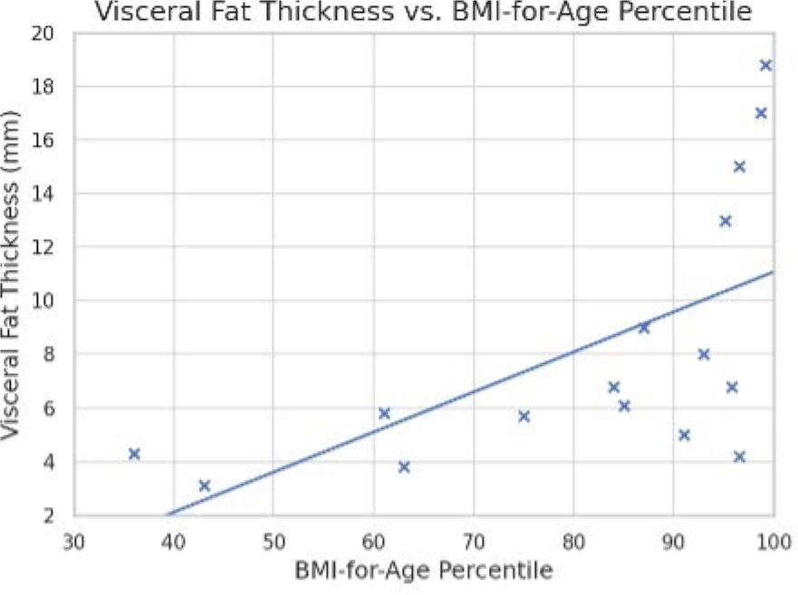
Figure 3
VFT vs. BMI-for-Age Percentile
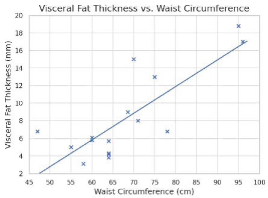
Figure 4
VFT vs. Waist Circumference
Age and Variability in Visceral Fat Thickness
A weak correlation was observed between VFT and age (r = 0.23, 95% CI –0.30 to 0.65, p = 0.383). VFT values ranged from 3.1 mm to 18.8 mm. The mean BMI-for-age percentile was 81.2 (± 20.1), and the mean age was 9.8 (± 2.7) years.
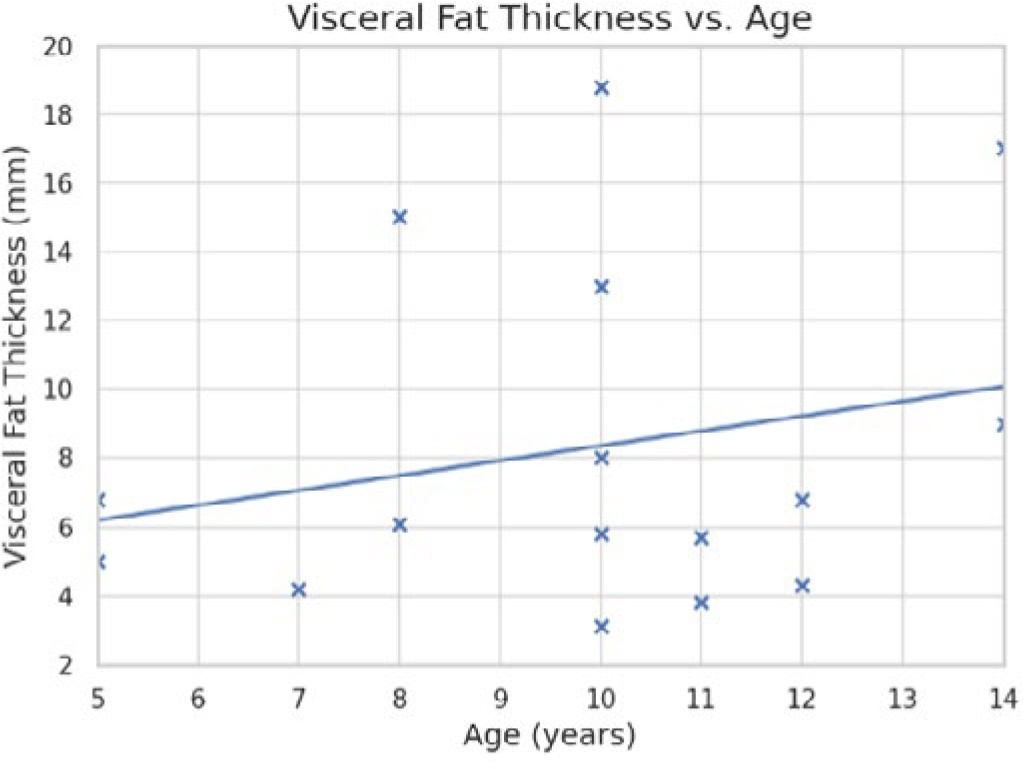
Figure 5
VFT vs. Age
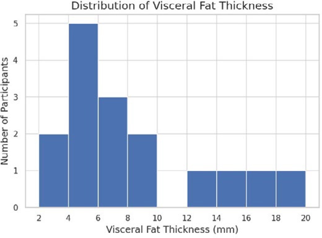
Figure 6
Range of Visceral Fat Thickness in study participants
Discussion
Implications of Ultrasound-Measured VFT
Our findings indicate that ultrasound-based VFT correlates well with traditional anthropometric measures like BMI and WC but may offer additional clinical value by specifically assessing visceral adiposity, a key contributor to cardiometabolic risk.2-5 The variability observed in VFT despite similar BMI percentiles suggests that traditional anthropometrics may overlook differences in metabolically active fat. Due to the strong association between visceral fat and metabolic and cardiovascular diseases, ultrasound-based VFT measurement has potential as a valuable clinical tool, particularly in settings where advanced imaging like MRI or biochemical testing is impractical or cost prohibitive.
In pediatric populations, ultrasound-assessed preperitoneal fat has demonstrated clinical relevance beyond standard anthropometrics. Hacihamdioglu, et al. observed that preperitoneal fat was the sole independent predictor of arterial stiffness in obese adolescents, even after adjusting for age, BMI, and blood pressure.5 Similarly, Jung, et al. established a VFT threshold of 34.3 mm as a predictor of NAFLD in children, with a sensitivity of 84.6% and specificity of 71.2%, closely linked to elevated ALT levels and insulin resistance.2 Such findings underscore VFT’s utility as an early indicator of pediatric metabolic dysfunction.
In adult cohorts, VFT measured via ultrasound has shown robust predictive value for cardiovascular diseases. Hamagawa reported significantly greater VFT among patients with CAD compared to healthy controls (8.8 ± 3.6 mm vs. 6.4 ± 2.8 mm), establishing a VFT cutoff of ≥6.9 mm as an independent predictor of CAD severity.3 Likewise, Kim, et al. demonstrated that ultrasound-derived VFT independently predicted metabolic syndrome and cardiovascular risk beyond BMI and WC, highlighting its potential as a low-cost, noninvasive screening tool.4 Additionally, Oh, et al., introduced the abdominal visceral-to-thigh muscle thickness ratio (AVTMR), which independently predicted carotid intima-media thickness, emphasizing the broader clinical utility of ultrasound-based adiposity measurements.8 These collective insights reinforce the potential role of VFT screening as an accessible and effective means of identifying early cardiometabolic and vascular risk, particularly impactful when coupled with culturally appropriate dietary and physical activity interventions.
Strengths and Limitations
Key strengths of this study include the use of a direct, validated, ultrasound-based method (Vmax) for VFT measurement. It also affords minimal interference with clinical workflows, and adherence to a standardized pediatric ultrasound protocol previously validated by Hamagawa, et al.3 The successful integration of VFT assessments into routine clinical visits demonstrates feasibility, particularly in resource-constrained community settings.10 In simple terms, acquiring the ultrasound images only takes roughly 2 minutes of time in a primary care visit setting. Additionally, once an ultrasound device is available, there is minimal cost involved to obtain VFT images. Many portable ultrasound devices will also continue to work locally without ongoing subscription fees. The ability to obtain a device through donation or grant funding is feasible. This is in contrast with other metabolic studies which require reference laboratory testing, cost prohibitive in many resource limited settings. Even wholesale prices for point-of-care glycosylated hemoglobin testing cost about $8 per test at the time of this study.
To be complete, several limitations must be noted. Foremost is the small sample size (N = 16), limiting generalizability and statistical power. This pilot study was not designed to evaluate feasibility or cost-effectiveness; follow-up with larger cohort sizes will provide enhanced capacity for data collection and analysis. The cross-sectional design limits causal inference and prediction of future risk. Furthermore, the absence of metabolic biomarkers, such as fasting glucose, insulin, or liver enzymes (ALT), constrains our ability to thoroughly assess the metabolic implications of elevated VFT in isolation. Additionally, lifestyle variables, including dietary patterns, physical activity levels, and genetic predispositions (e.g., family history), were not controlled for. This may further influence adiposity distribution independent of VFT measurements. A single, medical-student operator obtained scans; intra- and inter-operator reliability were not estimated. We used a 7.5-MHz linear probe and did not compare across transducer types; generalizability of measurements across different probes and vendors was not evaluated.
Future Directions
To extend and validate the clinical utility of ultrasound-based VFT, several directions are recommended:
Multicenter Validation: Conduct larger, multicenter studies across various clinics in Mexico and comparable international settings to establish standardized pediatric VFT cutoffs applicable across diverse populations.
Longitudinal Monitoring: Investigate longitudinal changes in VFT throughout key developmental stages, particularly during puberty, to identify critical periods for visceral fat accumulation and associated metabolic risks.
Biochemical Correlations: Incorporate comprehensive laboratory markers, such as fasting insulin, lipid profiles, and liver function tests (ALT) to better elucidate the metabolic and cardiovascular risks associated with increased VFT.
Community Health Worker Training: Develop structured training programs to empower community health workers to use portable ultrasound devices for VFT measurement, enabling accessible screening in resource-limited settings. Emerging automated tissue discrimination in handheld ultrasound could further reduce training burden for allied health workers; evaluating these tools with supervised image review is a logical next step.14
Nutritional and Exercise Interventions: Ultimately the health of those being measured in this pilot and future studies should be the greatest goal. As such, pursuing nutritional and exercise programming to provide early and effective intervention is of utmost importance. This will be briefly discussed below.
Targeted Nutritional Programming
Early identification of children with elevated VFT via ultrasound or other screening, could strategically inform nutritional policies and guide behavioral interventions towards those most susceptible to obesity-related metabolic complications. Consumption of sugar-sweetened beverages (SSBs) has consistently been associated with increased body weight and visceral fat accumulation. A recent meta-analysis by Nguyen, et al., demonstrated that higher SSB intake is significantly linked to greater weight gain across both pediatric and adult populations.15 Specifically, among adolescents, elevated SSB consumption correlates strongly with increased VFT and poorer overall dietary quality.16 Addressing this critical public health challenge, Mexico implemented a national excise tax on sugary beverages in 2014, resulting in an initial 6.1% decline in purchases that subsequently rose to a 12% reduction by year-end, most markedly among lower income households.17
Structured Exercise Regimens
Structured physical activity interventions effectively reduce visceral adiposity in pediatric populations. A recent meta-analysis by Wang indicated significant reductions in visceral fat with aerobic exercise and particularly high-intensity interval training (HIIT), with HIIT demonstrating superior effectiveness. Further subgroup analyses highlighted greater visceral fat reductions among adolescent and young adult males.18 Duncombe reported that a school-based HIIT intervention utilizing soccer-based drills significantly decreased waist circumference and body fat percentage among Mexican adolescents.19 In resource-constrained settings, combining early VFT measurement screening with structured, culturally appropriate, exercise interventions presents a practical pathway for early metabolic risk mitigation.
Policy Implications and Scalability
Implementation and Scalability
Routine VFT assessments using portable ultrasound devices could be incorporated into school-based screenings or pediatric clinic visits. The cost per scan depends on device utilization and local implementation strategies. In the context of rural Mexico, where devices may be shared across services and used for multiple programs, scan volumes of 300-500 per year are feasible. Given that handheld ultrasound devices are predominately a one-time cost (with options both with and without subscription fees), the amortized equipment cost remains relatively low. Donated equipment or grant funding further lowers barriers since there is not a per scan cost associated with VFT measurement, other than the time involved for training and acquiring images.
Still, the broader value of bedside ultrasound depends on thoughtful integration into local health systems. As emphasized by Bertram, et al., cost-effectiveness in low-resource contexts must also consider feasibility, equity, and long-term sustainability.20 Programs like RAD-AID have demonstrated the viability of task-shifting models and telemedicine to support diagnostic services in underserved areas.10
National Growth Chart Integration
Incorporating VFT percentiles into national pediatric growth charts could aid clinicians in tracking visceral adiposity alongside BMI and height-for-age. While the ENSANUT database has established population-level, obesity prevalence, large-scale studies are still needed to define VFT reference ranges stratified by age and sex for Mexican children.1 Integrating VFT metrics into national surveillance systems may enhance the precision of policy planning and allow for earlier, risk-targeted interventions.
Conclusion
Ultrasound-based measurement of VFT offers a practical and affordable method to identify children at elevated risk for obesity-related, metabolic complications. By directly assessing visceral adiposity rather than relying on BMI alone, VFT may better capture early markers of insulin resistance and related disorders. Despite the study’s small sample size, VFT showed strong correlations with established, obesity measures, supporting its potential utility in pediatric screening. The portability and accessibility of ultrasound enhance its feasibility in routine care, especially in resource-limited settings.
Future efforts should focus on larger cohorts, standardized protocols, and longitudinal tracking to evaluate how VFT-guided interventions affect long-term, cardiometabolic outcomes. Integrating VFT into pediatric obesity care may support earlier detection and more targeted prevention strategies.
Data Accessibility
The de-identified dataset and analytic code supporting this article are available from the corresponding author on reasonable request.
Funding
No external funding was received for this work.
Competing Interests
The authors declare no competing interests.
Author Contributions
Taaha Adamji (TA): co-conception and study design; ultrasound acquisition; data curation; formal analysis; co-drafting of the manuscript and subsequent revisions.
James A. Lindgren (JL): co-conception and study design; methodological guidance and supervision; co-drafting of the manuscript and critical revisions; interpretation of results.
Both authors read and approved the final manuscript.All authors read and approved the final version of the manuscript