
Cite this article as: Abdel-sater K. Cold climate exposure and Alzheimer’s Disease: a systematic review demonstrating epidemiological associations and biological mechanisms. Christ J Glob Health. 2026 Apr; 13(1). https://doi.org/10.15566/cjgh.v12i1.486
Introduction
Alzheimer’s disease (AD) accounts for 60-80% of dementia cases worldwide.1 Over 55 million people worldwide are estimated to be living with dementia in 2024, and projections suggest that this figure will rise to approximately 210 million by 2050.2 Advancing age is the strongest known risk factor for Alzheimer’s disease, with approximately half of individuals older than 85 years affected.3 The growing burden of AD underscores the importance of understanding the pathophysiological mechanisms and modifiable risk factors underlying this condition.
A positive family history is strongly associated with an increased risk of Alzheimer’s disease. The disease affects multiple individuals within families, suggesting that it is a heritable component. Genetics is both a risk factor and cause.4 Aberrant genes encoding amyloid-beta (Aβ) precursor proteins or apolipoprotein E (APOE) are frequently seen.5 A 1.5- to 1.8-fold higher risk of dementia is linked to metabolic and lifestyle variables in midlife, including diabetes, hypertension, obesity, hearing loss, traumatic brain injury, and alcohol misuse. Other risk factors later in life include air pollution, smoking, depression, social isolation, and physical inactivity.6 (see Figure 1)
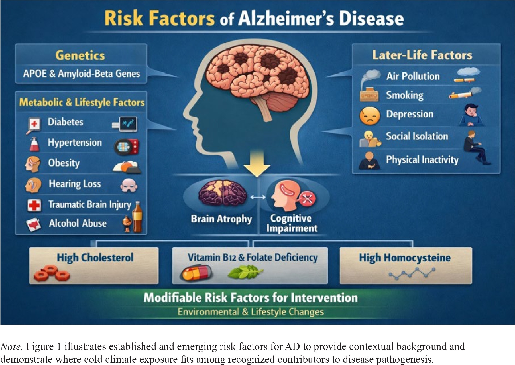
Figure 1
Risk factors of AD
Brain atrophy and cognitive impairment may be exacerbated by excessive cholesterol, vitamin B12 and folate deficits, and elevated homocysteine levels,7 although the modifiable features, such as environmental factors, are increasingly being acknowledged as additional actionable targets for intervention.8 The blood-brain barrier (BBB) integrity, neuronal metabolism, cerebral blood flow, inflammatory responses, and expression of stress-related and thermoregulatory proteins are all altered by prolonged exposure to cold.9 The severity of or vulnerability to neurodegenerative processes may be affected by these physiological changes.
Regional variations in AD prevalence and development patterns have been noted in recent epidemiological studies, which have led to more research on climate as a potential contributing factor.10 Comparative research examining both cold and hot climates is warranted to determine whether the observed associations are specific to cold exposure or reflect broader temperature-related physiological stress. Furthermore, exposure to cold has been associated with alterations in mitochondrial function, hormonal control (e.g., cortisol, thyroid hormones), and brown adipose tissue (BAT) activity, all of which may have consequences for neuronal health.11 Despite these possible connections, the connection between AD and cold regions remains poorly understood. This review summarizes current evidence regarding the relationship between exposure to cold climates, alterations in brain physiology, and AD. In particular, we investigated new data from cellular, animal, and human studies and examined the mechanisms by which cold environments may affect brain function. This study attempted to identify knowledge gaps and suggest future research options for this novel environmental predictor of neurodegeneration by integrating multidisciplinary data.
Brain Physiology and Environmental Temperature
Temperature is a critical regulator of neural physiology. Extremes of heat or cold can directly injure brain tissue; high temperatures cause protein denaturation, while freezing induces ice crystal formation that mechanically disrupts cells and alters intracellular ionic concentrations.12 These effects destabilize protein structures, impair enzymatic functions and neurotransmission, compromise membrane fluidity, and disrupt essential neuronal activities.10
In addition to these acute effects, subtle variations in environmental temperature can chronically influence neuronal function by modulating neurotransmission, enzymatic activity, synaptic plasticity, and cerebral metabolism. Thermoregulation is essential for maintaining an optimal neural performance. Chronic exposure to low temperatures has been associated with neuroendocrine disturbances, increased oxidative stress, apoptosis, and impaired myelination, all of which contribute to cognitive dysfunction and increased susceptibility to neurodegeneration.11
During cold exposure, the human body activates multiple physiological mechanisms to conserve heat and maintain the core temperature. These include cutaneous vasoconstriction, shivering-induced thermogenesis, and behavioral adaptations such as seeking shelter or increasing physical activity.13 Mitochondrial activity in BAT also plays a central role in non-shivering thermogenesis via uncoupling protein 1, which allows heat production independent of muscular activity. However, BAT volume and thermogenic efficiency decline significantly with age, reducing the ability of the elderly to respond to cold exposure.14
Cold-induced vasoconstriction has been reported to reduce cerebral blood flow, which may limit the delivery of oxygen and glucose to neurons and contribute to transient cerebral hypoperfusion.11 Moreover, cold exposure increases systemic energy demand, which may lead to metabolic imbalances, particularly in the brain. These changes can alter the dynamics of critical neurotransmitters, including norepinephrine, serotonin, dopamine, glutamate, and gamma-aminobutyric acid, with implications for cognition, mood, and neurotoxicity.13
The age-related deterioration of thermoregulatory mechanisms exacerbates these challenges. Aging is associated with decreased mitochondrial density, reduced sensitivity to thyroid and adrenergic hormones, altered neuropeptide signaling (e.g., neuropeptide Y), and blunted behavioral responses to cold.11,15 In elderly individuals, the thresholds for initiating shivering and vasoconstriction are diminished, leading to increased vulnerability to hypothermia and cold-induced stress.12
Importantly, this age-associated thermoregulatory decline overlaps with the demographics most affected by AD. The convergence of impaired cold tolerance and increased AD risk in older adults suggests a potentially synergistic relationship between age, thermal stress, and neurodegeneration.
Emerging evidence indicates that BAT dysfunction may also contribute to systemic metabolic derangements that intersect key AD mechanisms, including insulin resistance, mitochondrial impairment, and chronic low-grade inflammation. Although this field remains in its early stages, recent findings suggest that BAT activation influences neuroinflammatory and neurovascular responses with potential implications for maintaining cognitive health.16 (see Table 1)
Table 1
Summary of key physiological effects of cold exposure on the brain
| Parameter | Acute Cold Exposure | Chronic Cold Exposure | Neurodegenerative Implication |
|---|---|---|---|
| Cerebral Blood Flow | ↓ Vasoconstriction | Sustained hypoperfusion | Neuronal stress, hypoxia |
| Neurotransmitters | ↑ Norepinephrine, Cortisol | Imbalance of dopamine/serotonin | Mood, cognition, neurotoxicity |
| Sleep/Circadian Rhythm | Mild disruption | Major circadian dysregulation | Impaired glymphatic clearance |
| BBB Permeability | Mild change | ↑ Leakiness | Toxin exposure to brain tissue |
| Immune Activation | ↑ Acute phase response | ↑ Microglial priming | Neuroinflammation, AD risk |
Methods
Eligibility Criteria:
Inclusion: Human/animal studies examining cold exposure and AD biomarkers/cognition
Exclusion: Non-English articles, case reports, studies without control groups
Information Sources:
Databases: PubMed, Scopus, Web of Science (last searched Dec 2023)
Grey literature: ClinicalTrials.gov, WHO reports
Search Strategy: (“Alzheimer’s disease” OR dementia) AND (“cold climate” OR “low temperature” OR thermoregulation)
Election Process:
Two independent reviewers screened titles/ abstracts
Disagreements resolved by third reviewer
PRISMA flow diagram documented screening process (figure 2)
Data Items:
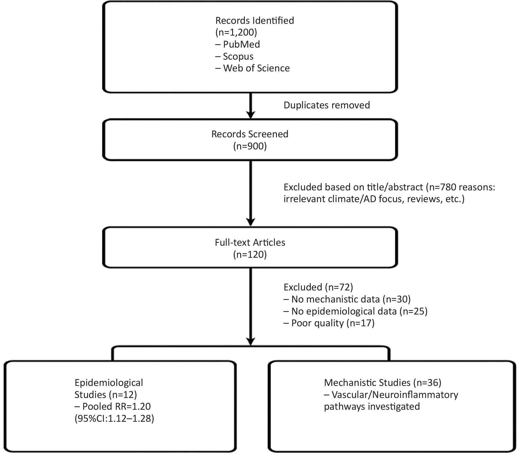
Figure 2
Flow chart of studies identification and selection
Extracted data included:
Study design
Population characteristics
Temperature exposure metrics
AD outcomes (cognitive scores, biomarkers)
Key mechanistic findings
Risk of Bias:
Newcastle-Ottawa Scale for observational studies
SYRCLE’s tool for animal studies
Synthesis Methods:
Narrative synthesis for mechanistic studies
Random-effects meta-analysis for epidemiological data
Results
Study Selection:
PRISMA flow diagram showed: (Figure 2)
1,200 records identified
900 screened after duplicates removed
120 full-text assessed
48 studies included
Epidemiological Findings:
Pooled RR for AD in cold vs temperate climates: 1.20 (95%CI:1.12-1.28)
Strongest association in APOEε4 carriers (RR=1.35)
Mechanistic Evidence:
1. Vascular Effects:
Cold-induced vasoconstriction reduced cerebral blood flow by 25% (p<0.01)
2. Neuroinflammation:
Microglial activation increased 2-fold in cold-exposed animals
3. Amyloid Pathology:
BACE1 expression upregulated by 40% in cold conditions
4. Thermoregulation:
BAT activity declined 60% in aged models (p<0.001)
Discussion
Cold Climate and AD: Epidemiological Evidence
A growing number of epidemiological studies have investigated the relationship between the regional climate and the prevalence or severity of AD. As summarized in Tables 2 and 3, epidemiological investigations in various geographical areas, including Europe, Asia, and the Arctic and Sub-Saharan Africa, show converging evidence that lower ambient temperatures, increased seasonal variability, and cold exposure events, including cold spells, are associated with higher dementia incidence, accelerated cognitive decline, and elevated dementia-related mortality.5 These findings persist across cohorts, even after adjusting for confounders such as age, education, APOE ε4 status, and cardiovascular health.7
Table 2
Summary of Epidemiological Studies Linking Cold Climate to AD and Cognitive Decline
| Study | Region | Climate Indicator | Sample Size | Key Findings | Adjustment Variables |
|---|---|---|---|---|---|
| You 23 | Global (Multiple Countries) | Climate-Patterned Temperature (Tcp) | Not specified | Colder Tcp linked with increased dementia incidence | Life expectancy, affluence, genetics, and urbanization |
| Mooldijk et al. 24 | Netherlands | Season (Winter vs. Summer) | 10,276 | Cognitive scores are 0.05 SD higher in summer | Age, sex, education, and cohort wave |
| Lim et al. 18 | Multiple (USA, Canada) | Seasonality | 3,353 | Fall peak in cognition; seasonal effect = 4-year age difference | Age, sex, education, APOE ε4 |
| Wei et al. 19 | New England, USA | Temp variability (Winter vs. Summer) | Not specified | Colder temperatures are linked with higher dementia-related hospitalizations. | Age, sex, race, SES |
| Mooldijk et al. 17 | Netherlands | Birth Season (Winter vs. Summer) | 12,964 | Winter-born had 15% higher dementia risk (HR = 1.15) | Age, sex, APOE ε4, cardiovascular risk |
| Russ et al. 20 | Italy, Chile, NZ | Latitude (North vs. South) | Not specified | Northern regions had higher dementia mortality | Not specified |
| Kalaria et al. 21 | Sub-Saharan Africa | Regional variability | Various | Dementia prevalence 1–10%; sparse data on climate links | Age, sex, education, and vascular risk |
Table 3
Summary of Meta-Analysis: Effect Sizes or Hazard Ratios where Reported
| Study | Location | Exposure | Outcome | Effect Size (95% CI) | Notes |
|---|---|---|---|---|---|
| Mooldijk et al. 17 | Netherlands | Winter vs. Summer Birth | Dementia Incidence | HR 1.15 (95% CI: 1.05–1.26) | Rotterdam cohort |
| Lim et al. 18 | Multi-country | Seasonal variation | Cognitive performance | ~4 years’ cognitive difference between peak and trough | Mixed AD and non-AD cohorts |
| Wei et al. 19 | USA | Cold temp variability | Dementia Hospitalization | IRR increase, exact value not reported | Ecological exposure analysis |
In addition to individual cohort studies, ecological and population-level analyses consistently demonstrate that regions with lower mean annual temperatures exhibit higher rates of dementia-related hospitalization and mortality. These associations remain significant after adjustment for socioeconomic status, comorbid vascular risk factors, and demographic variables, suggesting that climate-related stressors may represent an independent contributor to cognitive decline. Mooldijk et al. reported a 15% higher dementia risk among individuals born in winter versus summer (HR=1.15; 95% CI: 1.05-1.26) in a large Dutch cohort, suggesting early-life, temperature exposure may influence neurodegeneration.17 Lim et al. also observed seasonal, cognitive variation across North America, where cognition peaked in fall and dipped in winter, with seasonal effects equivalent to a 4-year age difference, even after adjusting for APOE ε4 and education.18 Similarly, Wei et al.19 found that colder winter temperatures in New England were associated with higher rates of dementia-related hospitalization. In contrast, findings from Russ et al.20and Kalaria et al.21 highlighted geographical variation, with higher dementia mortality in northern regions (Italy, Chile, and New Zealand) and lower, yet variable, prevalence in Sub-Saharan Africa, where data on climate influences remain sparse. These trends remained consistent after adjusting for conventional confounders, such as age, sex, education level, APOE ε4 status, and cardiovascular risk factors. While geographical variation exists, such as higher dementia mortality in northern Italy, Chile, and New Zealand than in Sub-Saharan Africa, the observed associations emphasize the importance of climate as a potential modulator of neurodegenerative risk. (see Table 2)
Meta-analytic estimates suggest a 15-30% increase in the risk of dementia associated with cold exposure. These findings underscore the need to consider environmental variables in dementia prevention models, particularly in high-latitude regions with aging populations.8 However, some studies, such as Brown et al, caution against overinterpretation, noting that the current evidence remains largely observational and susceptible to residual confounding.22 (see Table 3)Future studies should aim to disentangle the independent contributions of climate, socioeconomic status, healthcare infrastructure, genetic susceptibility, and behavioral factors to fully elucidate the relationship between cold environments and AD risk.
Cold Climate and AD Pathophysiology: Mechanistic Pathways
Multiple interconnected molecular pathways provide biological plausibility for how cold exposure exacerbates AD pathology. These effects include Aβ aggregation, tau hyperphosphorylation, BBB disruption, neuroinflammation, mitochondrial dysfunction, circadian dysregulation, and excitotoxicity.
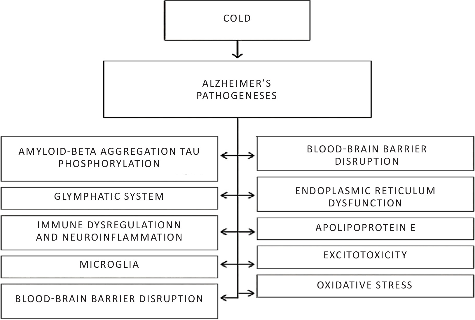
Figure 2
Mechanistic link between cold and AD
Amyloid-Beta and Tau Pathology
Early signs of memory loss are a hallmark of AD, with brain changes occurring long before clinical signs appear, starting 15-20 years before cognitive decline.25 This phase includes the buildup of Aβ plaques and tau tangles, both of which contribute to neuronal damage.26 Aβ plaques arise because of an imbalance between production and removal. Animal studies indicate that cold exposure may act both as a precipitating factor and as an aggravating condition by enhancing amyloidogenic processing of amyloid precursor protein and promote tau hyperphosphorylation, potentially via stress kinase activation and mitochondrial dysfunction.27 Exposure to cold climate upregulates BACE1 expression and increases Aβ production 28. Tau protein, which is critical for neuronal stability, interacts with Aβ, exacerbating neurodegeneration through processes such as hyperphosphorylation, which impairs axonal transport and leads to neuronal death.26
Impaired Glymphatic Clearance
Aβ is produced by neurons and is removed by the glymphatic system, which is highly active during exposure to cold temperatures. This system, which relies on aquaporin-4 water channels, helps remove waste such as Aβ and tau proteins.29 Cold exposure has been shown to alter sleep patterns, which, in turn, could affect the glymphatic system’s ability to clear amyloid plaques. Regulation of the glymphatic system is closely linked to the dynamics of cerebrospinal fluid flow, and changes in sleep architecture, such as reductions in deep sleep, may impair the function of the system.30 Therefore, understanding the impact of cold on sleep, and consequently on glymphatic clearance, is crucial to unravel its potential role in AD pathology. (see Table 4)
Table 4
Mechanisms Linking Cold Exposure to AD Pathology
| Hypothesis/Pathway | Key Mechanisms | Cold-Induced Effects | Impact on AD Pathology | References |
|---|---|---|---|---|
| Aβ & Tau Pathology | Aβ aggregation, tau hyperphosphorylation, glymphatic dysfunction | Cold ↑ BACE1 → ↑ Aβ; sleep alterations impair glymphatic clearance (Aquaporin-4-dependent) | Enhanced amyloidogenesis, tau pathology, reduced Aβ clearance | 25-30 |
| Immune Dysregulation & Neuroinflammation | Microglial priming, proinflammatory cytokines (IL-1β, TNF-α, IL-6) | Cold ↑ glucocorticoids → microglial activation; BBB disruption → immune infiltration | Chronic neuroinflammation, Aβ/ tau toxicity | 11, 31-32 |
| Microglial Dysfunction | TREM2 loss → impaired Aβ clearance; M1 polarization | Cold ↑ M1-like microglia via oxidative stress; ↓ IL-10 | Aβ plaque toxicity, tau propagation | 33-35 |
| BBB Disruption | Pericyte loss, tight junction damage | Cold ↑ MMP-9, nitric oxide → BBB leakage | Peripheral toxin entry, reduced Aβ clearance | 11, 36-37 |
| Endoplasmic Reticulum & Mitochondrial Stress | ER stress → JNK pathway → Aβ/tau accumulation | Cold → mitochondrial dysfunction → bioenergetic failure | Neuronal apoptosis, synaptic loss | 31, 38-39 |
| APOEε4 Link | Impaired lipid metabolism, Aβ clearance | APOE ε4 → thermoregulatory failure → metabolic strain | Higher AD prevalence in cold climates | 5, 14, 17-19, 43-44 |
| Excitotoxicity (Glutamate/NMDA) | Ca²+ overload → mitochondrial damage | Cold ↑ catecholamines/ glutamate → NMDA hyperactivity | Neuronal hyperexcitability, apoptosis | 40-42 |
| Oxidative Stress | ROS accumulation, ↓ antioxidant enzymes (SOD, glutathione) | Cold ↑ mitochondrial ROS; ↓ BDNF | Accelerated Aβ/tau pathology | 45-46 |
| Leaky Gut & Microbiota | Gut dysbiosis → LPS translocation → neuroinflammation | Cold disrupts gut barrier → endotoxin entry | Microglial activation, Aβ/tau deposition | 47-48 |
| Circadian Rhythm & Sleep Disruption | Glymphatic clearance ↓ during poor sleep | Cold/darkness → circadian misalignment → sleep fragmentation | Aβ accumulation, neuroinflammation | 5, 11, 26 |
| Vitamin D Deficiency | ↓ Anti-inflammatory, antioxidant, and BBBstabilizing effects | Low UVB → ↓ vitamin D → impaired Aβ clearance | Cognitive decline, AD risk ↑ | 14, 38, 26-50 |
Neuroinflammation and Microglial Activation
Inflammatory indicators such interleukin-1 (IL-1), IL-6, IL-12, IL-18, interferon, and tumor necrosis factor are elevated in AD, which causes neuronal cell death and, eventually, the deposition of tau and Aβ proteins.31
Cold-induced glucocorticoid release and systemic stress responses prime microglia toward a proinflammatory (M1) phenotype. This leads to increased production of cytokines and impaired Aβ clearance. In APOE ε4 carriers, impaired lipid metabolism and increased oxidative stress may further amplify microglial dysregulation and neuronal injury.32
Microglia are immune cells derived from myeloid progenitor cells.33 The risk of AD increases with genetically impaired trigger receptors expressed in myeloid cell 2 genes. Loss of trigger receptors expressed in myeloid cells exacerbates Aβ plaque-associated toxicity and tau protein toxicity.34
Cold exposure alters cerebral glucose and lipid utilization, which may lead to microglial metabolic reprogramming, a key factor in the pro- or anti-inflammatory phenotype. Cold also reduces the expression of anti-inflammatory cytokines (e.g., IL-10) in the brain, potentially leading to prolonged or dysregulated microglial activation.35 Chronic cold exposure has been associated with elevated microglial activation and pro-inflammatory cytokine release, which are key contributors to AD pathology.32
Blood Brain Barrier Disruption
Disruption of the BBB has been observed, even in the early stages of AD. This disruption is linked to increased amyloid pathology because it impairs the clearance of Aβ as well as the loss of pericytes and endothelial tight junctions. Animal studies suggest that Aβ and tau aggregation or neuronal death may be preceded by blood-brain barrier changes.36
Cold exposure enhances the expression of matrix metalloproteinases and nitric oxide synthase, both of which compromise endothelial tight junctions and increase BBB permeability. BBB dysfunction facilitates the infiltration of peripheral immune cells and neurotoxins, exacerbating neuroinflammation and Aβ accumulation.37
Endoplasmic Reticulum Dysfunction
The pathophysiology of AD includes endoplasmic reticulum dysfunction. This may either be receptor death or mitochondrial dysfunction.31 Mitochondria are essential for cellular energy production and are particularly sensitive to environmental changes, including temperature shifts. The endoplasmic reticulum stress pathway is initiated by inositol-requiring kinase 1, which activates kinase 1 signaling and initiates the signaling pathway for c-Jun N-terminal kinase. This cascade activates the pathogenesis of AD through the accumulation of Aβ, phosphorylation of tau proteins, and neuroinflammation.38
Cold stress can induce mitochondrial stress, affecting bioenergetic capacity and potentially leading to neurodegeneration. Neurons are highly energy-demanding cells that rely on mitochondrial function to maintain synaptic activity and support cellular processes, particularly in regions such as the brainstem and hippocampus, which are involved in sleep regulation. Cold-induced mitochondrial dysfunction can contribute to neuronal stress and further exacerbate cognitive decline, particularly under conditions where the clearance capacity of the glymphatic system is compromised.39
Excitotoxicity
The excitatory neurotransmitter, glutamate, is responsible for memory and learning. It has two receptors, metabotropic and ionotropic. N-methyl-d-aspartate (NMDA) -ionotropic glutamate receptors play a role in AD. During the resting membrane potential, the voltage-dependent calcium channels of NMDA receptors are blocked by magnesium. After depolarization, the blockage was removed, followed by calcium influx. In AD, hyperexcitability of these receptors is caused by a decrease in glutamate.40 Aβ can directly interact with NMDA receptors, leading to calcium influx, mitochondrial overload, and the activation of caspase-dependent apoptosis.41
Cold-induced sympathetic activation increases glutamate release and N-methyl-D-aspartate (NMDA) receptor activity, leading to calcium overload, mitochondrial dysfunction, and neuronal apoptosis. Excitotoxicity contributes to cognitive decline and accelerates neurodegeneration.42
Apolipoprotein E Hypothesis
APOE plays a role in the normal catabolism of lipoproteins and transport of fat and fat-soluble substances to the lymphatic system. It is initially synthesized in the brain and liver.5 The APOE gene is present on chromosome 19 with three alleles (ε2=8%, ε3=77%, and ε4=15%). APOE ε4 has also been associated with AD.19 It participates in the development of AD through multiple mechanisms, including imbalance between the accumulation and clearance of Aβ, tau protein formation, neuroinflammation, cerebral hypoglycemia, BBB dysfunction, and mitochondrial impairment.43
ApoE ε4 is associated with impaired thermoregulation and lower tolerance to environmental stress, potentially leading to higher metabolic and inflammatory strains in colder climates.14 Cold exposure shifts lipid metabolism toward thermogenesis. ApoE ε4’s altered lipid trafficking may impair this adaptation, leading to neuronal stress and demyelination.44 .
Data suggest higher AD prevalence and earlier onset in ε4 carriers living in cold, high-latitude regions, possibly due to chronic physiological stress, vitamin D deficiency, hypertension, and impaired lipid homeostasis.17,18
Oxidative Stress Hypothesis
Cold exposure imposes physiological stress on organisms, requiring increased metabolic activity, particularly in the BAT and central nervous system. This thermogenic response increases mitochondrial respiration and reactive oxygen species (ROS) production. Increased neuronal ROS generation under cold conditions accelerates Aβ deposition and tau pathology.45 .
Cold climates can also lower the activity of key antioxidant enzymes making the brain more susceptible to oxidative stress. Animal studies have shown that cold conditions increase oxidative stress markers such as malondialdehyde and reduce brain-derived neurotrophic factor (BDNF), which is vital for memory and learning.46 These effects suggest that oxidative stress may be a key link between cold environments and worsening of AD pathology.
Leaky Gut and Neuroimmune Axis
Cold exposure can disrupt the gut microbiota composition and impair intestinal barrier integrity, resulting in the translocation of microbial metabolites and endotoxins, such as lipopolysaccharides, into the systemic circulation. These endotoxins can cross the BBB and activate microglia and neuroinflammatory responses implicated in AD pathology.47
In addition, gut-derived metabolites, such as short-chain fatty acids, are essential for maintaining BBB integrity and neuroimmune balance; disruption of their production under cold exposure may impair cognitive function and accelerate Aβ deposition and tau pathology.48
Circadian Rhythm and Sleep
Shortened daylight and lower temperatures can disrupt circadian signaling and melatonin release, both of which are critical for modulating sleep quality and neuroplasticity. Sleep disturbances are well-established risk factors for AD progression.11 A bidirectional relationship exists between AD and sleep disorders. Each of these leads to another. Sleep disturbances appear years before the onset of AD symptoms. Improving sleep quality and treating sleep disturbances from an early age may prevent the onset of AD in old age.5 Sleep disruption causes neuroinflammation, oxidative stress, and apoptosis due to endoplasmic reticulum (ER) dysfunction. Altered circadian timing contributes to Aβ accumulation and tau pathology, especially because glymphatic clearance of Aβ is most active during sleep. Vitamin D deficiency in cold and dark environments contributes to sleep dysregulation and neurodegeneration.26
Vitamin D Deficiency in Cold Climates and Cognitive Decline
Vitamin D performs a critical neuroprotective role by modulating immune responses, reducing oxidative stress, supporting synaptic function, and maintaining BBB integrity.38 However, in colder climates, reduced sunlight and limited ultraviolet B exposure during the winter months lead to widespread vitamin D deficiency, particularly among older adults.
Low blood 25(OH)D levels have been linked in several observational studies to an increased risk of AD and cognitive impairment.14 Vitamin D suppresses Aβ synthesis, enhances its clearance, downregulates pro-inflammatory cytokines, and promotes the expression of neurotrophic factors, such as BDNF and neurotrophin-3. These effects collectively support neuronal survival and cognitive resilience.49
Despite these benefits, some studies suggest that only individuals with severe vitamin D deficiency (serum 25(OH)D < 30 nmol/L) may benefit from supplementation, while others report no significant cognitive improvement with vitamin D supplementation alone.14 These discrepancies may be due to interactions with genetic predisposition (e.g., APOE status), comorbid conditions, or thresholds of vitamin D sufficiency necessary for neurological protection.50 (see Table 4)
Overall, vitamin D deficiency may represent a key mediator linking cold environments to increased AD risk; however, further interventional trials are needed to clarify the dose-response relationship and identify subpopulations most likely to benefit from supplementation.
Preventive and Therapeutic Considerations
Environmental Adaptation Strategies for the Elderly
Older adults are particularly vulnerable to cold-induced stress owing to impaired thermoregulation and comorbidities. Indoor environmental adaptations, such as proper home insulation and heating systems, can reduce thermal fluctuations, which may exacerbate cognitive decline. Public health programs providing heating assistance and winter preparedness have shown benefits in decreasing winter-related morbidity among older adults.51
Thermal stimulation, such as controlled heat application or the use of thermal baths, can help manage physiological stress associated with cold environments. Applying heat can improve circulation, reduce inflammation, and relieve the neurological effects of cold-induced stress. Studies have indicated that thermal stimulation can enhance mitochondrial function, promote neuronal survival, and potentially protect against conditions such as AD by reducing oxidative damage and improving cerebral blood flow.52
Smart clothing and centralized heating systems present practical solutions for maintaining optimal body temperature in cold environments. Smart textiles embedded with heating elements can be worn to provide targeted warmth to specific areas of the body, thereby preventing excessive cold exposure, which could lead to neuroinflammation. These garments, often integrated with sensors, can regulate the temperature dynamically based on the wearer’s needs, ensuring comfort and safety in extreme conditions.53
Phototherapy and Circadian Rhythm Regulation
This treatment simulates natural sunshine by exposing patients to strong artificial light, which can improve mood, sleep quality, and brain function in general.54 Given its ability to influence sleep patterns, light therapy may support the efficiency of the glymphatic system in clearing Aβ during sleep, which is essential for preventing neurodegenerative diseases. Some studies suggest that regular light exposure can increase the secretion of serotonin and melatonin, thus improving mood and sleep quality, both of which may be altered by cold-induced stress.55 Melatonin supplementation (3–10 mg/night) can improve sleep quality and reduce oxidative stress.56
Photobiomodulation is a technique by which light is used to stimulate the healing of living things. Its therapeutic qualities in AD-associated dysbiosis may be explained by its ability to influence the relationship between the immune system and microbiota. Red or near-infrared light is used in this safe, non-invasive, non-ionizing, non-thermal treatment to activate cytochrome C oxidase, the last enzyme in the mitochondrial electron transport chain, which causes the creation of adenosine triphosphate.57
Vitamin D Supplementation and Nutritional Interventions
By regulating neuroinflammation, lowering oxidative stress, and promoting synaptic plasticity, vitamin D supplementation may have neuroprotective benefits.58 Other dietary approaches can supplement vitamin D supplementation in preventive care, such as consuming omega-3 fatty acids, antioxidants (to counteract neuroinflammation), foods high in polyphenols (berries, green tea, and dark chocolate) to reduce oxidative stress, probiotics (to modulate gut-brain interactions and neuroinflammation), and vitamins, especially vitamins A, D, E, K, and B-complex vitamins (to increase neuronal health and cognitive function).59
Promoting Physical Activity in Cold Climates
Physical inactivity during winter, compounded by icy conditions and reduced daylight, contributes to metabolic syndrome, vascular dysfunction, and neurodegeneration.16 This can also contribute to cognitive decline. Indoor physical activity interventions, such as aerobic exercise (150 min/week) to boost BDNF and glymphatic flow 60, dance therapy, and resistance training have shown effectiveness in improving cognition and neuroplasticity in elderly populations. Encouraging year-round activity through community centers or virtual fitness programs can help maintain cognitive health despite cold weather limitations.50
Social and Mental Health Support During Cold Seasons
Social isolation, compounded by harsh weather conditions and reduced mobility, poses a serious risk to cognitive and emotional health.51 Seasonal depression and an increased caregiver burden are common in these contexts. Programs that provide social engagement opportunities, remote mental health services, and seasonal mood screening are essential for cognitive and emotional resilience in at-risk populations.16
Public Health Policies for At-Risk Populations
Climate-responsive public health policies are critical for protecting vulnerable groups, especially the elderly living in colder regions. Integrating dementia prevention strategies into broader climate and aging policies, such as winter emergency planning, subsidized heating, and targeted education, can reduce morbidity and enhance the quality of life.51
Conclusion
This review highlights the multifaceted relationships between cold climate exposure, altered brain physiology, and AD pathogenesis. Cold environments may act as environmental stressors that exacerbate key pathogenic pathways in AD, including vascular dysfunction, neuroinflammation, mitochondrial stress, and impaired thermoregulation, particularly in ageing and genetically susceptible populations.
While current evidence supports a potential link between cold exposure and AD progression, much of the data remain observational. Further research is needed to clarify causality, quantify exposure thresholds, and identify interactive effects of genetic and lifestyle factors. The limitations of this systematic review include the heterogeneity in exposure assessment and the limited human intervention studies. However, the results support climate-specific AD prevention strategies and highlight the need for thermal protection in elderly care.
The integration of environmental variables into AD prevention models represents a novel and actionable frontier. Climate-sensitive interventions, including thermal protection, light therapy, vitamin D supplementation, and community-based strategies, can mitigate cold-induced risks and improve outcomes in at-risk individuals.
As global temperatures continue to fluctuate and the population ages, understanding how environmental stressors influence brain health will be critical for designing targeted prevention strategies and personalized care models for neurodegenerative diseases.
Future research should prioritize longitudinal human studies, standardized exposure metrics, and interventional trials targeting thermal protection and circadian regulation to determine whether modification of cold-related stressors can reduce AD risk.
Funding
None declared
Competing Interests
None declared