
Background
Due to global population growth and the influence of industrial expansion on the ecosystems, natural habitats face escalating pressures, and a significant portion of living organisms is regularly exposed to environmental pollutants.1 These pollutants, including heavy metals, are reported to have harmful consequences on various organs and systems of mammals, notably the immune system.2 Heavy metals are known by a density greater than 5 g/cm³ and include elements including lead (Pb), copper (Cu), etc.3 Lead, a metal that occurs naturally with a distinctive bluish gray colour, is omnipresent throughout the environment, contaminating food, water, air, and soil.4 Soil contaminated with lead deposits can lead to the uptake of this metal by crops, thus entering the food chain.5
Pb possesses enzymatic toxic potential with neurotoxic, immunotoxic, carcinogenic, hematotoxic, teratogenic, mutagenic, and cardiovascular toxic effects.6 It inflicts damage on cellular organelles, disrupts genetic material, and gives rise to antioxidant-oxidant imbalance by promoting the overproduction of reactive oxygen species (ROS), which in turn depletes antioxidant reserves and exacerbates cellular damage.7 Pb toxicity has a profound impact on cellular function, interfering with the activation of enzymes, trace mineral uptake, and protein synthesis. By binding to sulfhydryl proteins, lead disrupts structural integrity and alters calcium balance. Furthermore, Pb exposure compromises antioxidant defenses by depleting sulfhydryl reserves, rendering cells vulnerable to oxidative damage.8 The extent of Pb toxicity is influenced by several factors, including developmental stage, gender, administration method, dose, solubility and redox potential, absorption efficiency, period of contact, rate of ingestion, bioavailability, and the efficacy of elimination mechanisms. Pb exposure is reportedly linked to different cancers, kidney damage, neurotoxicity, and cardiac dysfunction in humans.9
Furthermore, lead exposure has been shown to impair immune function by altering parameters such as immune defense and response of antibodies and inducing T-helper cells (type 2) and the macrophages within the spleen.10 At the cellular level, lead poisoning involves mechanisms such as oxidative stress (OS) through secondary pathways by interacting with glutathione and antioxidants, hypoglycemia, as well as disruption in the regulation of the endoplasmic reticulum. Studies indicate that disequilibrium between antioxidants and oxidants constitutes the primary toxicity pathway underlying organ or system damage triggered by Pb, as it mediates oxidative stress, resulting in ROS production.11-12 Research has also highlighted the detrimental impacts of Pb on reproductive indices, mediated through dysfunctions in the neuroendocrine region and the impairment of cells in the interstitial and Sertoli regions.13
Pb exposure disrupts the serum volume of FSH, LH, in addition to testosterone, and is associated with inflammation and oxidative damage, both of which are implicated in reproductive disorders.14-15 The mitochondrial effects of Pb include the depletion of cellular energy16 and the induction of glucocorticoid secretion while suppressing gonadotropin release.17 Given that free radical production, cell suicide, and cytotoxicity caused by Pb are major contributors to male infertility, antioxidants have been proposed as therapeutic agents to mitigate these effects by reducing the expression of apoptosis-inducing genes.18 Vitamins, essential nutrients readily obtainable from the diet, have shown a protective role against damage from heavy metal exposure.19
Vitamin C (VC), a potent non-enzymatic antioxidant, has been shown to exhibit potent antioxidant properties, efficiently neutralizing ROS, resulting in the prevention of oxidative degradation of lipids.20-21 The protective roles of VC against Pb-induced oxidative damage have been extensively documented in various organs, including hepatoprotection, nephroprotection, and reproductive protection.20 VC exerts its antioxidative properties via neutralizing water-soluble ROS, thereby protecting cellular membranes.21 Recent advances in pharmaceutical delivery systems, including microencapsulation and nanotechnology, have further enhanced the efficacy of VC in mitigating Pb toxicity.22 The medicinal capability of VC in lead poisoning has been shown across human beings, animals, and different marine species.23-24 At the molecular level, the therapeutic effects of VC may also involve inhibiting Pb absorption in the intestinal tract and binding to Pb, reducing its toxic effects.25
Given the widespread nature of lead pollution and its well-documented toxicity, there is an urgent need for effective strategies to mitigate its deleterious effects. Therefore, this research was conducted to explore the modulatory potentials of VC on Pb-induced organotoxicity and histopathology in male Wistar rats. While VC has been shown to counter heavymetal toxicity, most existing studies use short exposure durations. This study addresses that gap by using a prolonged, 65-day, Pb exposure to model sub-chronic, real-world conditions, particularly in industrial and mining-affected areas. Our findings provide new insight into testicular micro-architecture and Pb accumulation over extended exposure, supporting VC as a low-cost protective option for vulnerable populations
Methods
Materials collection
Pb acetate was procured from SigmaAldrich Limited (St Louis, Missouri, USA). VC was procured from Emzor Pharmaceutical Industries Limited, Calabar, Nigeria.
Experimental animals
Twenty-four sexually viable male albino rats, aged 12 weeks, which weighed 160-170g, were housed at the Animal House, Department of Genetics & Biotechnology, University of Calabar-Nigeria. The male Wistar rat, a popular choice for scientific research, has its roots in a wild rat population from the early 20th century. Male Wistar rats are selected for research based on specific age and weight criteria, typically between 8-12 weeks. Wistar albino rats are renowned for their docility and ease of handling, making them an ideal choice for research. Their stable characteristics and consistent reproductive data make them a popular choice for toxicology, pharmacology, and nutritional studies.
The inclusion criteria were the sex of the animals, age, and body weight. These experimental animals were housed and accommodated in metal cages with breathable barriers, maintained under an animal-friendly environment (Standard laboratory conditions) with free access to standard feed (Top Feed Nig. Limited) and water throughout the duration of the research. The rats were acclimatized to the laboratory surroundings for fourteen days before the commencement of treatments. Helsinki’s protocols were strictly adhered to in handling the animals. Humane endpoints for this study include clinical symptoms of distress and disease and behavioural changes, which were monitored regularly. No adverse effect was observed during the duration of the treatment.
Experimental design and protocol
Animals were randomized into 4 treatment groups, each comprising of 6 rats, following a completely randomized design (CRD). Randomization protocol utilized the GraphPad randomization software. Researchers handling the administration of treatments and laboratory analysis were blinded to the allocation of the treatment groups. Only the Principal Investigator, Prof. Utip Ekaluo, and the corresponding author were aware of group allocation and played supervisory roles. Other researchers, especially those who handled treatment and laboratory analysis, were not. Sample size was determined based on our previous research and literature in line with the 3Rs guideline in animal research. The groups were designated as follows: Group I: Control; Group II: Administered 60 mg kg-1 BW of Pb acetate; Group III: treated 100 mg kg-1 BW of VC; Group IV: Administered a combination of 60 mg kg-1 BW of lead acetate and 100 mg kg-1 BW of VC. Vitamin C was freshly prepared daily in distilled water to prevent oxidation, stored in amber-covered containers, and administered within 30 minutes of preparation to ensure chemical stability and maximal antioxidant activity. A dose of 60 mg/ kg BW of Pb was chosen to mimic human environmental Pb exposure, a common approach in rat models of Pb poisoning.26-28 Meanwhile, a 100 mg/kg BW dose of VC was chosen based on existing research showing its effectiveness in reducing oxidative stress caused by heavy metal toxicity.29 The treatment was done daily for a period lasting for 65 days between FebruaryApril 2024. The 65-day duration was intentionally extended beyond the commonly used 21-45 day exposures to simulate sub-chronic, low-grade, environmental intake typical in industrial and mining-exposed populations. Twenty-four hours post-treatment, experimental animals were euthanized using cervical dislocation after Isoflurane anaesthesia. The testes were surgically removed and processed for histopathology. Vital organs were weighed and processed for lead residue assay.
Body weight and weight of organs
Body weight, weight of liver, kidney, brain, and heart were determined using an electronic weighing balance (Scout Pro SPU 601).
Detection of lead residue in the testes, brain, liver, and kidney
Tissue samples were meticulously collected and homogenized to ensure uniformity, followed by digestion using a wet digestion method with concentrated nitric acid (HNO3) and hydrochloric acid (HCl). The digested samples were then diluted with deionized water. Lead acetate concentration in the digested tissue samples was quantified using a Perkin-Elmer 2380 atomic absorption spectrophotometer (AAS) at a wavelength of 283.3 nm. Calibration curves were generated using five standard Pb concentrations (0.1-1.0 ppm), with blank runs and certified reference standards included for instrument validation.30 The lead concentrations were expressed in ppm of tissue with all safety protocols adhered to during chemical handling and sample processing.
Histology of testes
The testicular tissues were stabilized in a 10% neutral buffered formalin solution to preserve their structural integrity. Subsequent dehydration was achieved through a sequential ethanol gradient, followed by clarification in xylene. The tissues were then fixed in melted paraffin wax at 58°C to facilitate sectioning. Using microtomy sections of 5 micrometer thickness embedded in paraffin tissue blocks were mounted on clean glass slides and subjected to routine histological staining with H&E (Hematoxylin and Eosin) stains and examined in a light microscope to assess tissue morphology and architecture.
Statistical analysis
The data obtained were tested for normality, homogeneity, and independence of observations, and the assumptions of the statistical approach were met. The data obtained on body weight, weight of organs, and lead residue were analyzed using one-way ANOVA and subsequently, post hoc Tukey test at p<0.05 using Statistical Package for the Social Sciences (SPSS) 27 (IBM, New York, USA). All results obtained were presented as mean ± standard error of mean (SEM) (n=6 replicates).
Results
Lead residue concentration
Table 1 shows the results obtained on Pb residue concentration. Results showed a significantly high level of Pb residue in the brain, kidney, liver, and testes of rats administered Pb only in comparison to the control and other groups. In brain samples, the control, lead, VC, and Lead + VC groups had 0.21 ± 0.06, 0.34 ± 0.01, 0.16 ± 0.08, and 0.26 ± 0.04ppm, respectively. For the kidney, the control, Pb, VC, and Pb + VC recorded 0.23 ± 0.07, 0.96 ± 0.03, 0.22 ± 0.04, and 0.33 ± 0.06 ppm, respectively. For the liver, results revealed a concentration of 0.17 ± 0.01, 0.30 ± 0.02, 0.17, and 0.21 ± 0.01ppm for the control, Pb, VC, and lead + VC groups, respectively. For the testes, the control and VC groups had 0.22 ppm, while the lead and lead + VC groups had 0.33 ± 0.01 and 0.24 ± 0.02ppm, respectively. Results revealed the modulatory potential of VC on Pb residue levels in all the organs assayed.
Table 1
Modulatory effect of VC on lead residue concentration
| Organs | Control | Lead | VC | Lead + VC |
|---|---|---|---|---|
| Brain (ppm) | 0.21c±0.06 | 0.34a±0.01 | 0.16d±0.08 | 0.26b±0.04 |
| Kidney (ppm) | 0.23c±0.07 | 0.96a±0.03 | 0.22c±0.04 | 0.33b±0.06 |
| Liver (ppm) | 0.17c±0.01 | 0.30a±0.02 | 0.17c±0.04 | 0.21b±0.01 |
| Testes (ppm) | 0.22c±0.04 | 0.33a±0.01 | 0.22c±0.01 | 0.24b±0.02 |
[i] Notes. Values are presented as mean ± standard error. Means with similar case letters across the horizontal array are not significantly different at p>0.05
Body weight and weight of organs
Results obtained showed that there were no statistically significant differences (p=0.18) in the initial mean body weights of animals across groups before treatment commencement, indicating uniformity at baseline. The mean body weights reduced in a significant manner (p=0.012, η²=0.41) in Pb-treated rats (172.55 ± 9.86g) when compared with the control (229.00 ± 8.82g) and VC-treated animals (234.50 ± 10.12g). Lead + VC-treated animals had a mean body weight of 180.90 ± 6.89g, indicating the attenuating effect of VC (Table 2). Figure 1 presents the effect of VC on the body weights of animals obtained weekly.
Table 2
Effect of VC on lead-induced toxicity on body weights and weights of organs
| Body/organ weight | Control | Lead | VC | Lead + VC |
|---|---|---|---|---|
| Pre-treatment body weight (g) | 163.43a±1.98 | 166.87a±2.65 | I65.12a±3.76 | 164.22a±3.59 |
| Post-treatment body (g) | 229.00c±8.82 | 172.55a±9.86 | 234.50d±10.12 | 180.90b±6.89 |
| Liver (g) | 7.45a ±1.02 | 7.78 a ±0.49 | 6.95a ±0.56 | 7.32a ±1.08 |
| Heart (g) | 0.75c ±0.08 | 0.50 a ±0.02 | 0.73c ±0.04 | 0.73c ±0.04 |
| Kidney (g) | 0.65a ±0.09 | 0.65 a ±0.09 | 0.65a ±0.09 | 0.65a ±0.09 |
| Lungs (g) | 1.37a ±0.04 | 1.62 a ±0.05 | 1.35a ±0.06 | 1.68a ±0.03 |
| Testes (g) | 1.10c ±0.10 | 0.68 a ±0.09 | 1.10c ±0.11 | 0.70b ±0.02 |
| Epididymes (g) | 0.35c ±0.06 | 0.18 a ±0.04 | 0.38d ±0.12 | 0.20b ±0.04 |
[i] Notes. Values are presented as mean ± standard error. Means with similar case letters are not significantly different.
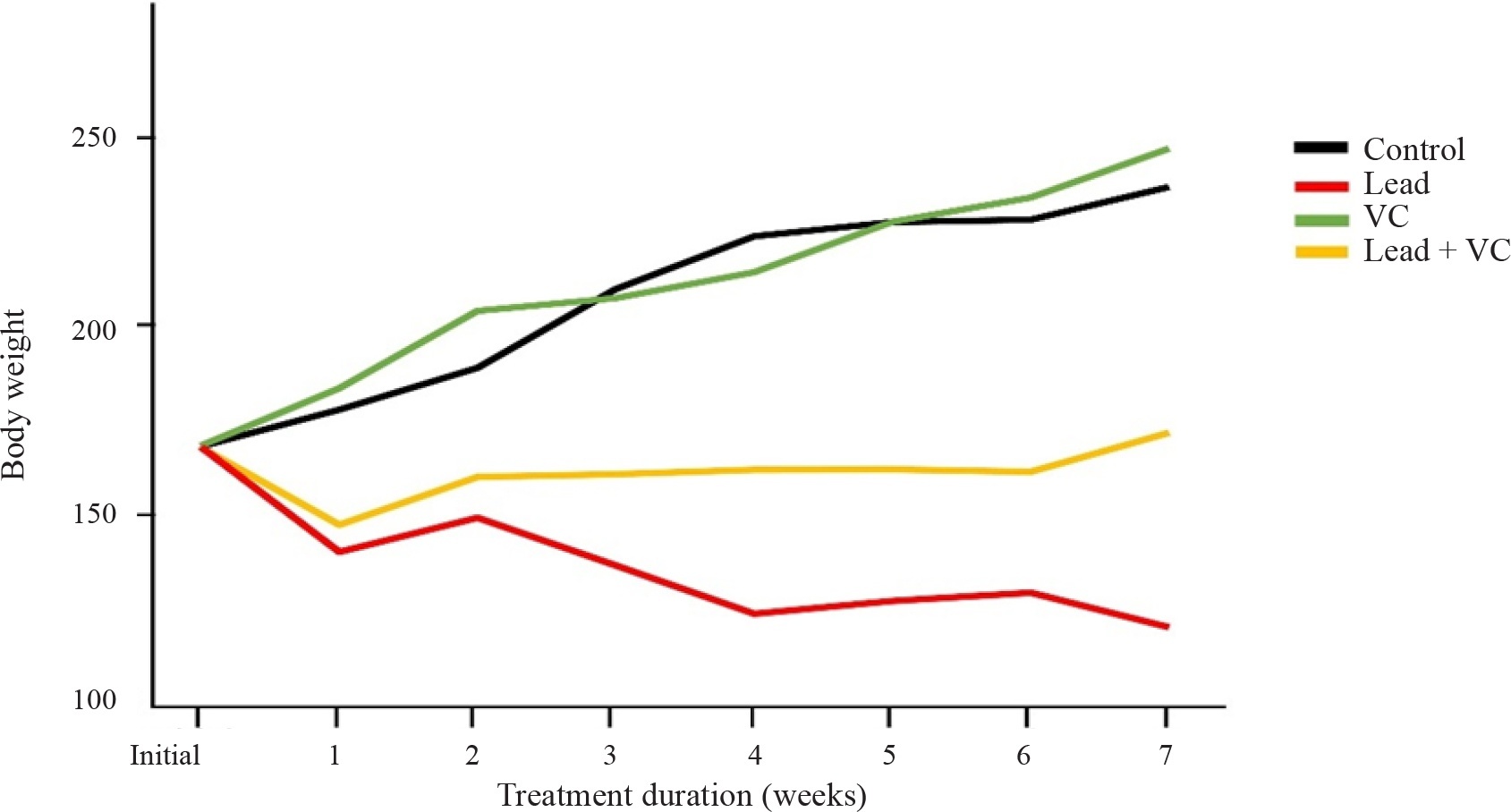
Figure 1
Effect of VC on weekly body weights of rats treated with lead
Results obtained on the weight of organs are also presented in Table 2. There was no significant (p=0.08) difference in the weights of the liver, kidney, and lung across the different groups. The weight of the heart was significantly higher in the control (0.75 ± 0.08 g) when compared with the lead group (0.50 ± 0.02 g). VC and lead + VC groups mean weights of 0.73 ± 0.04g and 0.50 ± 0.02g. The weight of the testes significantly reduced (p=0.008, η²=0.53) in the Pb-treated animals (0.68 ± 0.09g) when compared with the control (1.10 ± 0.10g) and VC group (1.10 ± 0.11g). The weight of the testes in the lead + VC group was 0.70 ± 0.02. Similarly, the weight of the epididymis reduced significantly in the lead group (0.18g) when compared with the control (0.35 ± 0.06g) and other treatment groups (0.38 ± 0.12g and 0.20± 0.04g for VC and lead + VC groups, respectively). Results also indicated an ameliorative effect of VC on the lead-induced reduction in weight of the heart, testes, and epididymis in animals treated with both Pb and VC.
Histology of the testes
Results showed that testes from control animals (Figure 2A) displayed well-organized, densely packed, seminiferous tubules with intact basement membranes. The germinal epithelium comprised 3–4 orderly layers of spermatogonia and successive cell generations, each with distinct nuclei and fine chromatin detail. Sertoli cell counts exceeded nine per tubule, and the lumina contained a moderate quantity of mature spermatozoa. The interstitial spaces were normal in width and contained more than three clusters of Leydig cells per field. In contrast, the testes of Pb-treated rats (Figure 2B) exhibited loosely arranged seminiferous tubules with reduced germ cell density and noticeable depletion of spermatogenic layers, indicating early atrophic changes. The epithelium was thinner, with fewer spermatocytes and spermatids present, and luminal spermatozoa were markedly reduced. Sertoli cell numbers remained above nine per tubule, but the interstitium appeared narrowed, with fewer Leydig cell clusters relative to control tissue.
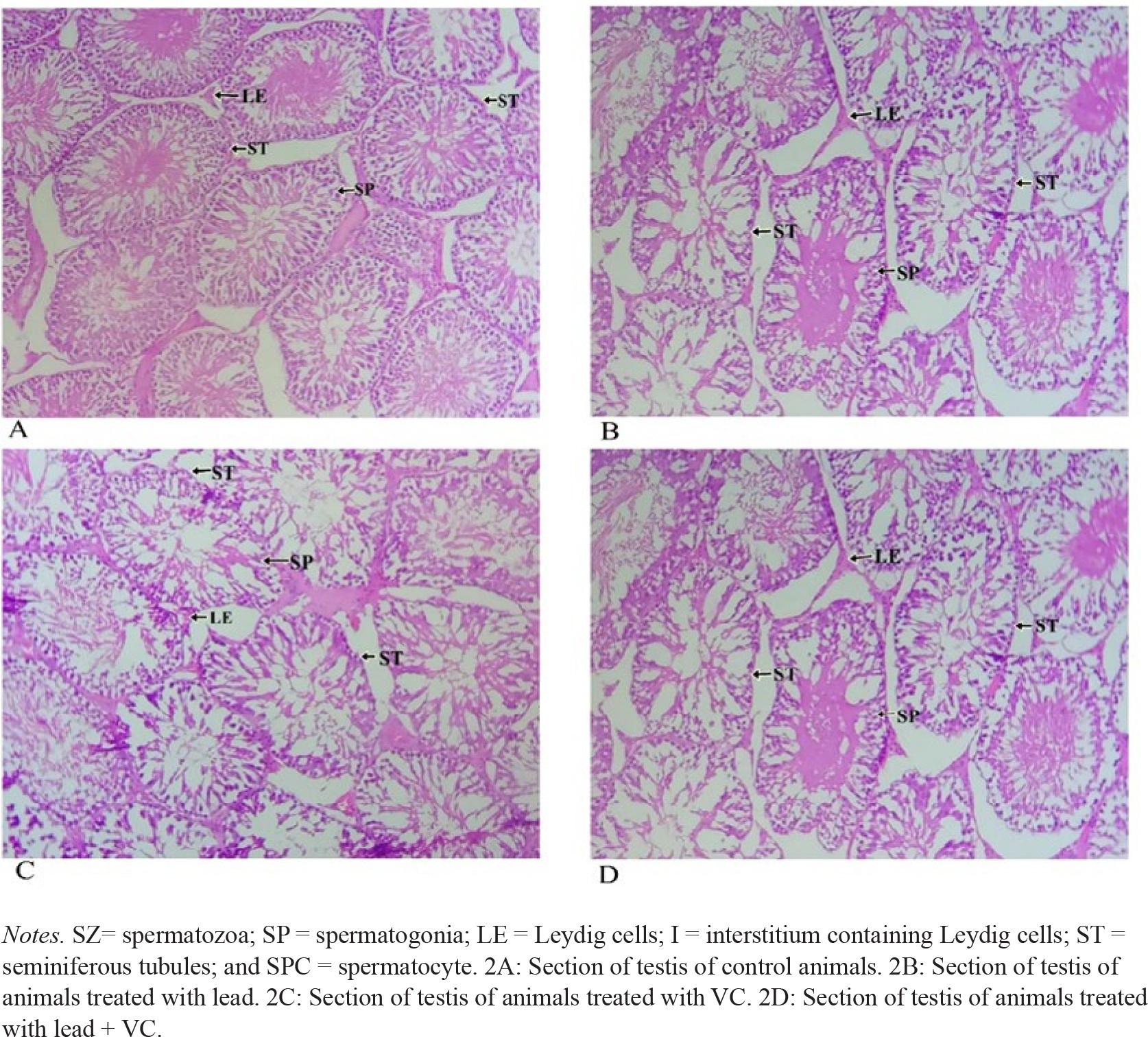
Figure 2
Modulatory effect of VC on lead-induced testicular toxicity of rats. (H&E X400)
VC-only animals (Figure 2C) maintained near-normal, testicular architecture. The seminiferous tubules were closely packed with intact basement membranes and 3-4 layers of predominantly early germ cells (spermatogonia A & B and primary spermatocytes) while spermatid and spermatozoa populations were present but lower than the control. Sertoli cells remained abundant (>9 per tubule), and interstitial Leydig cells were adequately distributed. In the Pb+VC co-treated group (Figure 2D), testicular morphology showed partial recovery. Seminiferous tubules were more compact than in Pb-only animals, with moderately repopulated germinal layers and improved luminal sperm presence, though not fully restored to control levels. The basement membrane appeared thickened but intact, and Leydig cells were present in 3-4 clusters per field, indicating amelioration of Pb-induced testicular injury.
Discussion
Findings of our study indicated a substantial rise in Pb residue concentrations in brain, kidneys, liver, and testes samples of the Pb-administered rats when compared to animals in the control group, aligning with previous research on the bioaccumulation of lead in various organs. Pb is known to distribute widely in the body, accumulating particularly in organs and viscera, including the brain, kidney, liver, and reproductive organs, because of its high attraction for sulfhydryl groups in proteins.31 This accumulation can result in severe toxic effects, as lead interferes with the normal physiological functions of these organs. For instance, the brain is highly susceptible to lead toxicity, which can result in neurobehavioral deficits and cognitive impairments.32 The level of Pb in brain cells is reportedly associated with OS and disruption of neurotransmitter systems.33 In the kidneys, lead accumulation can cause nephrotoxicity by damaging the proximal tubules, leading to impaired renal function.34 The liver, being a primary detoxification organ, also accumulates lead, which can interfere with metabolic processes and cause hepatotoxicity.35 In the testes, lead accumulation disrupts the endocrine system and spermatogenesis, contributing to reproductive toxicity.36
These findings underscore the systemic nature of lead toxicity, where its accumulation in critical organs leads to a cascade of adverse health effects. The differences in lead concentration between the exposed and control groups highlight the significant risk posed by environmental lead exposure and the need for protective measures to prevent lead accumulation and its associated toxicities. The results of this study indicate that VC effectively reduces Pb residues in lead-exposed animals, a finding consistent with prior research demonstrating the chelating and antioxidant properties of VC.20,37 The ability of VC to decrease lead accumulation in tissues is likely due to its capacity to chelate Pb ions, thus, enhancing their excretion from the body.38 Additionally, the role of VC as a potent antioxidant helps mitigate antioxidant-oxidant imbalance induced by exposure to Pb, which is known to exacerbate lead retention in tissues. Patrick40 highlighted the ability of VC to bind lead ions and prevent their absorption in the gastrointestinal tract, thereby, reducing systemic lead levels. Hashem40 further confirmed that VC administration lowers lead concentrations in the liver, kidneys, and brain, attributing this effect to the antioxidant properties of VC, which counteract oxidative damage and stabilize cell membranes.
The effectiveness of VC in reducing lead residues may also involve its interaction with glutathione, a key antioxidant in detoxification processes. VC enhances glutathione synthesis, which in turn facilitates the conjugation and excretion of lead.37 This mechanism is supported by Hashem et al.,39 who identified oxidative stress as a critical factor in lead toxicity and highlighted the protective role of antioxidants like VC. Moreover, studies such as Carocci et al.11 indicate that VC supplementation not only reduces lead-induced OS but also brings back the action of antioxidant enzymes that are typically inhibited by lead exposure. This restoration is crucial in reducing the overall lead burden in the body.
The observed significant reduction in the weights of vital organs and body in Pb-exposed animals aligns with previous studies and can be attributed to disruptions in metabolic processes and nutrient absorption, leading to significant weight loss.41 This weight reduction is often connected to oxidative stress and cellular damage due to Pb exposure, which impairs the optimum performance of organs such as the liver, kidney, and heart.39 In addition, Pb interferes with the endocrine system, disrupting hormone production and regulation, which can further contribute to the observed decrease in organ weights like testicular weight.42 For instance, the reduction in liver weight may be due to hepatotoxicity caused by oxidative damage and the inhibition of protein synthesis.43 Similarly, the reduction in kidney weight may be linked to nephrotoxicity, characterized by glomerular and tubular damage, which compromises renal function.44 VC modulated the effect of Pb on the weight of vital organs and body weight in animals treated with a combination of VC and lead in comparison to those treated with Pb alone. This is likely due to the antioxidant properties of VC, which mitigate the oxidative stress caused by lead, thereby improving metabolic functions and reducing tissue damage.45
Histopathological assessment of the testes in Pb-treated rats revealed pathological changes, specifically loosely packed seminiferous tubules with atrophic spermatogenic cells. These findings reflect the adverse impact of lead exposure on testicular tissue and function. The observed loosening of seminiferous tubules in Pb-treated rats indicates a disruption of the normal testicular architecture.46 Seminiferous tubules, with crucial significance in spermatogenesis, are typically tightly packed to support efficient sperm production.47 The loosening of these tubules suggests a loss of structural integrity and organization, which can compromise spermatogenic processes. This finding corroborates those reported by Gandhi et al.36 and Adetunji,48 who reported disordered arrangements of spermatogenic cells, degenerative changes, and significant alterations in the testes of lead-treated animals. Sertoli cells are essential for testicular development, while Leydig cells are important for testosterone synthesis and spermatogenesis.49-50 Atrophic spermatogenic cells further highlight the detrimental effects of Pb. Spermatogenic cells, including spermatogonia, spermatocytes, and spermatids, are essential to produce sperm. Atrophy of these cells indicates a severe impairment in spermatogenesis, which gives rise to a reduction in sperm quantity and quality.51 This atrophy may result from direct toxic effects of Pb on germ cells or secondary effects such as ROS production and inflammatory endpoints induced due to Pb exposure.52
Conclusion
Results revealed the modulatory potential of VC on Pb-induced organotoxicity and histopathology in male albino rats as a mammalian model. This highlights the apparent use of VC as a therapeutic or protective measure in cases of Pb toxicity, particularly in environments where lead contamination is common. By demonstrating the protective role of VC in modulating the effects of Pb on organ weight and testicular histology, this study underscores its importance in maintaining overall health. This is especially relevant for populations at risk of lead exposure, including industrial workers and people living in contaminated areas. The findings of this study open new strategies for reducing reproductive and organotoxic risks associated with Pb toxicity. Moreover, the use of male albino rats as a mammalian model in the present study enhances the significance of our findings to human health, given the biological similarities in organ systems and responses to toxicants between rats and humans, thus, providing a possible countermeasure to the toxic effects of Pb as a crucial environmental pollutant.
Acknowledgements
Dr. Francis Awusha is deeply appreciated for his immense assistance during the laboratory analysis. The Late Mr. Emmanuel Ejesi is not forgotten for his hard work and resilience throughout the duration of the experiment before his untimely demise.
Ethical approval and consent to participate
The research obeyed the principles outlined in the Declaration of Helsinki. The Faculty of Biological Sciences Ethical Committee issued approval with the approval number: BIOSC22-10. This manuscript adheres to the guidelines set by ARRIVE in conducting studies with animals.
Availability of data and materials
This article contains all relevant data produced or analyzed during our research and available upon reasonable request from the corresponding author.
Competing interests
No competing interests were reported by the authors.
Funding
This research received no external funding.
Author contributions
UUU and UBE contributed to the conceptualization of the study, methodology, formal analysis, and manuscript preparation (writing, reviewing, and editing). EEE and RBA participated in the study methodology, treatment administration, formal analysis, and drafting the original manuscript. ISE, ULE and EE reviewed and edited the manuscript. UUU verified the authenticity of all raw data. All contributors gave consent for the publication of the manuscript after review.