
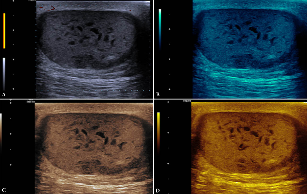
Fig. 1.
Grayscale (A) and various tints (B–D) of an epidermal inclusion cyst
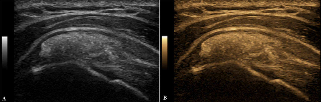
Fig. 2.
Grayscale (A) and tint (B) of supraspinatus calcific tendinopathy
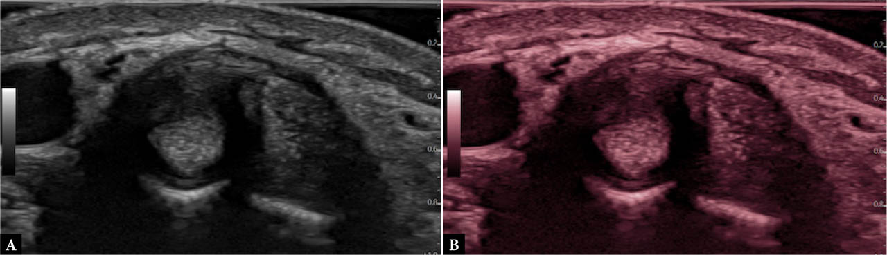
Fig. 3.
Grayscale (A) and tint (B) of de Quervain tenosynovitis
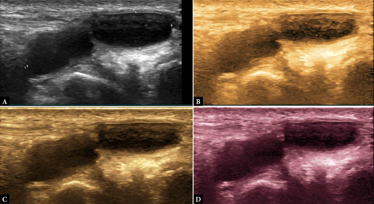
Fig. 4.
Grayscale (A) and various tints (B–D) of a ganglion