
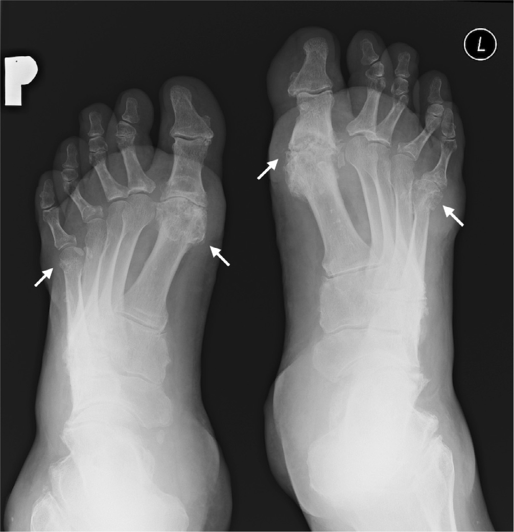
Fig. 1.
Radiography of feet, AP projection, year 2015. Advanced degenerative changes, with multiple geodes in the 1st MTP joints, geodes and erosions in head of 5th metatarsal bilaterally, arrows
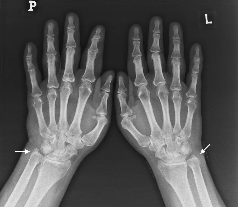
Fig. 2.
Radiography of hands in pronation, PA projection, year 2015. Geodes in styloid process of ulna bilaterally, narrowing of intra-articular spaces in the radiocarpal joint bilaterally, degenerative changes in the interpha-langeal joints and a small contracture in the 2nd proximal interphalangeal (PIP) joint of the right hand, arrows
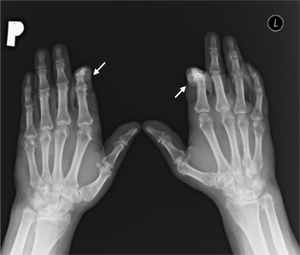
Fig. 3.
Radiography of hands in pronation, PA projection, year 2019. Thickening of soft tissues and contractures in 2nd PIP joints bilaterally, arrows
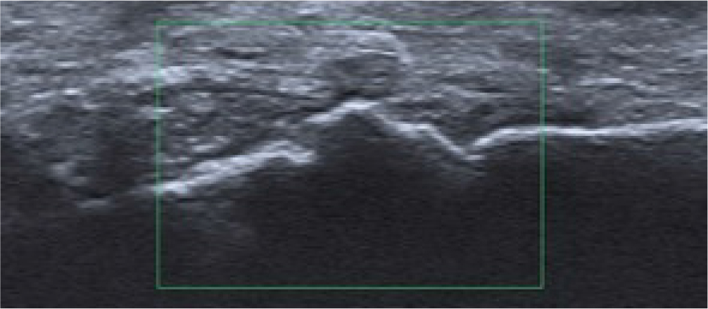
Fig. 4.
Ultrasound of the right hand shows thickening of the synovial membrane in the radiocarpal joint, with crystals and without hyperemia
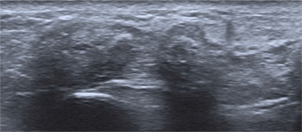
Fig. 5.
Ultrasound of the right hand shows hyperechoic content with tiny calcifications in the tenosynovium of the 3rd and 4th extensor compartments
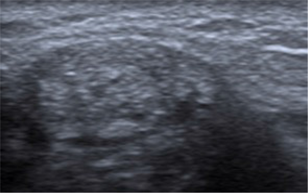
Fig. 6.
Tophus of high echogenicity, with tiny echogenic foci representing crystals (a “starry sky” sign) in the 6th compartment of the extensors
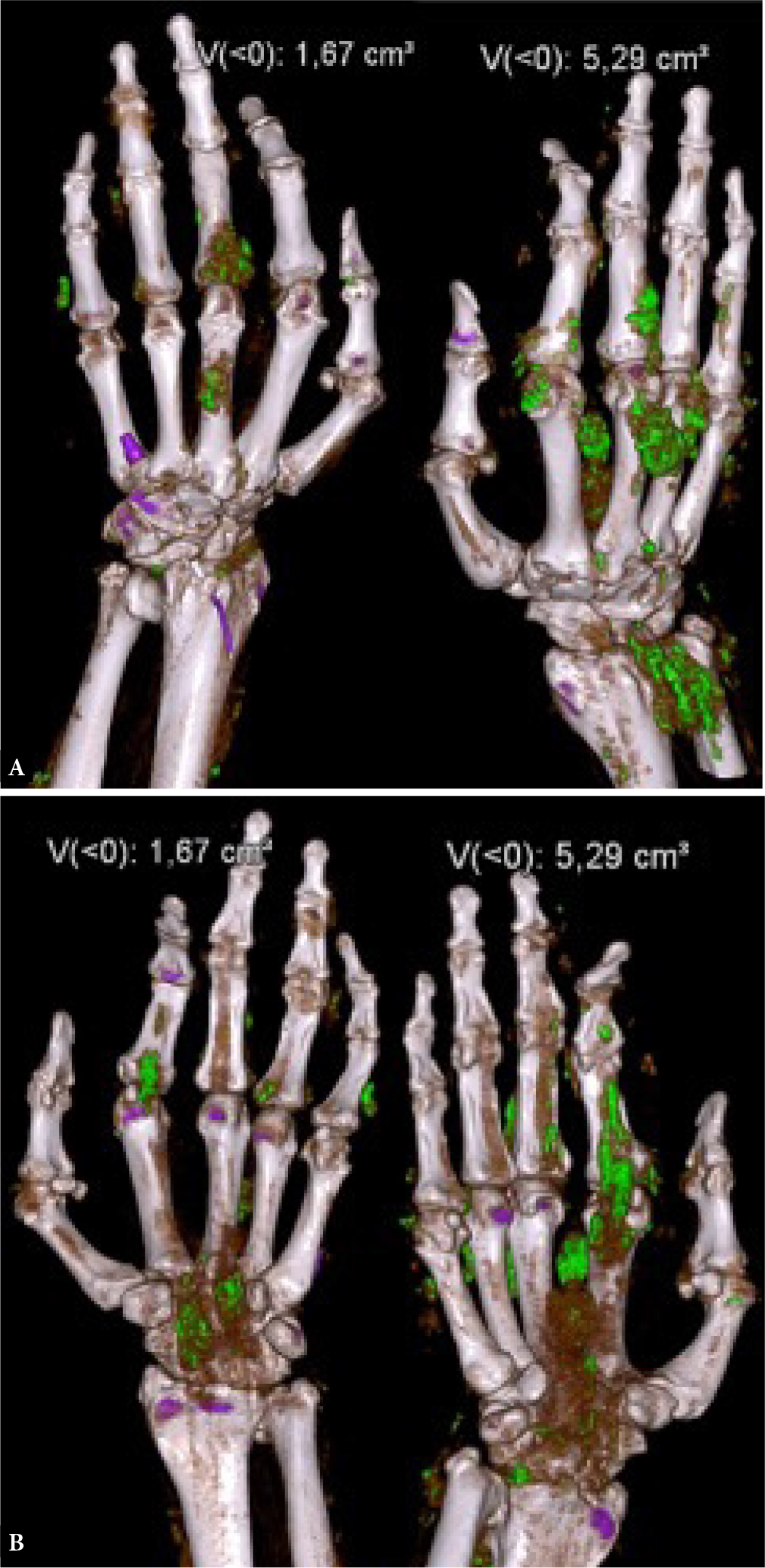
Fig. 7.
DECT of the hands in pronation (A) and in supination (B), year 2019. MSU crystal deposits are color-coded green
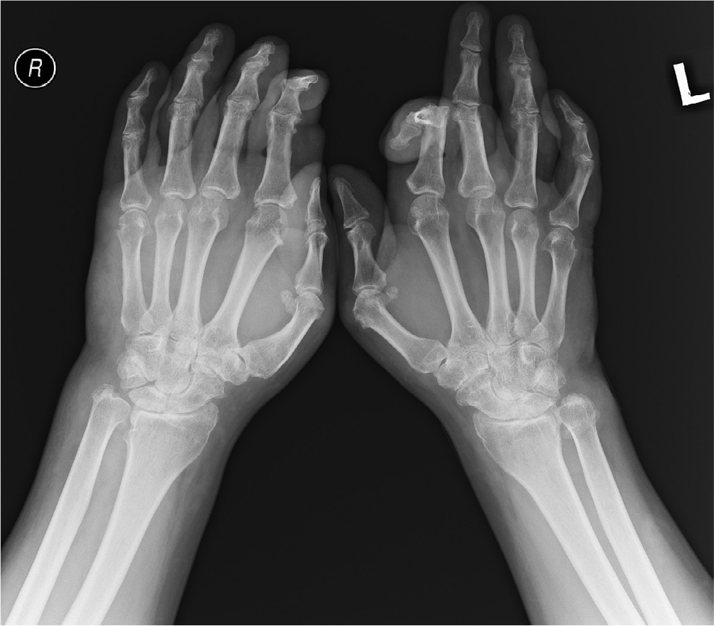
Fig. 8.
Radiography of the hands in pronation, PA projection, year 2021. Progression of contractures in the 2nd fingers
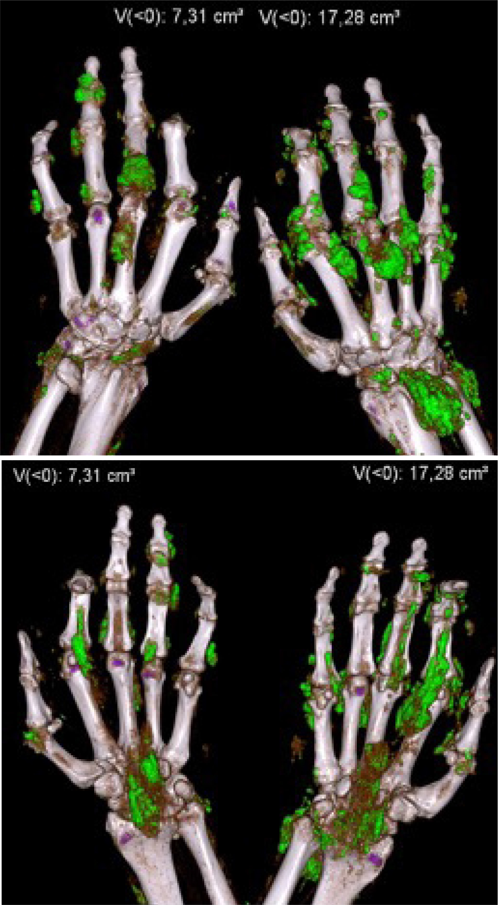
Fig. 9.
DECT of the hands in pronation (A) and in supination (B), year 2021. MSU crystal deposits are color-coded green
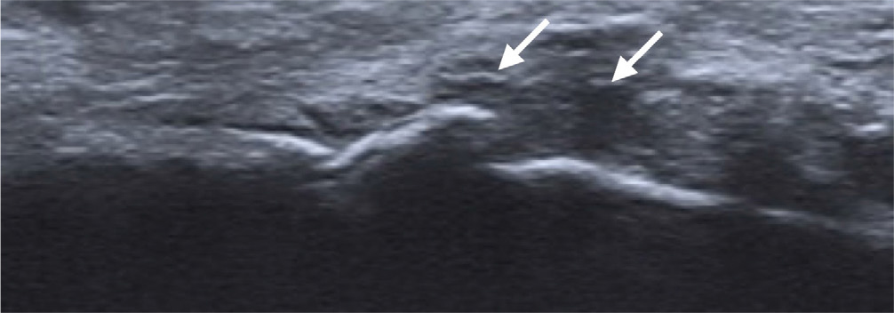
Fig. 10.
Ultrasound of the right 1st MCP joint, longitudinal view. Effusion and thickening of synovium with embedded small calcifications (a “starry sky” sign), arrows
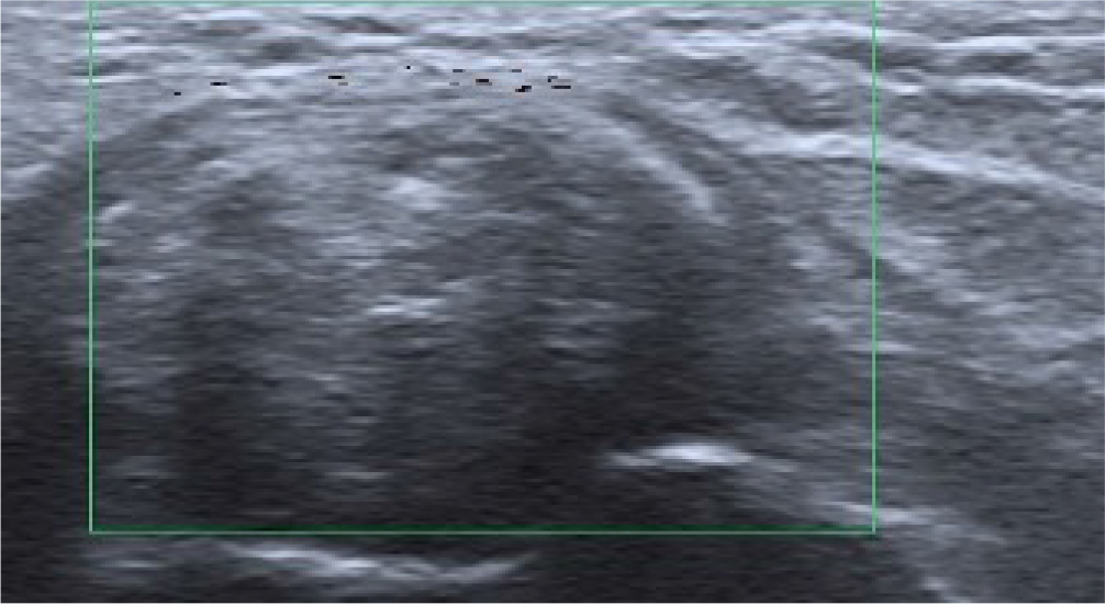
Fig. 11.
Hyperechoic tophus with tiny calcifications in the 5th MCP joint of the right hand, without increased vascularity, longitudinal view
Tab. 1.
Radiological features of gout based on the imaging modality in early and chronic stages of the illness(2)
| Early stage | Chronic stage | |
|---|---|---|
| Ultrasound | effusion synovial/tenosynovial hypertrophy bursitis starry sky sign (MSU deposits <1 mm) snowstorm sign/micro tophi (MSU aggregates >1 mm) double contour sign | tophi tendinopathy, tendons tears erosions |
| DECT | MSU crystal deposits detection and quantification | MSU crystal deposits detection and quantification |
| Radiography | asymmetric soft tissue swelling normal periarticular bone mineralization preservation of joint space rarely periostitis | erosions tophi pseudotumor pathological fracture normal periarticular bone mineralization preservation of joint space secondary osteoarthritis |
Tab. 2.
Specificity and sensitivity of ultrasound and DECT in the diagnosis of gout based on Ogdie et al.(17)
| Specificity | Sensitivity | |
|---|---|---|
| Ultrasound: Tophus | 0.80 | 0.65 |
| Ultrasound: Double contour sign | 0.76 | 0.83 |
| DECT: MSU crystal deposition | 0.84 | 0.87 |