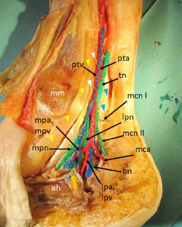
Fig. 1.
Fig. 2.
Fig. 3.
Fig. 4.

Fig. 5.
Fig. 6.

Fig. 7.
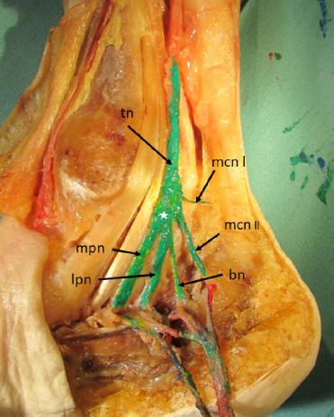
Fig. 8.

© 2023 Nežka Harej, Vladka Salapura, Erika Cvetko, Žiga Snoj, published by MEDICAL COMMUNICATIONS Sp. z o.o.
This work is licensed under the Creative Commons Attribution-NonCommercial-NoDerivatives 4.0 License.