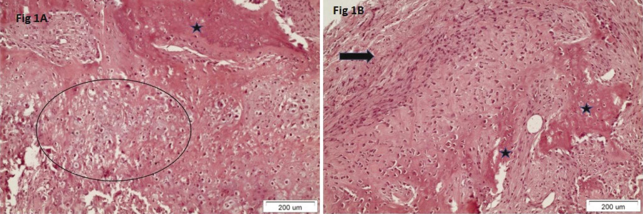
Fig. 1.

Fig. 2.
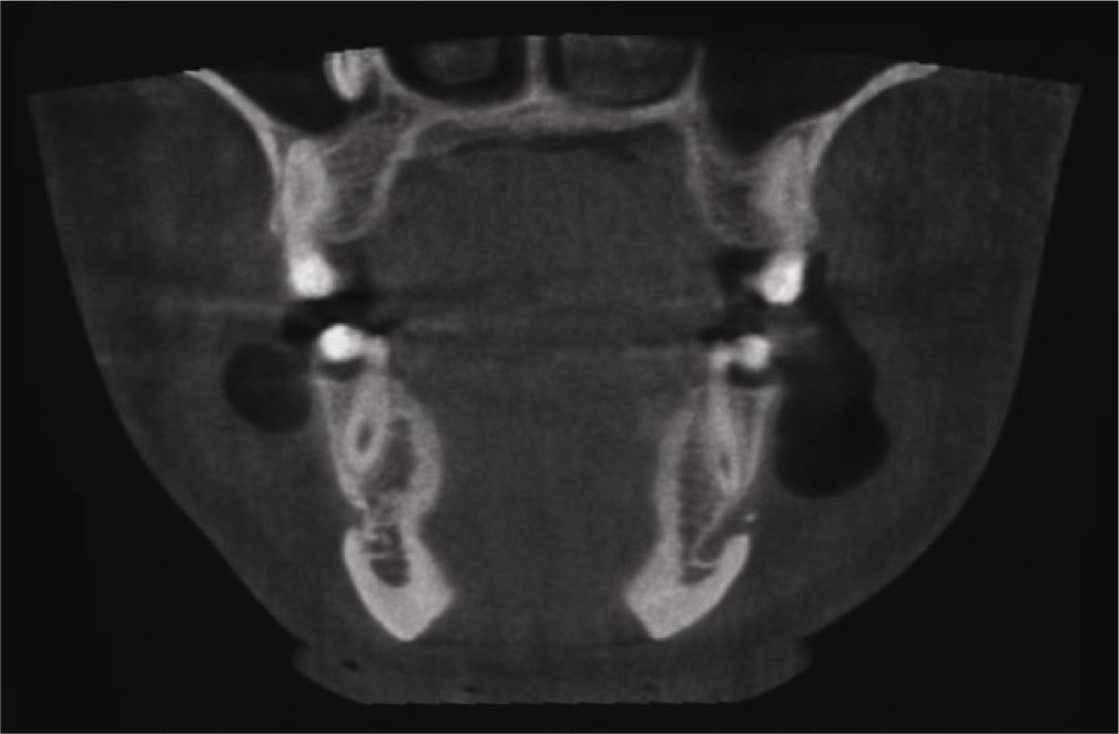
Fig. 3.
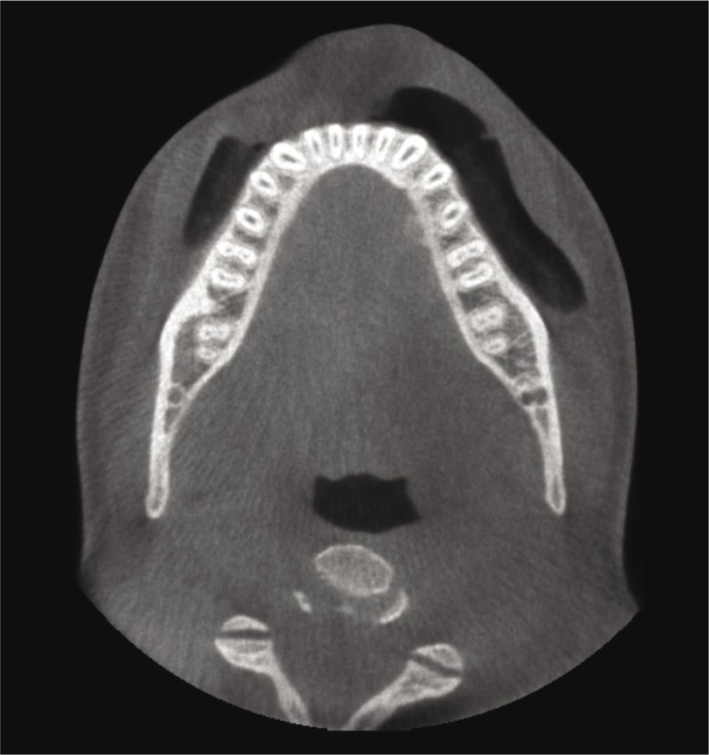
Fig. 4.
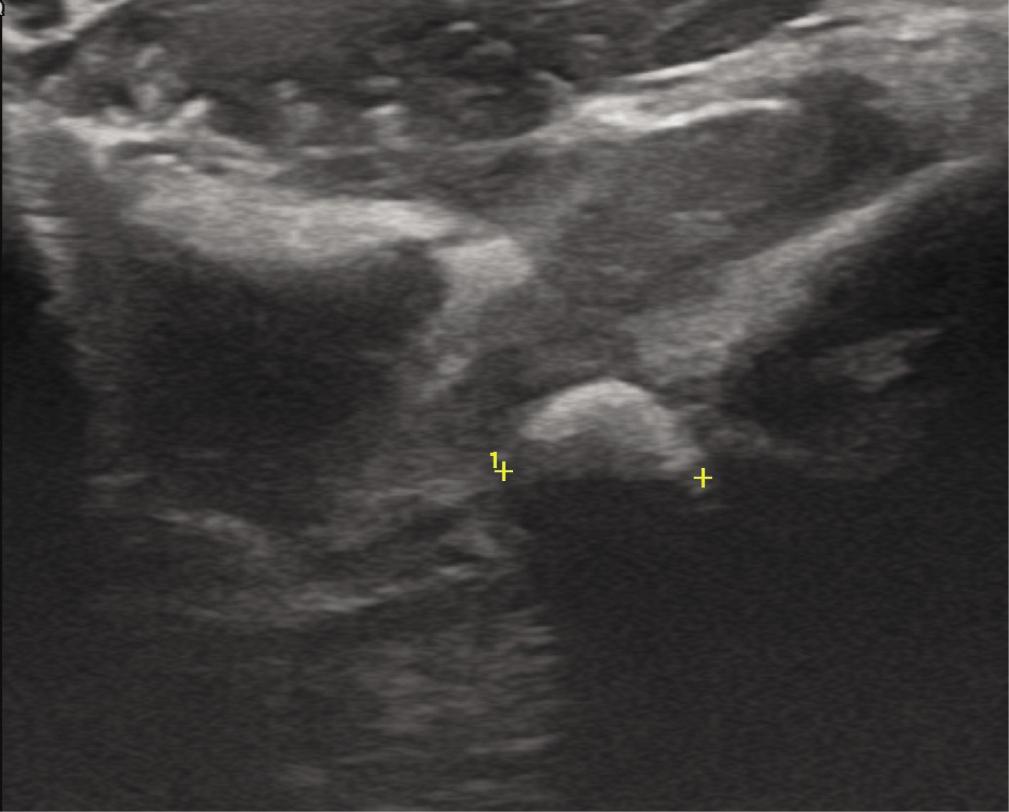
Fig. 5.
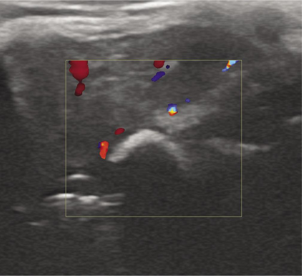
Fig. 6.
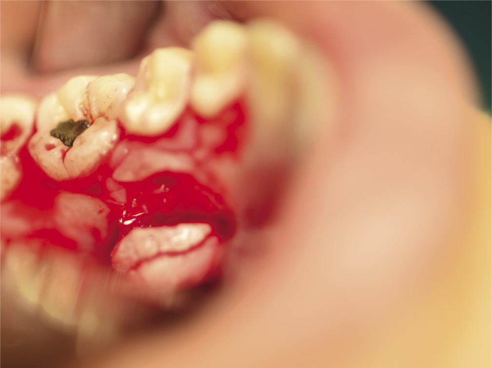
Fig. 7.

© 2022 Antigoni Delantoni, Apostolos Matiakis, Dimitrios Andreadis, Athanasios Poulopoulos, published by MEDICAL COMMUNICATIONS Sp. z o.o.
This work is licensed under the Creative Commons Attribution-NonCommercial-NoDerivatives 4.0 License.