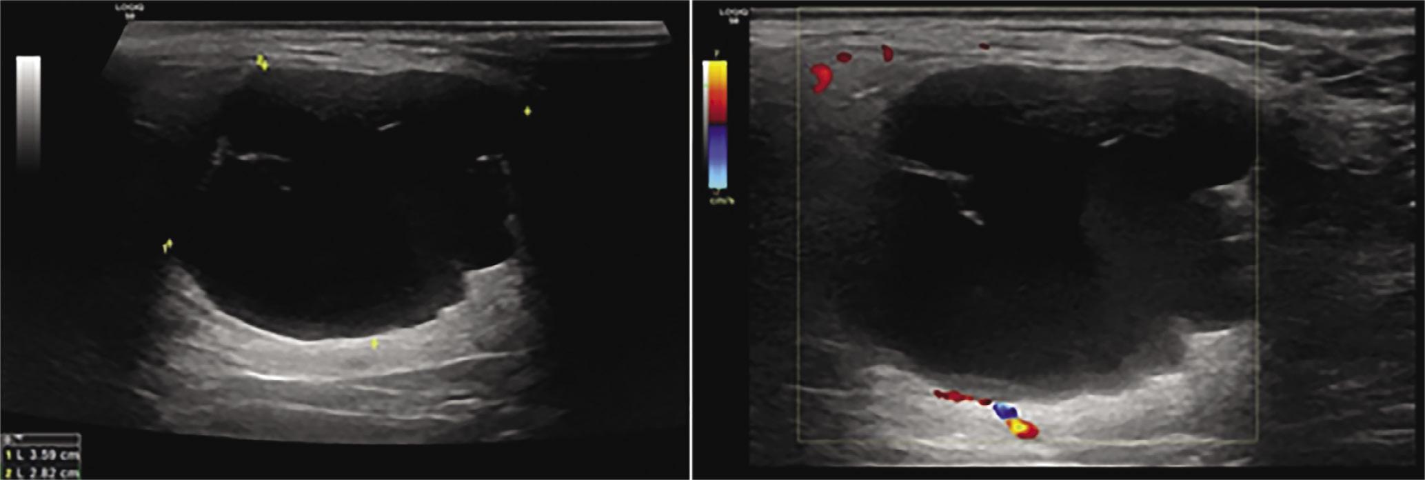
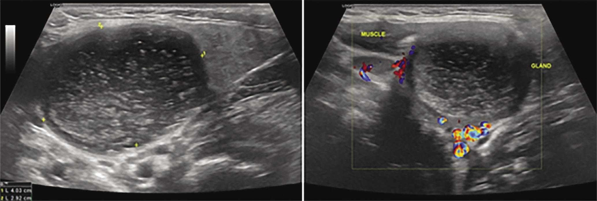
Have a personal or library account? Click to login
B-mode and color Doppler imaging of different types of branchial cleft cysts in children. A multicenter study and review of the literature Cover

B-mode and color Doppler imaging of different types of branchial cleft cysts in children. A multicenter study and review of the literature

Open Access
|Sep 2022

Figures & Tables

Fig. 1.

1st cleft cyst: in the inferior pole of the parotid gland one can observe a cystic-like lesion of multilobular borders, septa that are of low vascularity on color Doppler, and thick content, attached to the lateral borders

Fig. 2.

2nd cleft cyst: located in the submandibular region with thick mucous content of high echogenicity and without increased vascularity on color Doppler

Fig. 3.

2nd cleft cyst: typical location of the lesion with anterior displacement of the submandibular gland, posterior displacement of the parotid gland, and inner pressure on the large vessels of the area. The presence of a small stem connecting the lesion to the carotid space is pathognomonic of the lesion

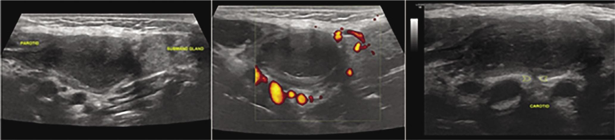
Table showing the differential diagnosis according to BCC types

Cleft cystDifferential diagnosis
1stAbscessesLymphatic malformations e.g. necrotic lymph nodesParotid mucoceleBenign lymphoepithelial cystWarthin tumor
2ndLymphatic malformationsNodal metastasisLymphadenopathyAbscessSchwannoma
3rdLymphatic malformationsAbscessInfrahyoid thyroglossal duct cystNodal metastasisLaryngoceleThyroid ectopic nodules
4thNecrotic lymph nodesAbscessCystic schwannomaThyroid colloid cystThymic cystLymphatic malformations

1st cleft cyst: in the inferior pole of the parotid gland one can observe a cystic-like lesion of multilobular borders, septa that are of low vascularity on color Doppler, and thick content, attached to the lateral borders

TypeTotal numberMale to female ratioDensityDoppler ultrasound (internal or peripheral vasculature increase/no flow)
1st cleft95/4All hypoechoic2/7
2nd cleft167/9Hypoechoic/hyperechoic (1 case)/mixed (1 case)1 (1 inconclusive)/14
3rd cleft32/1All hypoechoic0/3
4th cleft21/1All hypoechoic0/2
Inconclusive31/2All hypoechoic0/3
DOI: https://doi.org/10.15557/jou.2022.0028 | Journal eISSN: 2451-070X | Journal ISSN: 2084-8404
Language: English
Page range: e174 - e178
Submitted on: Nov 20, 2021
Accepted on: Feb 25, 2022
Published on: Sep 1, 2022
Published by: MEDICAL COMMUNICATIONS Sp. z o.o.
In partnership with: Paradigm Publishing Services
Publication frequency: 4 issues per year

© 2022 Antigone Delantoni, Merve Onder, Kaan Orhan, published by MEDICAL COMMUNICATIONS Sp. z o.o.
This work is licensed under the Creative Commons Attribution-NonCommercial-NoDerivatives 4.0 License.