Fig. 1
Fig. 2
Fig. 3
Fig. 4
Fig. 5
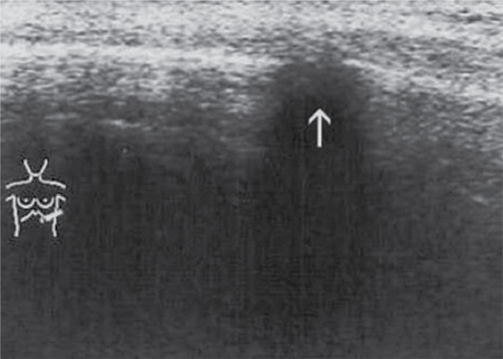
Fig. 6
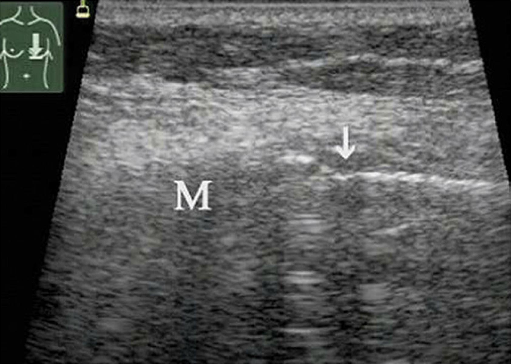
Fig. 7
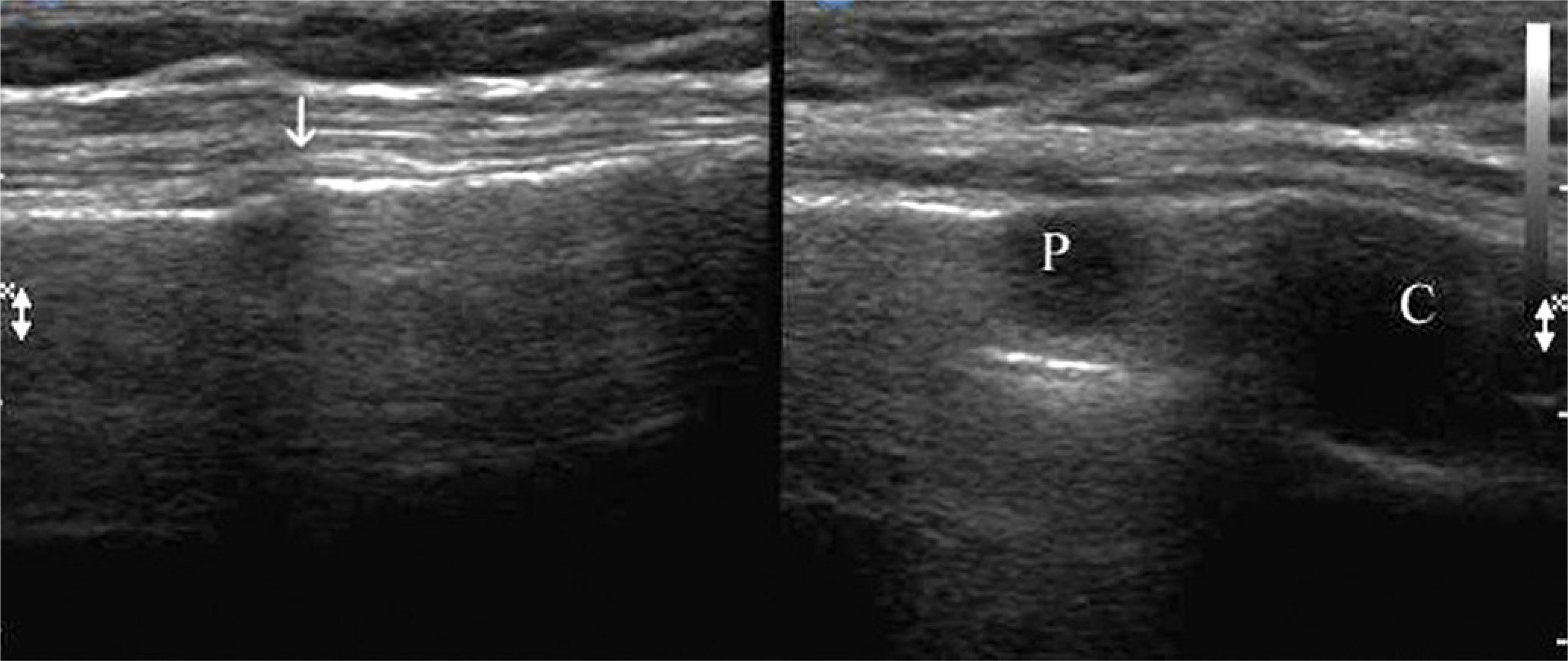
Fig. 8
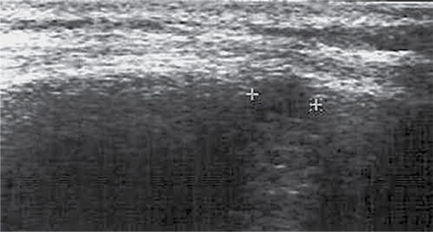
Fig. 9
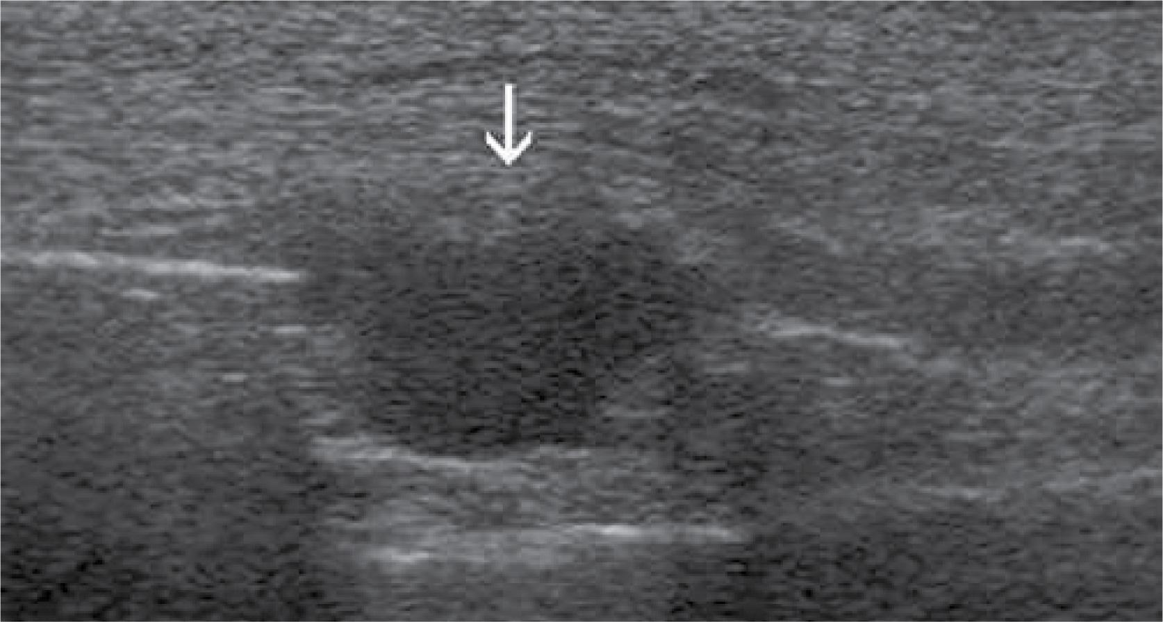
Fig. 10
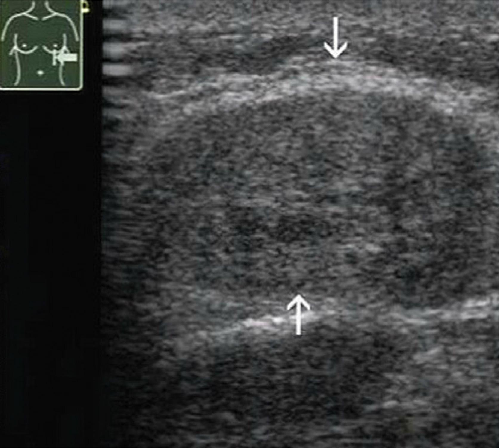
Fig. 11

Fig. 12
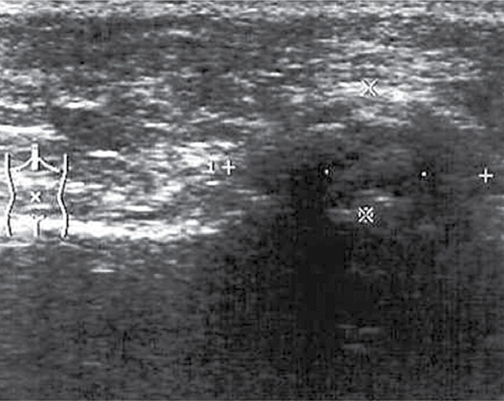
Fig. 13
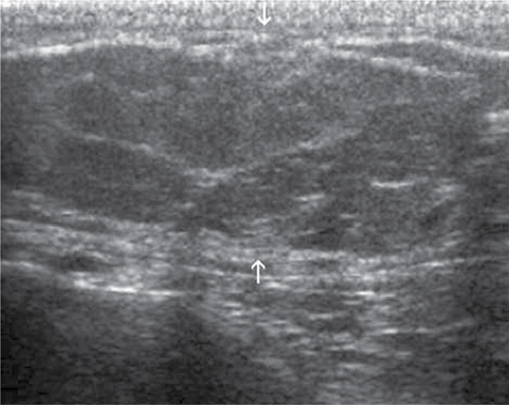
Fig. 14
Fig. 15
Fig. 16
Fig. 17
Fig. 18
Fig. 19
Fig. 20

© 2017 Andrzej Smereczyński, Katarzyna Kołaczyk, Elżbieta Bernatowicz, published by MEDICAL COMMUNICATIONS Sp. z o.o.
This work is licensed under the Creative Commons Attribution-NonCommercial 3.0 License.