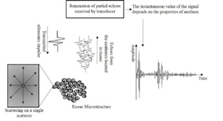
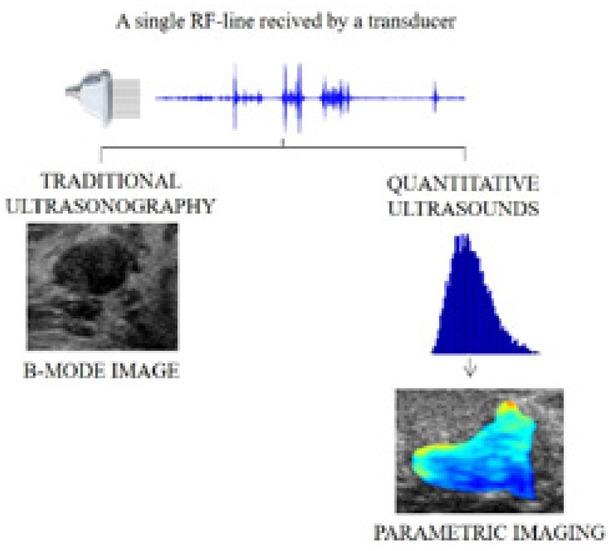
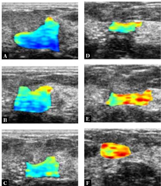
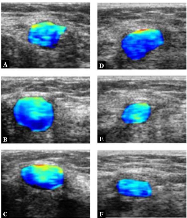
Have a personal or library account? Click to login
Quantitative ultrasonography as a tool for the evaluation of breast tumor response to neoadjuvant chemotherapy Cover

Quantitative ultrasonography as a tool for the evaluation of breast tumor response to neoadjuvant chemotherapy

Open Access
|Apr 2022

Figures & Tables

Fig. 1

Diagram of ultrasonic RF signal formation as a result of scattering on inhomogeneities present in the tissue

Fig. 2

Principle of operation of traditional ultrasonography (B-mode imaging) and quantitative ultrasound. In the case of a parametric map (generated based on the analyzed biomarker value – the effective number of diffusers), red denotes high values of the parameter (indicating a large number of identical small scatter structures), while blue represents low values (indicating a small number of large scatterers, e.g., clusters of cancer cells)

Fig. 3

B-mode ultrasound images with overlaid parametric IBC images determined for a patient responding to NAC before (A) and one week after each chemotherapy cycle (B–F). Blue indicates low IBC values; red indicates high values

Fig. 4

B-mode ultrasound images with overlaid parametric IBC images determined for a non-responder to NAC before (A) and one week after each chemotherapy cycle (B–F). Blue indicates low IBSC values; red indicates high values

Characteristics of the ultrasonic parameters discussed in the article

Determination methodParameter name and definitionTissue features affecting its value
Spectral parameters – determined directly from the signal spectrum in the frequency range corresponding to the transducer frequency bandMid-band fit (MBF) [dB]Size, shape, quantity, and elastic properties of scatterers
0-MHz intercept (SI) [dB]:Size, shape, quantity, and elastic properties of scatterers
Spectral slope (SS) [dB/MHz]:Size, shape of scatterers
Backscatter scattering parameters – determined on the basis of the backscattering coefficient in the frequency range corresponding to the transducer frequency bandAverage scatterer diameter (ASD) [μm]Average size of the scatterers (e.g., single cells or clusters of cells)
Average acoustic concentration (AAC) [dB/cm3]Spatial density, organization, elastic properties of scatterers
Integrated backscatter coefficient (IBC) [dB]Size (AND), shape, quantity, organization, and elastic properties of scatterers
Statistical properties
First-order statistical properties of the RF echo envelope – basic concept relies on modeling the magnitude of speckle with probability density functions, shape parameters of the K homodyne distributionENS – effective numbers of scatterers the scatterer clustering parameterQuantity, organization, and elastic properties of scatterers
k– the structure parameterSize, elastic properties of diffusing structures
DOI: https://doi.org/10.15557/JoU.2022.0015 | Journal eISSN: 2451-070X | Journal ISSN: 2084-8404
Language: English
Page range: 86 - 92
Submitted on: Nov 11, 2021
Accepted on: Jan 25, 2022
Published on: Apr 27, 2022
Published by: MEDICAL COMMUNICATIONS Sp. z o.o.
In partnership with: Paradigm Publishing Services
Publication frequency: 4 issues per year

© 2022 Hanna Piotrzkowska-Wróblewska, Katarzyna Dobruch-Sobczak, Jerzy Litniewski, published by MEDICAL COMMUNICATIONS Sp. z o.o.
This work is licensed under the Creative Commons Attribution-NonCommercial-NoDerivatives 4.0 License.