
Table 1
Epitope sequences in Thioredoxin Transglutaminase Multi Antigen Peptide (TT MAP)
| Antigens | Epitope Sequences |
|---|---|
| Transglutaminase peptide | KEFLLHETNGLVGIRTSENRYQFD |
| Thioredoxin peptide 1 | ADLLANINLKKADGTVKKGSDALANK |
| Thioredoxin peptide 2 | SEIEKLKNKYEVAGIP |
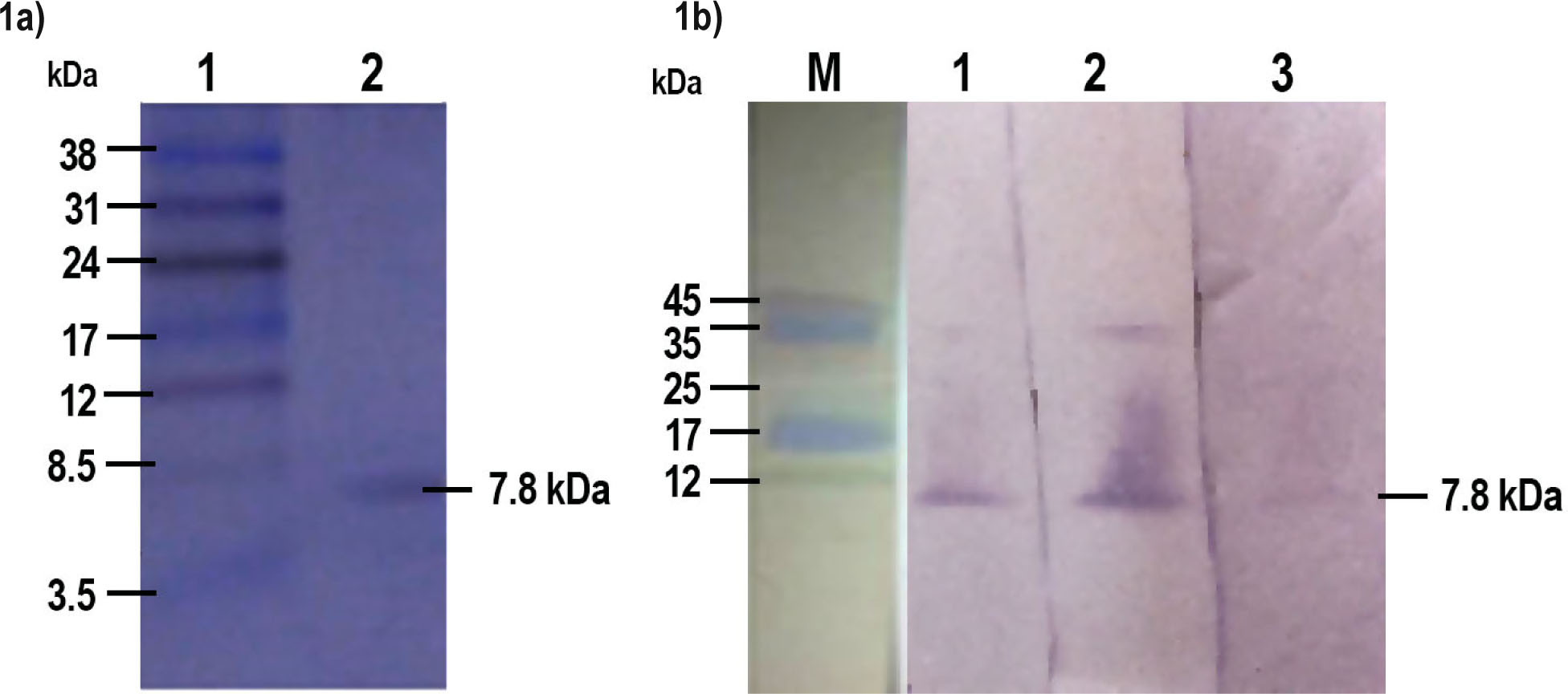
Fig. 1
Thioredoxin-Transglutaminase Multi Antigen Peptide (TT MAP):
1a. SDS-PAGE analysis of the synthetic TT MAP antigen: Lane 1 - Molecular marker, Lane 2 -TT MAP.
1b. Immunoblot analysis of TT MAP peptide: Lane M - Molecular marker, Lane 1-3 - Cross-reactivity of TT MAP with antibodies raised in mice against individual peptides (Bm TGA P1, Bm TRX P1 and Bm TRX P2, respectively)
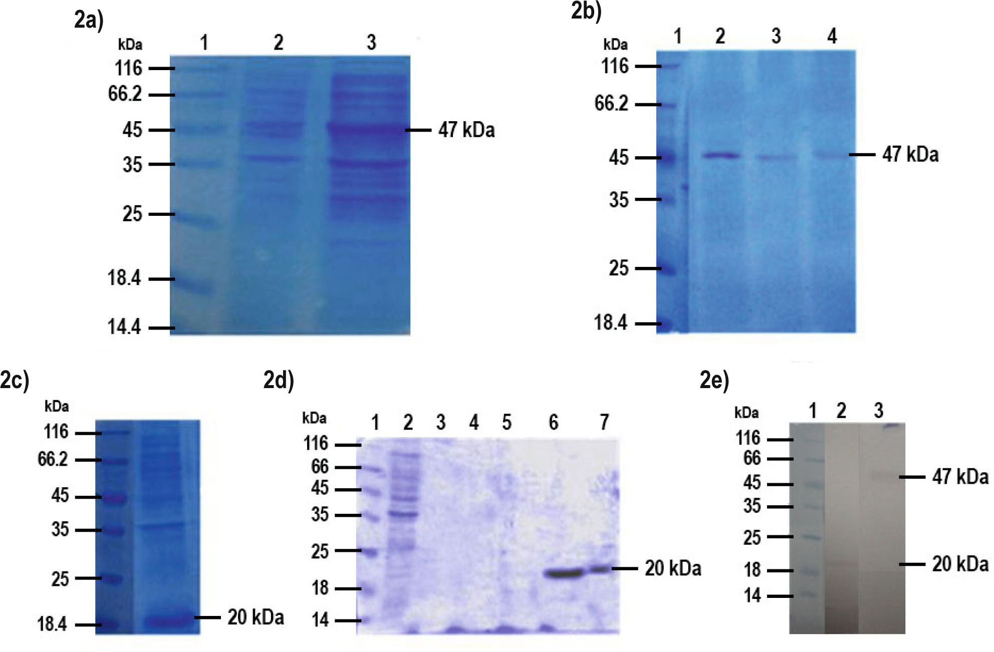
Fig. 2
Expression and purification of recombinant proteins:
(2a) Expression of the rTGA analyzed by 12 % SDS gel: Lane 1- Molecular marker, Lane 2 - pRSETB TGA uninduced, Lane 3 - pRSETB TGA induced
(2b) Purification of rTGA analyzed by 12 % SDS gel: Lane 1 - Molecular marker, Lanes 2-4 Pure TGA elutes
(2c) Expression of rTRX analyzed by 12 % SDS gel. Lane 1 - Molecular marker, Lane 2 - Expression of the 20 kDa rTRX produced from E. coli (GJ1158)
(2d) Purification of rTRX analyzed by 12 % SDS gel: Lane 1- Molecular marker, Lane 2- Flow through elute of TRX, Lanes 3-7 - Pure TRX elutes
(2e) Immunoblot cross-reactivity of purified rTRX and TGA with anti filarial mouse serum: Lane 1 - Molecular marker, Lane 2 - immunoblot with anti-TRX sera, Lane 3 - immunoblot with anti-TGA sera
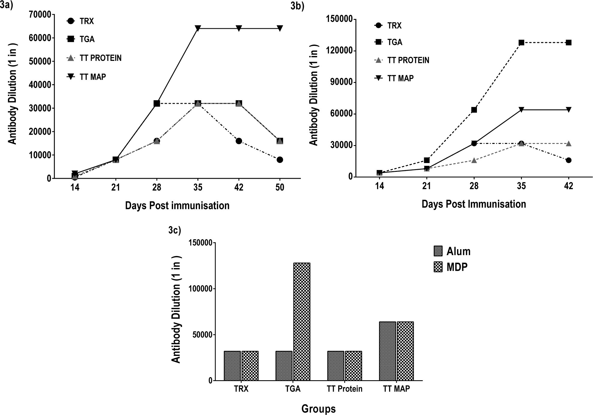
Fig. 3
Humoral immune response of TT MAP with Alum and MDP
3a. Antigen specific antibody titers (in thousands) in mice immunised with alum as adjuvant at day 14, 21, 35 and 42 post-immunization.
3b. Antigen specific antibody titers (in thousands) in mice immunised with MDP as adjuvant at day 14, 21, 35 and 42 post-immunization.
3c. Comparison of peak antibody titers followed by immunization with TRX, TGA, TT protein and TT MAP with alum/MDP as adjuvants. Individual points represent mean peak titers determined on day 35 (n=5 mice per experimental group)
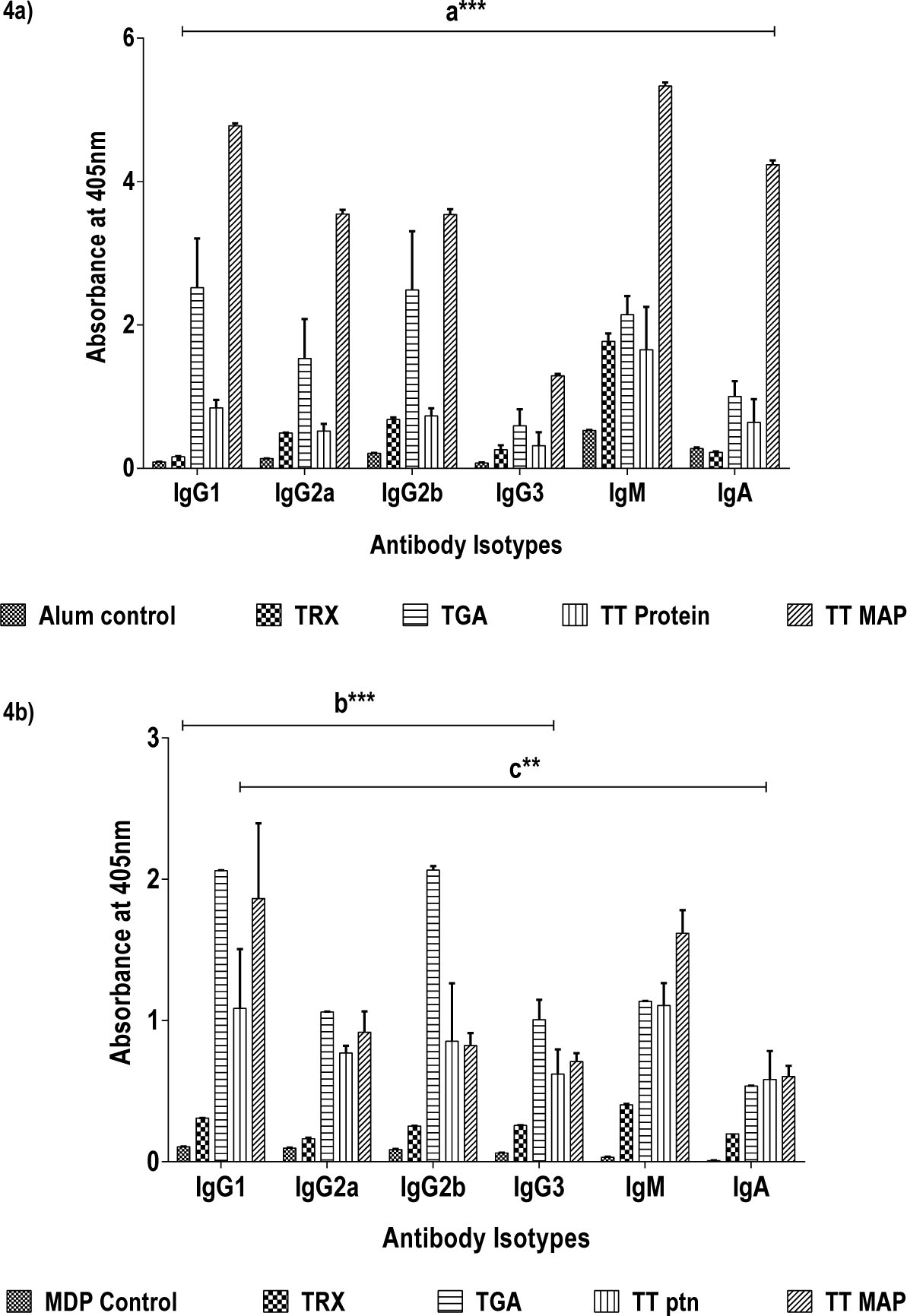
Fig. 4
Immunglobulin isotype profiles detected after the immunizations with Alum or MDP adjuvant
4a. Immunoglobulin isotype profile (IgG1, IgG2a, IgG2b, IgG3, IgM, IgA) followed by immunization with TRX, TGA, TT protein and TT MAP with alum as adjuvant.
4b. Immunoglobulin isotype profile (IgG1, IgG2a, IgG2b, IgG3, IgM, IgA) followed by immunization with TRX, TGA, TT protein, TT MAP with MDP as adjuvant. The antibody isotypes measured in 1:500 diluted sera are shown in bars. Data represents mean titer ± SD (n=5 mice per experimental group). Asterisks represents level of significance between control (adjuvant) and immunised groups (*** P < 0.001; ** P<0.01)
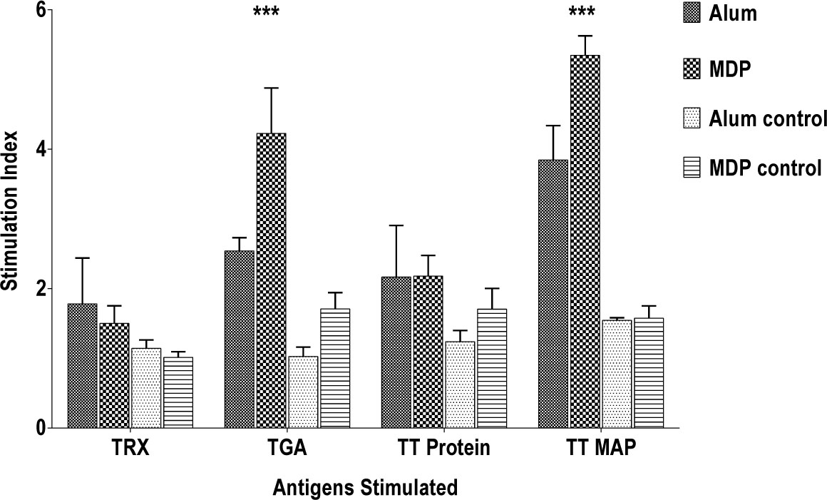
Fig. 5
Splenocyte proliferation
Splenocyte proliferation in different mice groups stimulated with corresponding antigens TRX, TGA, TT protein, TT MAP, compared to MDP/Alum controls. Data are presented as mean (± SD) Stimulation Index (S.I.) of five mice per experimental group. Asterisks represents level of significance between the TGA and TT MAP group where the MDP was used as adjuvant (*** P < 0.001)
Table 2
Cytokine levels
| Groups | IL2 | IFN-G | IL-4 | IL-5 | IL-10 | |
|---|---|---|---|---|---|---|
| TRX | Alum | 98 ± 10.6 | 160.81 ± 12.21aa | 321 ± 3.11aa | 230.33 ± 6.55aa | 89.27 ± 2.29 |
| MDP | 144.25 ± 1.76aa | 100.81 ± 3.21 | 259.29 ± 0.41 | 116.53 ± 3.68 | 110.47 ± 1.45a | |
| TGA | Alum | 177.35 ± .88 | 375.81 ± 6.423 | 509 ± 2.49 | 216.85 ± 6.766 | 61.11 ± .66 |
| MDP | 199.25 ± 5.3aa | 367.45 ± 3.21 | 547.38 ± 1.35 | 336.30 ± 3.9aa | 90.64 ± 0.06aa | |
| TT Protein | Alum | 127.37 ± 0.88 | 230.54 ± 1.28aa | 329.88 ± 6.66aa | 283.88 ± 0.922 | 94.44 ± .54 |
| MDP | 159.25 ± 1.78aa | 150.81 ± 5.78 | 293.11 ± 4.57bb | 263.95 ± 6.55 | 132.62 ± 7.25aa,bb | |
| TT MAP | Alum | 149.25 ± 8.83 | 214 ± 2.86 | 341 ± 3.26aa | 293 ± 5.48 | 85 ± 4.32aa |
| MDP | 221 ± 8.87aa,bb | 412 ± 2.86aa,bb | 226.91 ± 6.86 | 309.92 ± 5.09bb | 44.4 ± 0.2 |
Cytokine levels (pg/ml) in culture supernatants of spleen cells, from mice, immunized with the TRX, TGA, TT Protein or TT MAP with MDP as adjuvant and stimulated in vitro with respective antigens. Experiments were done thrice and data represented as mean concentration±SD.