
Table 1 (Tabelle 1)
Kinetic properties of CDHs from various sources (Kinetische Eigenschaften von CDHs unterschiedlicher Herkunft). All values were determined with dichloroindophenol (DCIP) as terminal electron acceptor, with the exception of the N. crassa CDHs IIA and IIB, for which benzoquinone was used.
| Cellobiose | Xylobiose | Maltose | Glucose | ||||||||
|---|---|---|---|---|---|---|---|---|---|---|---|
| Source of CDH | kcat | KM | kcat | KM | kcat | KM | kcat | KM | pH | References | |
| Class I | |||||||||||
| Phanerochaete chrysosporium | 16 | 0.11 | 0 | 0 | 1.1 | 240 | 2.6 | 1600 | 4.5 | Henriksson et al., 1998 | |
| Trametes villosa | 24 | 0.21 | 17 | 7.7 | 2.1 | 350 | 1.9 | 1300 | 4.5 | Ludwig et al., 2003 | |
| Sclerotium rolfsii | 27 | 0.12 | 27 | 5.4 | 0.8 | 240 | 1.5 | 1250 | 3.5 | Baminger et al., 2001 | |
| Trametes hirsuta | 11 | 0.04 | 0 | 0 | 2.8# | n.d. | 1.0# | n.d. | 5.0 | Nakagame et al., 2006 | |
| Ceriporiopsis subvermispora | 25 | 0.14 | n.d. | 1.3 | 300 | 1.0 | 3300 | 4.5 | Harreither et al., 2009 | ||
| Schizophyllum commune | 13 | 0.03 | n.d. | 0 | 0 | 0 | 0 | 4.5 | Fang et al., 1998 | ||
| Cerrena unicolor | 19 | 0.29 | n.d. | n.d. | 0 | 0 | 4.5 | Sulej et al., 2015 | |||
| Coprinopsis cinerea | 97 | 0.16 | n.d. | n.d. | 34.4 | 354 | 5.0 | Turbe-Doan et al., 2013 | |||
| Class II | |||||||||||
| Humicola insolens | 14 | 0.05 | 3.0 | 7.1 | 1.7 | 7.6 | 0 | 0 | 7.0 | Schou et al., 1998 | |
| Neurospora crassa IIA§ | 46 | 0.09 | 45 | 3.6 | 0.4 | 17 | 55 | 3700 | 6.0 | Sygmund et al., 2012 | |
| Neurospora crassa IIB§ | 11 | 0.02 | 5.8 | 1.3 | 3.5 | 3.4 | 8.1 | 550 | 6.0 | Sygmund et al., 2012 | |
| Thielavia terrestris | 1.2 | 0.04 | n.d. | 0.2 | 2.5 | 1 | 54 | 6.0 | Langston et al., 2012 | ||
| Myriococcum thermophilum§ | 13 | 0.01 | n.d. | 1.2 | 11 | 14 | 250 | 6.0 | Zámocký et al., 2008 | ||
| Corynascus thermophilus | 13 | 0.08 | n.d. | n.d. | 13 | 93 | 7.5 | Harreither et al., 2012 | |||
| Podospora anserina | 8.6 | 0.13 | n.d. | n.d. | 1.6 | 329 | 6.0 | Turbe-Doan et al., 2013 | |||
estimated from rel. activity (cellobiose=100%)
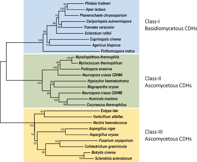
Unrooted phylogenetic tree comprising 27 cellobiose dehydrogenases (Ungewurzelter phylogenetischer Stammbaum von 27 Cellobiosedehydrogenasen). CDH sequences were derived from the NCBI database (http://www.ncbi.nlm.nih.gov/protein). Signal sequences were identified using the SignalP 4.1 server (http://www.cbs.dtu.dk/services/SignalP) and removed before further analysis. Evolutionary relationships were inferred with the minimum-evolution method (Rzhetsky and Nei, 1992) using the MEGA 7 software package (http://www.megasoftware.net). The tree was generated by the neighbor-joining method and the bootstrap consensus tree was inferred from 1000 replicates.
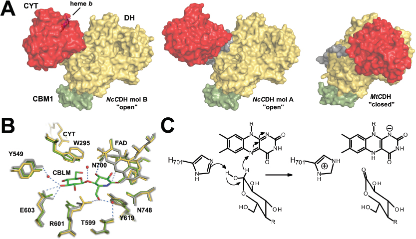
Structures and kinetic mechanism of cellobiose dehydrogenase (Strukturen und kinetischer Mechanismus von Cellobiosedehydrogenase). (A) Crystal structures of full-length CDHs from N. crassa (pdb: 4QI7) and M. thermophilum (pdb: 4QI6). Shown are the relative orientations of the cytochrome (CYT) domains (red), the dehydrogenase (DH) domains (yellow) and the linker peptides (grey) in the “open” state of two N. crassa CDHs (“mol A” and “mol B”) in comparison to the “closed” state of M. thermophilum CDH. Note the different orientations of the CYT domains in the structures of N. crassa. Family 1 carbohydrate binding modules (CBM1s) are shown in green. (B) Active--site architecture of M. thermophilum CDH (yellow) in comparison to the CBLM-bound DH (grey; pdb: 4QI5) and ligand-free DH (green; pdb: 4QI4). Figure modified from Tan et al. (2015). The propionate-A chain of CYT is approx. 9 Å away from the FAD. (C) Proposed catalytic hydride-transfer mechanism of CDH. Indicated is the suggested catalytic histidine. Bound substrates are eventually converted to a lactone. Numbering of amino acids is for M. thermophilum CDH. Figure adapted from Wongnate and Chaiyen (2013).
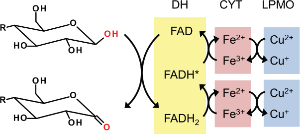
Electron-chain model for electron transfer in CDH (Das Elektronenketten-Modell für den Elektronentransfer in CDH). Following substrate oxidation, electrons are transferred sequentially from the reduced flavin to the heme cofactor and eventually to a suitable electron acceptor such as LPMO. FADH, flavin semiquinone. Figure adapted from Henriksson et al. (2000).
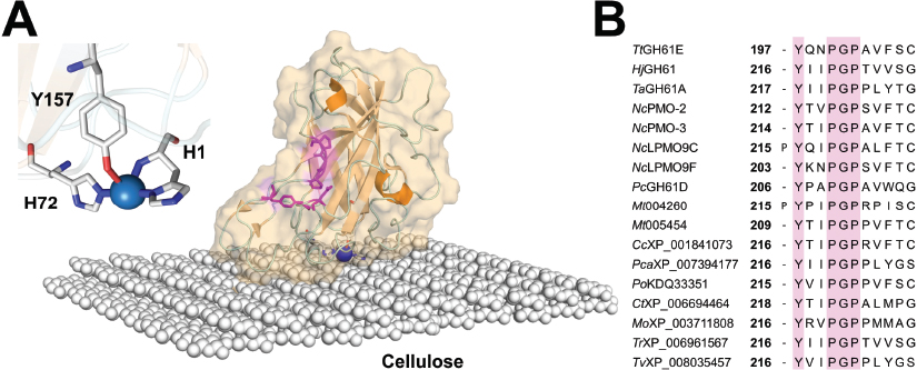
Structure of lytic polysaccharide monooxygenases (Struktur von lytischen Polysaccharid-Monooxygenasen, LPMOs). (A) N. crassa LPMO9F modeled onto an artificial cellulose surface. The active-site copper is shown in blue. Conserved aromatic amino acids on the surface are indicated in pink. The inset shows the copper coordination with the typical “histidine brace” formed by His1 and His72. (B) Sequence alignment of 18 crystallized and putative LPMOs from basidiomycetes and ascomycetes. The suggested “PGP” docking patch for CDH is highlighted in pink. The sequence alignment was generated using MUSCLE.
Table 2 (Tabelle 2)
Literature overview of in vitro assays demonstrating the synergy of LPMO and CDH (Literaturübersicht über in vitro Analysen, die den Synergismus zwischen LPMO und CDH verdeutlichen).
| CDH | LPMO | Substrate | Cellulase | Reference | ||
|---|---|---|---|---|---|---|
| Organism | Name | Organism | Name | |||
| H. insolens | HiCDH | T. aurantiacus | TaGH61A | PASC, MC | + | Langston et al., 2011 |
| T. terrestris | TtCDH | T. terrestris | TtGH61E | PASC,MC | + | Langston et al., 2011 |
| M. thermophila | MtCDH-2 | N. crassa | NCU01050 | PASC | - | Phillips et al., 2011b |
| M. thermophila | MtCDH-2 | N. crassa | NCU07898 | PASC | - | Phillips et al., 2011b |
| M. thermophila | MtCDH-2 | N. crassa | NCU08760 | PASC | - | Phillips et al., 2011b |
| T. terrestris | TtCDH | T. aurantiacus | TaGH61A | PASC | + | Langston et al., 2012 |
| N. crassa | CDH IIA | N. crassa | NCU01867 | MC | - | Kittl et al., 2012 |
| N. crassa | CDH IIA | N. crassa | NcLPMO9C | MC | - | Kittl et al., 2012 |
| N. crassa | CDH IIA | N. crassa | NcLPMO9F | MC | - | Kittl et al., 2012 |
| N. crassa | CDH IIA | N. crassa | NCU08760 | MC | - | Kittl et al., 2012 |
| P. cinnabarinus | PcCDH | P. anserina | PaLPMO9A | PASC | - | Bey et al., 2013 |
| P. cinnabarinus | PcCDH | P. anserina | PaLPMO9B | PASC | - | Bey et al., 2013 |
| M. thermophilum | MtCDH | N. crassa | NcLPMO9C | Cellopentaose | - | Isaksen et al., 2014 |
| M. thermophila | MtCDH-2 | N. crassa | NCU08746 | Amylopectin | - | Vu et al., 2014 |
| P. anserina | PaCDHB | P. anserina | PaLPMO9A | PASC | - | Bennati-Granier et al., 2015 |
| P. anserina | PaCDHB | P. anserina | PaLPMO9E | PASC | - | Bennati-Granier et al., 2015 |
| P. anserina | PaCDHB | P. anserina | PaLPMO9H | PASC | - | Bennati-Granier et al., 2015 |
PASC, phosphoric acid swollen cellulose; MC, microcrystalline cellulose
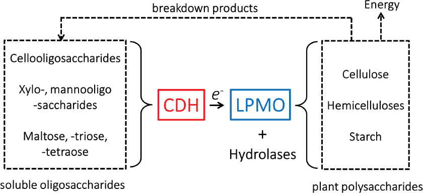
Suggested substrate cycling during oxidative cellulolysis by wood decaying fungi (Substratkreislauf während des oxidativen Zelluloseabbaus durch holzabbauende Pilze). Cellulose-derived oligosaccharides, hemicellulose-derived xylo- and manno-oligosaccharides and starch-derived malto-oligosaccharides are substrates for a number of CDHs, and can be released by the action of LPMO and hydrolases.