
Table 1
Laboratory findings during hospitalization and follow-up of an acutely febrile patient with suspected dengue infection†
| Day of fever | 2 | 4 | 5 | 7 | 2nd wk | 5th wk | 6th wk | 8th wk |
|---|---|---|---|---|---|---|---|---|
| Body temperature (°C) | 39.8 | 39.4 | 37.2 | 36.8 | 36.5 | 36.2 | 36.5 | 36.5 |
| Hematocrit (%) | 29.4 | 21.9 | 22.0 | 27.5 | 37.3 | 43.7 | 41.3 | 44.3 |
| WBC (×103/μL) | 4.1 | 3.7 | 5.1 | 11.4 | 11.9 | 8.4 | 3.6 | |
| Neutrophils (%) | 64.0 | 59.0 | 71.0 | 79.0 | 75.0 | 62.0 | 41.0 | |
| Lymphocytes (%) | 14.0 | 20.0 | 22.0 | 20.0 | 20.0 | 28.0 | 50.0 | |
| Atypical lymphocytes (%) | 6.0 | 2.0 | 4.0 | 0 | 0 | 0 | 0 | |
| Monocytes (%) | 3.0 | 4.0 | 3.0 | 0 | 4.0 | 9.0 | 8.0 | |
| Eosinophils (%) | 10.0 | 10.0 | 0 | 1.0 | 0 | 1.0 | 2.0 | |
| Basophils (%) | 0 | 0 | 0 | 0 | 1.0 | 0 | 0 | |
| Platelet count (×103/μL) | 59 | 43 | 58 | 194 | 275 | 179 | 202 | |
| AST (U/L) | 28 | 16 | ||||||
| ALT (U/L) | 16 | 24 | ||||||
| LDH (U/L) | 523 | 769 | ||||||
| NS1 Ag | Negative | |||||||
| Dengue virus IgM and IgG antibodies | Positive |
[i] WBC, white blood cells; AST, aspartate transaminase; ALT, alanine aminotransferase; LDH, lactate dehydrogenase; Ig, immunoglobulin; NS1, dengue virus nonstructural protein 1 was negative and the †ultimate diagnosis was systemic lupus erythematosus
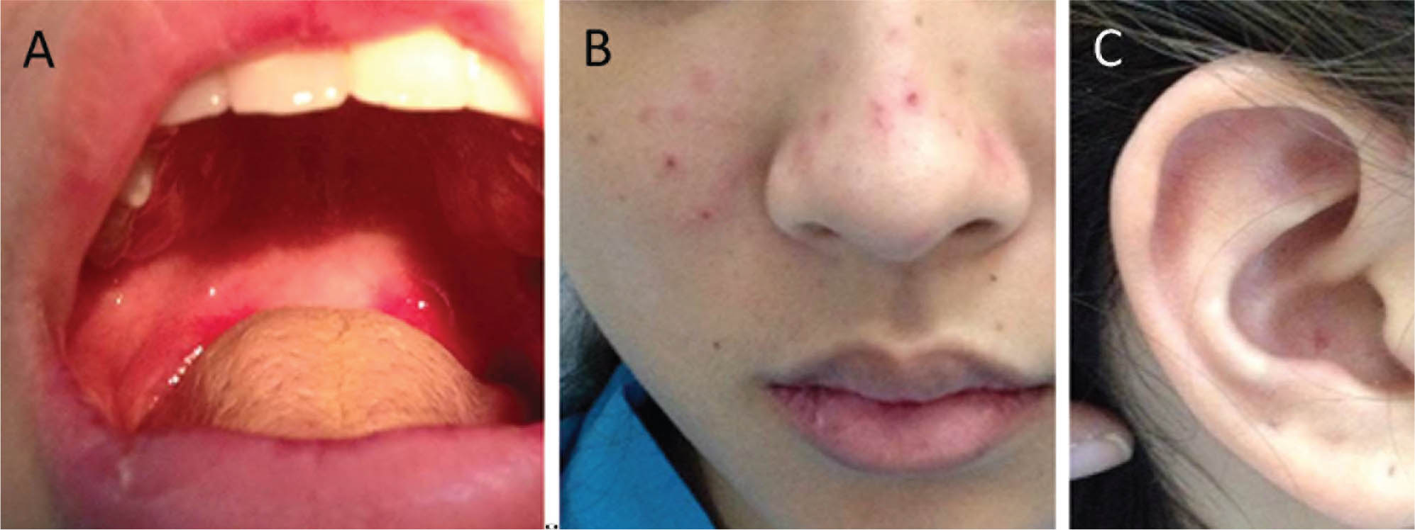
Figure 1
Presentation of a 19-year-old acutely febrile patient from a region endemic for dengue. A. Shallow clear painless ulcer on the soft palate area. B. Erythematous macular rash on the malar area. C. Discoid rash on the ear. With consent for publication from the patient.
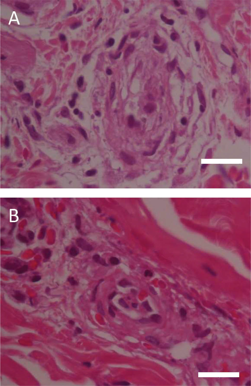
Figure 2
A and B. Skin biopsy from an erythematous nonblanchable patch on the abdomen of the acutely febrile patient showing a pattern of vasculitis. Dense superficial and mid-perivascular and interstitial mixed inflammatory cell infiltrate composed of lymphohistiocytes, neutrophils, a few nuclear remnants, and extravasated red blood cells (hematoxylin and eosin staining; original magnification ×100; scale bars indicate 10 μm).