
Case Presentation
A 68-year-old African American man developed exertional dyspnea and volume overload three months before admission. Technetium-99m pyrophosphate scintigraphy was positive, and serum free light chains with serum and urine immunofixation were normal, establishing transthyretin amyloid cardiomyopathy (ATTR-CM). He had stage IV chronic kidney disease from focal segmental glomerulosclerosis and was already listed for kidney transplantation. He had never had carpal tunnel syndrome, lumbar spinal stenosis, or tendon rupture; sibling neuropathy was elicited only on directed questioning.
QRS voltage was not low. Echocardiography showed concentric hypertrophy with an ejection fraction of 55% to 59% but a global longitudinal strain of –11.9% with apical sparing, grade III restrictive filling, and a stroke volume index of 27 mL/m². Magnetic resonance imaging confirmed 1.5-cm wall thickening with diffuse late gadolinium enhancement and an extracellular volume fraction of 70%; N-terminal pro-B-type natriuretic peptide was 4,900 pg/mL.
Targeted TTR sequencing on whole blood was sent on the second hospital day for three reasons: It is recommended in every patient with confirmed ATTR-CM; he was 68 with West African ancestry and a family history of neuropathy, each raising the probability of hereditary disease; and the result would change both his relatives’ surveillance and his own management after transplantation. It identified the pathogenic p.Val142Ile variant, establishing hereditary rather than wild-type disease. He progressed to cardiogenic shock requiring an intra-aortic balloon pump and to dialysis, was listed status 2 for combined heart-kidney transplantation, and his relatives were referred for genetic counseling and cascade testing (Figure 1).
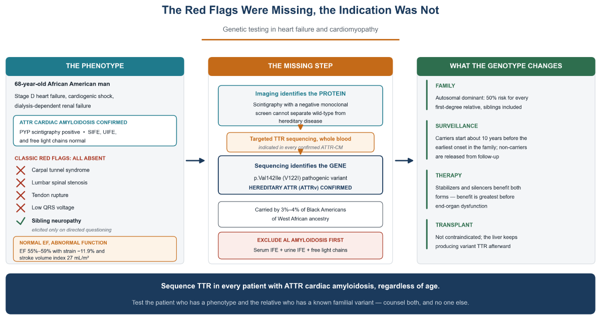
Figure 1
A fully characterized phenotype is not a complete diagnosis. Bone scintigraphy with a negative monoclonal screen identifies the amyloid protein but cannot separate wild-type from hereditary transthyretin amyloidosis; only TTR sequencing does. Every classic red flag was absent in this patient, yet the indication to test was the confirmed diagnosis itself. Testing is directed at the patient with a phenotype and at relatives of a known familial variant, with counseling before and after. AL: light chain amyloidosis; ATTR: transthyretin amyloidosis; ATTR-CM: ATTR cardiomyopathy; EF: ejection fraction; IFE: immunofixation electrophoresis; PYP: technetium-99m pyrophosphate; TTR: transthyretin
Points to Remember
(1) Characterizing the cardiomyopathy is only half the diagnosis; the genotype is the other half. Bone scintigraphy identifies the protein, not the gene; a positive scan with a negative monoclonal screen confirms ATTR but cannot separate wild-type from hereditary disease, which is why TTR sequencing is recommended in all patients with ATTR-CM regardless of age.1,2 Across cardiomyopathies, the 2023 European Society of Cardiology guidelines make testing with counseling Class I whenever the result would establish a diagnosis, refine prognosis, guide therapy, or enable family screening.3,4
(2) The family history is the cheapest genetic test you have and often the only clue. This patient’s sole heritable signal was sibling neuropathy, and it surfaced only because someone asked. Take a three-generation pedigree covering heart failure, unexplained hypertrophy, pacemakers, neuropathy, and sudden death, with ages. In dilated cardiomyopathy, family history is itself a testing indication, alongside conduction disease with ventricular dysfunction (consider LMNA), skeletal myopathy or raised creatine kinase, and unexplained sudden death before age 50.4,5
(3) Red flags, age, and ancestry shift pretest probability; they never remove the indication once disease is confirmed. Bilateral carpal tunnel syndrome, lumbar spinal stenosis, and tendon rupture precede cardiac presentation by 5 to 10 years, but their absence, as here, is a reason to look harder, not to test less.2 Low QRS voltage is a poor rule-out, and ejection fraction misleads equally: His was normal while strain and stroke volume index were not. Wild-type disease predominates after age 70, so older patients are labeled “senile” and never genotyped; ours was 68. The p.Val142Ile variant is carried by 3% to 4% of Black Americans of West African ancestry, one of the most common pathogenic cardiovascular variants in the country, with polyneuropathy in about 10% of carriers; failing to act on that is a health equity problem.6
(4) Exclude light chain amyloidosis first, then match the test to the phenotype. Order serum immunofixation, urine immunofixation, and serum free light chains, all three, as here. Electrophoresis alone is insufficient, and a positive scan with a monoclonal protein mandates tissue typing.1,7 In advanced kidney disease, use the renal free light chain reference range (approximately 0.37–3.1).8 When the amyloid protein is known, targeted TTR sequencing suffices; otherwise a multigene panel is first-line, capturing phenocopies such as Fabry, PRKAG2, and Danon disease.5,9 Turnaround is typically 2 to 4 weeks, and laboratories will often expedite when the result will change inpatient decisions, so ask rather than defer.
(5) Test the patient who has a phenotype, and count what a positive result costs. Testing belongs to the patient with a cardiomyopathy phenotype and, once a pathogenic variant is found, to at-risk relatives, not to the asymptomatic population and not to patients whose phenotype is explained by another cause. Relatives are tested for the single familial variant, not with a panel; a variant of uncertain significance must never be cascaded, and when proband testing is uninformative, relatives receive clinical surveillance instead. Wild-type ATTR confers no heritable risk, so family testing is not indicated. Counsel before and after. For adult-onset disease, defer testing of minors until the age at which surveillance would begin.4,5 Note that a patient with either phenotype (polyneuropathy or cardiomyopathy) can be tested; testing is not limited only to cardiomyopathy.
(6) The result belongs to the family, and it changes what you do for the patient. ATTRv is autosomal dominant: Every first-degree relative carries a 50% risk, siblings included—a group easily overlooked when the proband is elderly. Variant-positive, phenotype-negative relatives begin surveillance about 10 years before the earliest onset in the family; non-carriers are released from it.1,4 Genotype does not gate therapy since stabilizers and silencers benefit both forms, but benefit is greatest before end-organ dysfunction, and our patient reached stage D untreated.10,11 Hereditary disease is not a contraindication to transplantation, and because the liver keeps producing variant transthyretin afterward, TTR-directed surveillance remains relevant.
Conclusion
The difficulty here was not the diagnosis but the order of our reasoning. A phenotype exhaustively characterized by echocardiography, magnetic resonance imaging, and scintigraphy was still one blood test short of a complete answer, and that test was the only one that reached beyond the patient to his family. Test the patient who has a phenotype and the relative who has a known familial variant, counsel both, and leave everyone else alone.