
Introduction
Thoracic aortic aneurysm (TAA) is an abnormal widening of the aorta that predisposes to acute aortic syndromes with high morbidity and mortality, including aortic dissection and aortic rupture. TAA arises from medial degeneration of the aortic wall due to fragmentation and loss of elastic fibers, damage to vascular smooth muscle cells, deposition of proteoglycans, and increased extracellular matrix degradation by matrix metalloproteinases.1,2 Approximately 60% of TAAs occur in the aortic root and/or ascending aorta, 10% occur in the aortic arch, and 30% occur in the descending thoracic aorta.3
TAA with genetic causes can be due to monogenic heritable thoracic aortic disease (HTAD), familial disease without an identified causal variant, polygenic risk factors, and rare-variant modifiers. Historically, approximately 20% of thoracic aortic disease has been attributed to inherited, monogenic disease based on contemporary HTAD genetic testing panels.3,4,5 This includes both syndromic HTAD due to genetic vasculopathies, including Marfan syndrome (MFS), Loeys-Dietz syndrome (LDS), and vascular Ehlers-Danlos syndrome (vEDS), as well as non-syndromic HTAD, in which there is a positive family history of thoracic aortic disease and an identified monogenic variant without additional phenotypic features consistent with a known vasculopathy syndrome. There are additional patients with a family history of thoracic aortic disease suggestive of inherited disease but without identified causal genetic variants, likely reflecting patients with not-yet-described monogenic variants and/or polygenic risk factors.
Non-inherited contributors to thoracic aortic disease include cardiometabolic comorbidities (eg, hypertension, atherosclerosis), inflammatory or infectious causes (eg, immune-mediated vasculopathies, syphilis), and prior dissection or traumatic aortic injury.3,6 TAA is also associated with bicuspid aortic valve and Turner syndrome, which are outside of the scope of this review.
The recognition of HTAD has important implications for clinical management and may prompt more frequent imaging surveillance, the use of medical therapies for afterload reduction and impulse control, lower aortic diameter thresholds for prophylactic TAA repair, and cascade screening among first-degree relatives. HTAD comprises a heterogeneous group of disorders involving defective extracellular matrix integrity, as well as issues with mechanosensing and compliance, smooth muscle contraction, cellular differentiation, and genetic regulation.1,6,7 Clinically, risk stratification has traditionally focused on identifying patients with syndromic connective tissue disorders that carry elevated risk of acute aortic syndrome.3,8 However, more recently, genome-wide association studies (GWAS), exome or genome-wide sequencing, and modern machine learning techniques have enabled a more nuanced understanding of the polygenic architecture of TAA.
In this review, we discuss known monogenic aortopathies, the impact of polygenic risk variants, the association of TAA with other arteriopathies, and the role of genetic testing. We conclude by reviewing evidence gaps and future directions in the field of genetic aortopathy. A better understanding of the monogenic and polygenic mechanisms of TAA is crucial to reduce the burden of acute aortic syndromes, an important cause of sudden cardiac death and preventable cardiovascular morbidity and mortality.
Syndromic and Non-Syndromic Hereditary Thoracic Aortic Disease
Genetic variants causing HTAD disturb the TGFβ signaling cascade, the smooth muscle contraction apparatus, and components of the extracellular matrix, all of which are essential to maintain connective tissue homeostasis and vascular wall integrity. During physiologic smooth muscle signaling, a large latent complex containing inactive TGFβ is sequestered to the extracellular matrix by microfibrils containing fibrillin proteins (FBN1 and FBN2), which concentrate ligands in the extracellular matrix, potentiating activity of TGFβ and binding of ligand to the TGFβ receptor (TGFBR1 and TGFBR2). Receptor activation results in the phosphorylation and activation of SMAD transcription factors (SMAD2, SMAD3, and SMAD4) in the canonical pathway or other transcription factors such as ERK, JNK, and p38 in the non-canonical pathway. SMAD transcription factors translocate to the nucleus and regulate gene expression of connective tissue growth factors, collagens, and matrix metalloproteinases.9,10 A decrease in the potency of this signaling system results in vascular smooth muscle cell (VSMC) dysfunction and aortopathy.
Disruption of other gene networks also undermines aortic homeostasis. The integrity of the VSMC cytoskeleton is dependent on the contractile apparatus of the smooth muscle cell. In VSMCs, muscle contraction depends on contractile fibers that are composed of actin thin filaments (ACTA2), myosin heavy chain (MYH11), myosin light chains (MYL6), and additional regulatory proteins such as myosin light chain kinase (MYLK) and cGMP-dependent protein kinase (PRKG1), which promote VSMC contraction and relaxation, respectively.11 Genetic disruption of the smooth muscle cytoskeleton destabilizes the VSMC phenotype and aortic structural stability. Aortic structure and function are ultimately maintained by the extracellular matrix, which contains proteins necessary for elastin and collagen organization and signaling including type 3 collagen (COL3A1), fibrillin-1 (FBN1), elastin (ELN), and modifying enzymes such as lysyl oxidase (LOX).6 TAA can arise from genetic variants that disrupt any component of these pathways (Figure 1). Connective tissue disorders with syndromic features arise when the tissue distribution of the affected gene product extends beyond the aorta and can include diverse phenotypic findings (Table 1).
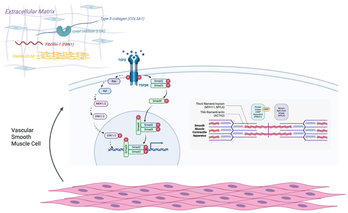
Figure 1
Pathogenic variants in the extracellular matrix, TGFβ signaling pathway, and smooth muscle contractile apparatus. Schematic of genetic variants associated with thoracic aortopathy. Genetic variants associated with thoracic aortopathy include those involved in the extracellular matrix, TGFβ signaling pathway, and smooth muscle contractile apparatus, all of which are essential to maintain connective tissue homeostasis and vascular wall integrity. During smooth muscle signaling, fibrillin proteins coordinate the co-localization of inactive TGFβ with ligands. Activation of the TGFβ receptor leads to the phosphorylation and activation of SMAD and other transcription factors, which leads to downstream signaling and activation of the smooth muscle contractile apparatus. In the extracellular matrix, variants affect elastin (ELN), fibrillin-1 (FBN), lysyl oxidase (LOX), and type 3 collagen (COL3A1). In the TGFβ signaling pathway, variants affect TGFβ isoforms, the TGFβ receptor, SMAD transcription factors (SMAD2, SMAD3, and SMAD4), and additional factors (ERK, JNK, and p38). In the smooth muscle contractile apparatus, variants affect the actin thin filaments (ACTA2), myosin heavy chain (MYH11), myosin light chains (MYL6), and additional regulatory proteins such as myosin light chain kinase (MYLK) and cGMP-dependent protein kinase (PRKG1). Created in BioRender. Wittenberg R. (2026) https://BioRender.com/mgpwapl.
Table 1
Syndromic heritable thoracic aortic disease.
| SYNDROME | GENE | AFFECTED PROTEIN | PREVALENCE | PRIMARY INHERITANCE PATTERN | OTHER PHENOTYPIC FINDINGS | THRESHOLD FOR PROPHYLACTIC AORTIC SURGERY3,* |
|---|---|---|---|---|---|---|
| Marfan Syndrome13,14,47,61 | FBN1 | Fibrillin-1 | 1:5,000 | Autosomal dominant | Long bone overgrowth and joint laxity, dolichostenomelia, pectus excavatum/carinatum, scoliosis, myopia, ectopia lentis, retinal detachment, glaucoma, early cataracts, mitral or tricuspid valve prolapse, pulmonary artery enlargement | 4.5–5.0 cm |
| Loeys-Dietz Syndrome16,17,19 | TGFB2 TGFB3 TGFBR1 TGFBR2 SMAD2 SMAD3 | TGFβ signaling proteins | 1:50,000 | Autosomal dominant | Pectus excavatum/carinatum, scoliosis, joint laxity, arachnodactyly, talipes equinovarus, cervical spine instability, hypertelorism, strabismus, bifid uvula/cleft palate, craniosynostosis, velvety/translucent skin, easy bruising, dystrophic scars, uterine rupture, extra-aortic aneurysms | 4.0–5.0 cm |
| Vascular Ehlers-Danlos Syndrome (Ehlers-Danlos Syndrome Type IV)20,21,62 | COL3A1 | Collagen type III alpha 1 chain | 1:50,000 | Autosomal dominant | Spontaneous pneumothorax, congenital hip dislocation, hypermobility, keratoconus, tendon and muscle rupture, talipes equinovarus, bowel and uterine rupture, sunken eyes, narrow nose, gingival recession, lobeless ears, acrogeria, easy bruising, translucent skin, varicose veins, extra-aortic aneurysms | 4.0–5.0 cm |
| Multisystem Smooth Muscle Dysfunction Syndrome24 | ACTA2 (Arginine 179 Variant) | Smooth muscle alpha actin | 1:1,000,000 | Autosomal dominant (majority de novo) | Congenital mydriasis, patent ductus arteriosus or aorto-pulmonary window, pulmonary artery hypertension, moyamoya-like cerebrovascular disease, intestinal hypoperistalsis and malrotation, hypotonic bladder | 4.2–4.5 cm |
| Shprintzen-Goldberg Syndrome25,26,63 | SKI | TGFβ signaling protein | Very rare | Autosomal dominant (majority de novo) | Developmental delay (motor and cognitive), craniosynostosis, dolichostenomelia, arachnodactyly, camptodactyly, pectus excavatum/carinatum, scoliosis, joint hypermobility or contractures, pes planus, foot malposition, cervical spine malformations, abdominal wall defects, myopia, mitral valve prolapse, atrial septal defects | Individualized recommendations |
| Arterial Tortuosity Syndrome27,28 | SLC2A10 | Glucose transporter in TGFβ signaling | Very rare | Autosomal Recessive | Pulmonary artery stenosis, soft or hyperextensible skin, joint hypermobility, inguinal or diaphragmatic hernias, pectus excavatum/carinatum, arachnodactyly, scoliosis, joint contractures, camptodactyly, cerebrovascular and abdominal infarction | Individualized recommendations |
| Ehlers-Danlos Syndrome with Periventricular Nodular Heterotopia29,30 | FLNA | Filamin A | Very rare | X-Linked | Seizures, subependymal gray matter peri-ventricular nodules, hyperextensible skin, joint hypermobility, tissue fragility, patent ductus arteriosus, bicuspid aortic valve, congenital lung disease | Individualized recommendations |
| Meester-Loeys Syndrome31,32,64 | BGN | Biglycan | Very rare | X-Linked | Hypertelorism, bifid uvula, cervical spine instability, pectus excavatum/carinatum, joint hypermobility or contractures, striae, extra-aortic aneurysms | Individualized recommendations |
| LOX-Related Thoracic Aortic Aneurysm33,34 | LOX | Lysyl oxidase | Very rare | Autosomal dominant | High-arched palate, pectus excavatum/carinatum, dolichostenomelia, scoliosis, joint hypermobility, skin striae, dural ectasia, bicuspid aortic valve | Individualized recommendations |
[i] *All surgical recommendations are individualized based on genotype- and phenotype-specific features, family history, and shared decision making.
Marfan Syndrome
MFS is inherited primarily through an autosomal dominant variant in the FBN1 gene, which encodes fibrillin-1, a glycoprotein important for the structural integrity and elasticity of the extracellular matrix and regulation of TGFβ signaling.12,13,14 Diagnosis is based on the 2010 Ghent nosology15 and requires a combination of phenotypic features including aortic root dilation, ectopia lentis, genetic testing, and other systemic features (eg, joint hypermobility, chest wall deformities, facial features, or mitral valve prolapse). Aortic dilation characteristically occurs initially at the level of the sinuses of Valsalva. Prophylactic aortic replacement is a cornerstone of reducing morbidity and mortality for patients with MFS and replacement is recommended for patients with an aortic root diameter ≥ 5.0 cm, typically with the modified Bentall procedure or a valve-sparing aortic root replacement (which obviates the need for lifelong anticoagulation). Surgery is recommended at a reduced diameter of 4.5 cm for patients with additional high-risk features, including a family history of aortic dissection, rapid aortic growth (≥ 0.3 cm/year), diffuse aortic root and ascending aortic dilation, marked vertebral arterial tortuosity, or patient preference at high-volume centers.3
Loeys-Dietz Syndrome
LDS is caused by loss of function of TGFβ signaling including variants in the TGFβ subunits (TGFβ2, TGFβ3) or receptor (TGFβR1, TGFβR2) or in the downstream effectors SMAD2 or SMAD3, which, upon activation by TGFβ, translocate to the nucleus and regulate gene expression.16,17,18 LDS is additionally associated with Marfanoid skeletal abnormalities as well as cranial features (eg, hypertelorism, bifid uvula or cleft palate, craniosynostosis) and cutaneous features (eg, translucent skin, easy bruising, dystrophic scars).16,19 Patients with LDS have a higher risk of extra-aortic vasculopathies across multiple vascular beds, including cerebral and peripheral aneurysms and dissections. LDS is also associated with rapid arterial dilation and can be associated with aortic dissection at smaller, non-aneurysmal aortic diameters, particularly in patients with genetic variants in TGFβR1 and TGFβR2. This is reflected in the current guidelines for prophylactic aortic root and ascending aorta replacement, which is recommended at diameters of 4.0 and 4.5 cm for TGFβR1/2 variants with or without high-risk features, respectively, 4.5 cm for SMAD3 and TGFβ2 variants, and 5.0 cm for TGFβ3 variants. Referral to a multidisciplinary aortic team and consideration of additional factors including patient age, phenotypic features, aortic growth rate, and family history are also important given the heterogeneity in dissection risk within LDS.3
Vascular Ehlers-Danlos Syndrome
vEDS (also known as Ehlers-Danlos syndrome type IV) arises due to a pathogenic variant in COL3A1, which encodes type III collagen, a major structural component of arterial walls and hollow organs.20 Phenotypic features include thin, translucent skin and characteristic facial features (eg, micrognathia and thin vermillion of the lips). vEDS is associated with widespread vasculopathy throughout multiple vascular beds with a high prevalence of carotid, vertebral, and visceral dissections with resultant aneurysm formation.20,21 vEDS is also associated with spontaneous coronary artery dissection (SCAD), with COL3A1 being the most commonly identified genetic cause of SCAD.22,23 Although less frequent, aortic dissections can also occur at smaller, non-aneurysmal aortic diameters. Because of the role of type III collagen in the walls of hollow organs, additional complications include uterine rupture and spontaneous bowel perforation.3
Multisystem Smooth Muscle Dysfunction Syndrome
Pathogenic variants in ACTA2, which encode the smooth muscle isoform of the actin thin filament, are associated with variable penetrance of aortic aneurysms. Variants in arginine 179 produce the most severe phenotype with a syndrome of multisystemic smooth muscle dysfunction (MSMDS) with widespread vasculopathy including TAA and dissections, large and atypical patent ductus arteriosus or aortopulmonary window, congenital mydriasis, hypotonic bladder, dysautonomia, gastrointestinal dysmotility, and chronic lung disease.24
Shprintzen-Goldberg Syndrome
Shprintzen-Goldberg Syndrome (SGS) is caused by a loss of function variant in SKI, which typically acts as a negative regulator of SMAD, a transcription factor in the TGFβ signaling pathway. Phenotypic changes include Marfanoid habitus, craniosynostosis, and intellectual disability.25 Aortic dilation is variably penetrant, with TAA occurring in approximately 40% of 29 patients in an international cohort.26 The fact that the resulting upregulation in TGFβ signaling produces a similar aneurysmal phenotype as occurs with loss of TGFβ signaling in LDS may be due to compensatory pathway upregulation or may reflect the detrimental impact of perturbations in either direction of TGFβ signaling on vascular smooth muscle homeostasis.6
Other Syndromic HTAD
Additional syndromic causes of TAA include arterial tortuosity syndrome (resulting from a pathogenic variant in SLC2A10 that encodes a glucose transporter involved in TGFβ signaling),27,28 Ehlers-Danlos syndrome with periventricular nodular heterotopia (caused by a variant in FLNA, which encodes filamin A, a regulator for cell cytoskeleton and cell mobility),29,30 Meester-Loeys syndrome (resulting from an X-linked loss-of-function variant in biglycan (BGN), a regulator of TGFβ signaling),31,32 and LOX-related TAA (resulting from a loss of function variant of LOX, which encodes a crosslinking enzyme, lysyl oxidase, in the extracellular matrix).33,34
Non-Syndromic HTAD
Numerous additional genes are associated with the development of TAA and dissection but do not occur along with syndromic features of connective tissue disorders such as MFS, LDS, or vEDS. For example, these include variants in ACTA2 (non-arginine 179 variants), MYH11, MYLK, PRKG1, ELN, and FBN2. MYH11 encodes smooth muscle myosin heavy chain 11 and MYLK and PRKG1 gene products involved in the phosphorylation and regulation of myosin changes, both of which are necessary for smooth muscle cell organization and contraction. ELN and FBN2 encode elastin and fibrillin-2, two components of the extracellular matrix.7,8,11,35
The ClinGen Genome Resource group used semiquantitative tools to evaluate 53 candidate genes with potential associations with syndromic and non-syndromic HTAD (Table 2). In 2018, definite or strong gene-disease associations were established for 11 genes, which are now all included on widely available aortopathy testing panels: ACTA2, COL3A1, FBN1, MYH11, MYLK, SMAD3, TGFβ2, TGFβR1, TGFβR2, LOX, and PRKG1.8
Table 2
Genes associated with heritable thoracic aortic disease.8
| GENE | PROTEIN | OMIM NUMBER | HUMAN SYNDROME | CLINICAL PATHWAY |
|---|---|---|---|---|
| Category A: Definite or Strong Gene-Disease Association | ||||
| ACTA2 | Alpha smooth muscle actin | 102620 | Multisystemic smooth muscle dysfunction syndrome (R179) | Cytoskeletal/smooth muscle contraction |
| COL3A1 | Collagen 3 alpha-1 | 120180 | Ehlers-Danlos syndrome, type IV | Collagen |
| FBN1 | Fibrillin-1 | 134797 | Marfan syndrome | TGFβ signaling |
| MYH11 | Smooth muscle myosin | 160745 | Cytoskeletal/Smooth Muscle Contraction | |
| MYLK | Myosin light chain kinase | 600922 | Cytoskeletal/smooth muscle contraction | |
| SMAD3 | SMAD family member 3 | 603109 | Loeys-Dietz syndrome, type 3/aneurysms-osteoarthritis syndrome | TGFβ signaling |
| TGFB2 | Transforming growth factor beta 2 | 190220 | Loeys-Dietz syndrome, type 4 | TGFβ signaling |
| TGFBR1 | Transforming growth factor beta receptor 1 | 190181 | Loeys-Dietz syndrome, type 1 | TGFβ signaling |
| TGFBR2 | Transforming growth factor beta receptor 2 | 190182 | Loeys-Dietz syndrome, type 2 | TGFβ signaling |
| Category B: Moderate or Limited Gene-Disease Association (Primarily Associated with Other Features; Do Not Carry Significant Risk of Progression to Aortic Dissection) | ||||
| ELN | Elastin | 130160 | Cutis laxa | Extracellular matrix |
| FBN2 | Fibrillin-2 | 612570 | Congenital contractures | Extracellular matrix |
| FLNA | Filamin A | 300017 | Periventricular nodular heterotopia | Cytoskeletal/smooth muscle contraction |
| NOTCH1 | Notch 1 | 190198 | Associated with bicuspid aortic valve | Developmental |
| SKI | v-SKI sarcoma oncogene homolog | 164780 | Shprintzen-Goldberg syndrome | TGFβ signaling |
| SLC2A10 | Glucose transporter type 10 | 606145 | Arterial tortuosity syndrome | TGFβ signaling |
| SMAD4 | SMAD family member 4 | 600993 | Juvenile polyposis syndrome/hereditary hemorrhagic telangiectasia syndrome | TGFβ signaling |
| EFEMP2 | Fibulin 4 | 604633 | Cutis laxa type 1B | Extracellular matrix |
| Category C: Limited Gene-Disease Association (Diagnosis Primarily Based on Non-Vascular Features) | ||||
| CBS | Cystathionine beta-synthase | 613381 | Homocystinuria | Miscellaneous/Unknown |
| COL4A5 | Collagen 4-alpha5 | 303630 | X-linked Alport syndrome | Collagen |
| PKD1 | Polycystin 1 | 601313 | Polycystic kidney disease with intracranial aneurysms | Miscellaneous/Unknown |
| PKD2 | Polycystin 2 | 173910 | Polycystic kidney disease with intracranial aneurysms | Miscellaneous/Unknown |
| Category D: No Clinical Evidence for Gene-Disease Association | ||||
| ACVRL1 | Activin receptor-like kinase 1 | 601284 | Hereditary hemorrhagic telangiectasia syndrome, type 2 | TGFβ signaling |
| ADAMTS10 | A disintegrin and metalloproteinase with thrombospondin motifs 10 | 608990 | Weill-Marchesani syndrome | Miscellaneous/Unknown |
| B3GAT3 | Galactosylgalactosylxylosylprotein 3-beta-glucuronosyltransferase 3 | 606374 | Larsen-like syndrome | Extracellular matrix |
| COL11A1 | Collagen 11 alpha-1 | 120280 | Stickler syndrome, type 2/Marshall syndrome | Collagen |
| COL18A1 | Collagen 18 alpha-1 | 120328 | Knobloch syndrome | Collagen |
| COL1A1 | Collagen 1 alpha-1 | 120150 | Ehlers-Danlos syndrome, type 7A, osteogenesis imperfecta | Collagen |
| COL1A2 | Collagen 1 alpha-2 | 120160 | Ehlers-Danlos syndrome, type 7B, osteogenesis imperfecta | Collagen |
| COL4A1 | Collagen 4 alpha-1 | 120130 | HANAC | Collagen |
| COL5A1 | Collagen 5 alpha-1 | 120215 | Ehlers-Danlos syndrome, type 1–2 | Collagen |
| COL5A2 | Collagen 5 alpha-2 | 120190 | Ehlers-Danlos syndrome, type 1–2 | Collagen |
| COL9A1 | Collagen 9 alpha-1 | 120210 | Multiple epiphyseal dysplasia | Collagen |
| COL9A2 | Collagen 9 alpha-2 | 120260 | Multiple epiphyseal dysplasia | Collagen |
| EMILIN1 | Elastin microfibril interfacer 1 | 130660 | Miscellaneous/Unknown | |
| ENG | Endoglin | 131195 | Hereditary hemorrhagic telangiectasia syndrome | TGFβ signaling |
| GATA5 | GATA binding protein 5 | 611496 | Associated with bicuspid aortic valve | Miscellaneous/Unknown |
| GJA1 | Connexin-43 | 121014 | Associated with hypoplastic left heart syndrome | Miscellaneous/Unknown |
| JAG1 | Jaggard-1 | 601920 | Alagille syndrome | Developmental |
| MED12 | Mediator of RNA polymerase II transcription, subunit 12 homolog | 300188 | Lujan-Fryns syndrome | Miscellaneous/Unknown |
| PLOD1 | Lysyl hydroxylase 1 | 153454 | Ehlers-Danlos syndrome, type VI | Extracellular matrix |
| PLOD3 | Lysyl hydroxylase 3 | 603066 | Bone fragility, contractures, arterial rupture, deafness | Extracellular matrix |
| SMAD6 | SMAD family member 6 | 602931 | TGFβ signaling | |
| UPF3B | Regulator of nonsense transcripts 3b | 300298 | Miscellaneous/Unknown | |
| VCAN | Versican | 118661 | Miscellaneous/Unknown | |
| LOX* | Lysyl oxidase | 153455 | Extracellular matrix | |
| PRKG1* | Protein kinase, cGMP-dependent, type 1 | 176894 | Cytoskeletal/smooth muscle contraction | |
| Recent Genes | ||||
| BGN | Biglycan | 301870 | Meester-Loeys syndrome | Extracellular matrix |
| FOXE3 | Forkhead box 3 | 601094 | Miscellaneous/Unknown | |
| HCN4 | Hyperpolarization activated cyclic nucleotide gated potassium channel 4 | 605206 | Miscellaneous/Unknown | |
| MAT2A | Methionine adenosyltransferase II, alpha | 601468 | Extracellular matrix | |
| MFAP5 | Microfibril-associated glycoprotein 2 | 601103 | Extracellular matrix | |
| SMAD2 | SMAD family member 2 | 601366 | TGFβ signaling | |
| TGFB3 | Transforming growth factor beta 3 | 190230 | Loeys-Dietz syndrome, type 5 | TGFβ signaling |
[i] *Subsequent accumulated genetic evidence since 2018 has strongly causally implicated LOX and PRKG1 in thoracic aortic disease.
Familial Disease without an Identified Causal Variant
Even among patients with an established family history of TAA, an identified causal genetic variant is identified in only a minority of cases using contemporary aortopathy genetic testing panels.3,5 Some of the remaining patients may still have a monogenic form of inheritance with a yet-undiscovered gene. Patients with thoracic aortic dissection with no known family history also have more variants of unknown significance (VUS) in genes related to HTAD than controls, likely reflecting genetic influence with more modest effects.36 Similarly, in a study of patients with thoracic dissection, patients with multiple VUS had a significantly younger age of dissection compared to patients with one VUS.37 Thus, multiple VUS may have an additive effect that is sufficient to generate a clinically significant aortopathy in some patients, depending on additional genetic and environmental risk factors.38 In other patients with familial disease, multiple polygenic risk factors may be inherited simultaneously, leading to recurrence of thoracic aortic disease through several generations. Nonetheless, the identification of VUS and polygenic genes of interest do not make these variants individually actionable and there is not currently evidence to support their use for cascade testing or management.
Polygenic Risk Factors
There is growing recognition of the role of polygenic risk factors in TAA, catalyzed by the use of GWAS and novel machine learning techniques to analyze large biobank datasets. Klarin et al.39 identified 21 loci of interest using the Million Veteran Program. Tcheandjieu et al.,40 Nekoui et al.,41 Pirruccello et al.,42 and Francis et al. (using the GWAS for maximum ascending aortic diameter)43 identified genetic loci of interest, all using the UK Biobank. A comparison of genes of interest to which loci are mapped from each of these GWAS studies is summarized in Table 3. Seven genes were identified in all five of these GWAS studies (ELN, FBN1, PRDM6, SMG6, TBX20, THSD4, ULK4), 13 genes were identified in four out of five studies (ADAMTS8, AIG1, ANGPT1, CBFA2T3, CDH13, EDN1, FGD5, FLNB, GATA2, HAS2, TNRC6B, USP15, WWP2), and 13 genes were identified in three out of five studies (ANO1, COL6A3, DCDC1, DLEU1, ESR1, FGGY, GNAO1, MASP1, NOC3L, OSR1, SLX4IP, SPSB1, ZNF827) (Table 3). These include genes that are involved in the structural integrity of the aortic wall (eg, ELN, WWP2), embryonic cardiac development (eg, OSR1), and congenital aortic defects such as aortic coarctation and patent ductus arteriosus (eg, TBX20, PRDM6).39 Together, these GWAS studies contribute to the understanding of the vascular biology of TAA and offer potential opportunities for risk stratification and screening, particularly among patients with HTAD with no causative gene identified on current aortopathy panels.
Table 3
Comparison of loci identified in three or more genome-wide association studies (GWAS) studies.
| NEAREST GENE | TCHEANDJIEU ET AL., 202240 | NEKOUI ET AL., 202241 | KLARIN ET AL., 202339 | PIRRUCCELLO ET AL., 202142 | FRANCIS ET AL., 202243,* |
|---|---|---|---|---|---|
| ELN | ✓ | ✓ | ✓ | ✓ | ✓ |
| FBN1 | ✓ | ✓ | ✓ | ✓ | ✓ |
| PRDM6 | ✓ | ✓ | ✓ | ✓ | ✓ |
| SMG6 | ✓ | ✓ | ✓ | ✓ | ✓ |
| TBX20 | ✓ | ✓ | ✓ | ✓ | ✓ |
| THSD4 | ✓ | ✓ | ✓ | ✓ | ✓ |
| ULK4 | ✓ | ✓ | ✓ | ✓ | ✓ |
| ADAMTS8 | × | ✓ | ✓ | ✓ | ✓ |
| AIG1 | ✓ | ✓ | × | ✓ | ✓ |
| ANGPT1 | ✓ | ✓ | × | ✓ | ✓ |
| CBFA2T3 | ✓ | ✓ | × | ✓ | ✓ |
| CDH13 | × | ✓ | ✓ | ✓ | ✓ |
| EDN1 | ✓ | ✓ | × | ✓ | ✓ |
| FGD5 | ✓ | ✓ | × | ✓ | ✓ |
| FLNB | ✓ | ✓ | × | ✓ | ✓ |
| GATA2 | ✓ | ✓ | × | ✓ | ✓ |
| HAS2 | ✓ | ✓ | × | ✓ | ✓ |
| TNRC6B | ✓ | ✓ | × | ✓ | ✓ |
| USP15 | ✓ | ✓ | × | ✓ | ✓ |
| WWP2 | ✓ | ✓ | × | ✓ | ✓ |
| ANO1 | ✓ | ✓ | × | ✓ | × |
| COL6A3 | × | ✓ | ✓ | ✓ | × |
| DCDC1 | × | ✓ | × | ✓ | ✓ |
| DLEU1 | × | ✓ | ✓ | ✓ | × |
| ESR1 | × | ✓ | × | ✓ | ✓ |
| FGGY | × | ✓ | × | ✓ | ✓ |
| GNAO1 | × | ✓ | × | ✓ | ✓ |
| MASP1 | ✓ | × | × | ✓ | ✓ |
| NOC3 L | ✓ | × | × | ✓ | ✓ |
| OSR1 | × | ✓ | ✓ | ✓ | × |
| SLX4IP | × | ✓ | × | ✓ | ✓ |
| SPSB1 | × | × | ✓ | ✓ | ✓ |
| ZNF827 | × | ✓ | ✓ | × | ✓ |
[i] *For Francis et al., 2022, multiple GWAS were conducted. Genes relating to loci identified in the GWAS for maximum ascending aortic diameter are considered in this table.
The identification and mapping of genetic variants conferring increased risk of TAA has enabled the development and deployment of polygenic risk scores (PRS). Patients in the top 5% of the novel PRS generated based on the Klarin et al. GWAS were 2.5 times more likely to experience an incident thoracic aortic event and > 4.0 times as likely to experience TAA- or dissection-related mortality. These results were validated in the Mass General Brigham, Cardiovascular Health Improvement Project, and UK biobanks.39 Similarly, an increase of one standard deviation in the PRS generated from Tcheandjieu et al. was associated with a 0.64 mm increase in ascending aortic diameter, 1.5 times higher odds of TAA, and 1.44 times higher odds of surgical intervention for TAA, with findings replicated across additional biobanks with diverse populations.40
In the AORTA Gene study, the addition of a 1.1 million-variant aortic polygenic score building on the TAA GWAS conducted by Pirruccello et al. improved the prediction of aortic diameter compared to clinical variables alone. In four major biobanks, the combined risk score accounted for 34.9% to 41.8% of the variation in ascending aortic diameter while a model with clinical variables alone accounted for 28.9% to 33.0% of the variation in ascending aortic diameter. The same PRS similarly predicted adverse aortic events including dissection in two major biobanks.44
Machine learning techniques have further allowed the incorporation of automated imaging measurements from large biobanks to more finely map PRS to predict aortic dilation in specific aortic segments. Nekoui et al. performed a machine learning analysis of cardiac MRI images from 43,317 UK Biobank participants to generate separate PRS to predict the diameter of the left ventricular outflow tract, the aortic root, and six segments along the ascending aorta. The authors identified 79 loci associated with dilation of at least one aortic segment, with previously identified genes such as OSR1, FLNB, PCSK1, ELN, FGF9, and WWP2 significant in all six aortic segments and additional genes including FIGN, EDN1, ANO1, DLEU1, and TNRC6B significant in at least half of the aortic segments.41
The development of PRS for TAA is part of a broader effort to understand predictors of inherited risk across common cardiovascular conditions, reflecting the growing role of cardiovascular genetics in clinical care. Misra et al. developed the first clinically available PRS for TAA as part of an integrated PRS panel including eight cardiovascular conditions, which was validated in a large institutional biobank. Patients with a PRS for TAA in the top 20% had 1.68 times higher odds of TAA compared to patients at average risk.45 Notably, it is important that these and future PRS are generated and validated in diverse populations, accounting for ancestry- and sex-specific differences in performance. Select recent PRS studies of interest for TAA are summarized in Table 4.
Table 4
Select thoracic aortic aneurysm polygenic risk factor studies. GWAS: genome wide association studies; MRI: magnetic resonance imaging; PRS: polygenic risk score
| STUDY | PRIMARY BIOBANK/DATASET | OUTCOME OF INTEREST | FINDINGS |
|---|---|---|---|
| High Heritability of Ascending Aortic Diameter and Trans-Ancestry Prediction of Thoracic Aortic Disease (Tcheandjieu et al., 2022)40 | UK Biobank | Ascending aortic diameter, TAA, surgical intervention for TAA |
|
| Spatially Distinct Genetic Determinants of Aortic Dimensions Influence Risks of Aneurysm and Stenosis (Nekoui et al., 2022)41 | UK Biobank | Left ventricular outflow tract, aortic root, and ascending aorta diameter |
|
| Genome-Wide Association Study of Thoracic Aortic Aneurysm and Dissection in the Million Veteran Program (Klarin et al., 2023)39 | Million Veteran Program | TAA/thoracic aortic dissection |
|
| The AORTA Gene Score for Detection and Risk Stratification of Ascending Aortic Dilation (Pirruccello et al., 2024)44 | UK Biobank | Ascending aortic diameter |
|
| Development and Validation of a Clinical Polygenic Risk Report in U.S.-Based Health Systems for 8 Cardiovascular Conditions (Misra et al., 2026)45 | Mass General Brigham Biobank | TAA |
|
Finally, “GWAS-by-subtraction” studies have attempted to isolate the genetic predictors of acute aortic events independent of aortic dilation and aneurysm. DePaolo et al. identified 43 genetic risk loci associated with aortic dissection and used these to create a dissection-PRS that predicted dissection even after adjusting for aortic diameter and other risk factors.46 This methodology may have particular utility in identifying patients at high risk of acute aortic events at smaller aortic diameters, a distinct population of interest.
Rare-Variant Modifiers
There is also emerging evidence that single nucleotide variants and copy number variants are risk modifiers in syndromic and non-syndromic HTAD. In MFS, the type of pathogenic FBN1 variant (dominant negative versus haploinsufficiency), modifying genes, copy number variants, and epigenetic regulation have been shown to influence FBN1 expression and aortic diameter.47 Similar effects may also impact the penetrance of TAA in patients with other TAA-associated gene variants, further modulating baseline monogenic genetic risk.
Association with Other Arteriopathies
Patients with syndromic HTAD are at risk for diverse arteriopathies in multiple vascular beds including SCAD, peripheral aneurysms, and intracranial aneurysms, reflecting a shared genetic overlap causing systemic arterial dysfunction. This is well-established in LDS48 and vEDS,21 which is reflected in the guideline recommendations for periodic cross-sectional imaging from head to pelvis to evaluate the entire aorta and its branches.3 There are not current guideline recommendations for screening for extra-aorta vasculopathy in MFS. However, two recent studies identified extra-aortic aneurysms in 26.7% and 32.1% of MFS patients, most commonly involving the iliac, carotid, and subclavian arteries. Extra-aortic aneurysms were also associated with the need for aortic surgery in MFS, independent of aortic diameter.49,50
In a retrospective analysis of 1,780 patients with genetic variants in FBN1, COL3A1, and the TGFβ pathway genes in the Montalcino Aortic Consortium, non-aortic arterial events (dissections, arterial ruptures, or aneurysms) requiring open or endovascular repair occurred in 1.5% of patients with pathogenic FBN1 variants, 5.6% to 7.7% of patients with pathogenic TGFβ variants, and 20.8% of patients with pathogenic COL3A1 variants. The frequency of abdominopelvic and head or neck vasculopathies were similar for pathogenic COL3A1 variants (10.4% and 8.8%, respectively), while head or neck vasculopathies comprised the majority of events for pathogenic FBN1 and TGFβ pathway variants.51
There are fewer data on the association of non-syndromic HTAD with other arteriopathies. A comparison of 17 retrospective and prospective studies of thoracic aortic disease and SCAD identified 15 overlapping genes associated with both TAA or dissection and SCAD, including genes not associated with syndromic HTAD (eg, MYLK, LOX, NOTCH1, ELN, COL5A1, COL5A2, COL1A2, MYH11, and TLN1).52 A study of genetic testing yield in a SCAD registry revealed 8.2% of patients had pathogenic variants in genes associated with both syndromic and non-syndromic TAA (COL3A1, LMX1B, PKD1, and SMAD3).22 Clinical genetic testing has also revealed pathogenic variants in patients with SCAD and peripheral aneurysms and dissections, both with and without concurrent TAA.53 More studies are needed to unveil the common genetic predictors of TAA and non-aortic arteriopathies and to understand predictors of disease penetrance among patients with causative variants.
The Role of Genetic Testing
Commercially available genetic testing is an important part of the multidisciplinary care of patients with TAA. The 2022 ACC/AHA Guideline for the Diagnosis and Management of Aortic Disease recommends genetic testing for patients with a personal history of TAA or dissection and risk factors for heritable disease including the presence of syndromic features, an age at presentation of less than 60 years, or a family history of aortic aneurysm or dissection, intracranial aneurysm, or sudden cardiac death at a relatively young age in first- or second-degree relatives.3 Recommended genes include those classified as having a “definitive” or “strong” causal association with hereditary thoracic aortic disease based on the semiquantitative ClinGen framework (Table 2).8 However, many commercially available panels include additional genes with proposed associations with HTAD—for example, the Invitae Aortopathy Comprehensive Panel and the TAADNext® Panel (Ambry Genetics) contain 29 and 35 genes, respectively, at the time of this publication. Of note, the identification of a pathogenic or likely pathogenic variant may alter surveillance, gene-specific management, and cascade testing, whereas a negative aortopathy panel does not exclude heritable disease, and family screening may still be warranted. A VUS may or may not guide predictive testing or management, generally depending on familial segregation.3
Among patients with non-syndromic HTAD, an estimated 20% have pathogenic or likely pathogenic variants,3,4,5 although real-world studies have identified a lower yield among patients presenting to large cardiovascular genetics clinics. This has included positive genetic testing yields of 2.6% among 426 patients with TAA who underwent guideline-driven genetic testing54 and 5.4% among 223 patients with TAA or dissection who were referred to a cardiovascular genetics clinic.53 In another study of 1,034 patients with thoracic aortic diameter greater than 4.0 cm, the overall yield for pathogenic variants was 4.0% but varied based on genetic testing criteria and was 13.2% among 197 patients with syndromic features, 5.5% among 438 patients less than 60 years, 4.6% among 432 patients with a positive family history, and 2.2% among 238 patients who met none of these genetic testing criteria.55 Patients with thoracic aortic dissection represent an overlapping but distinct population with a more severe phenotype and had a higher genetic testing yield at 9.3%36 and 12.5%37 in two recent studies of 355 and 368 patients, respectively.
Of note, there are currently no comparable guidelines for genetic testing in patients with SCAD or peripheral aneurysms or dissections. In a recent study of 699 patients presenting to a large cardiovascular genetics clinic with TAA or dissection, SCAD, or peripheral aneurysm or dissection who underwent genetic testing with a standard aortopathy panel, there was comparable diagnostic yield across vascular beds, suggesting that patients with non-thoracic arteriopathies may similarly benefit from genetic testing.53 Future research should validate the use of existing aortopathy panels and new genetic testing panels incorporating vasculopathy-specific variants of interest in these populations.
Future Directions
Although tremendous progress has been made in cardiovascular genetics, important evidence gaps remain. Further research on monogenic and polygenic predictors of TAA has the potential to inform aortopathy care including screening, medical management, and prophylactic aortic surgery.
PRS identifying the risk for aortic dilation and acute aortic events are starting to enter clinical practice and may enable more effective and nuanced genetic testing and risk stratification for TAA in the future. Screening is currently recommended only for probands and their relatives but, in the future, the deployment of cardiovascular PRS may enable population-wide screening with improved disease detection and early treatment. Moreover, current recommendations depend primarily on aortic diameter and growth to guide the timing of prophylactic aortic surgery. However, aortic diameter is an imperfect measure of dissection risk. PRS may inform individualized and genetics-based thresholds for prophylactic aortic replacement surgery. GWAS-subtraction methodology, which can be utilized to understand the predictors of dissection independent of aortic diameter, may be particularly important for patients with genetic variants that predispose to dissection at non-aneurysmal aortic diameters.46
Modern genetic research techniques including GWAS and machine learning have also catalyzed significant progress in the understanding of the vascular biology of TAA. Future research should focus on understanding the interplay between HTAD and other cardiovascular comorbidities as well as the association between TAA and other arteriopathies. A better understanding of polygenic risk also has the potential to inform the development of novel therapeutics. Angiotensin receptor blockers have been shown to reduce the rate of aortic dilation in MFS,56,57,58 in part due to their mechanistic effect in blocking the angiotensin II receptor, a mediator of TGFβ signaling. Identifying additional genetic determinants of HTAD may similarly inform our understanding of vascular biology and the development of new therapies.
Hereditary aortopathies such as MFS represent some of the most canonical genetic disorders in medicine. Novel discoveries including the CRISPR-Cas9 enzyme system have the capacity of directly modifying patient DNA, allowing the possibility of directly addressing the cause of genetic disease. Specifically, gene editing therapies for aortic disease are under development and have the potential to transform the treatment of syndromic HTAD such as MFS, LDS, and vEDS, which are associated with significant morbidity and mortality. In a first-in-kind approach, Ding et al. used CRISPR-Cas9 adenine base editing to correct the ACTA2 R179H variant in a humanized mouse model for MSMDS, which rescued the smooth muscle dysfunction phenotype that is normally associated with severe aortopathy including early TAA and dissection.59 Alves et al. designed a bespoke CRISPR-Cas9 enzyme with a smooth muscle-tropic adeno-associated virus for vascular localization that successfully corrected the ACTA2 R179H variant in a murine model of MSMDS. Mice treated with the CRISPR-Cas9 enzyme had ascending aorta and aortic root diameters that were comparable to controls.60
As novel genetic testing modalities and therapeutics are developed, it is important that they be validated and deployed in diverse populations. Understanding predictors of genetic testing yield among diverse populations will inform targeted testing and resource allocation. Ensuring broad and equitable access to multidisciplinary aortic teams is also an important priority.
Conclusion
HTAD has traditionally been attributed to monogenic risk factors and includes syndromic and non-syndromic presentations. However, there is emerging evidence for the role of polygenic risk variants in HTAD. A better understanding of these polygenic risk variants, the association between HTAD and other arteriopathies, and the utility of genetic testing in these populations has the potential to reduce preventable cardiovascular morbidity and mortality.
Key Points
An improved understanding of the genetic basis of thoracic aortic aneurysm has important implications for the medical and procedural care of patients and their relatives.
There is a growing recognition of polygenic risk factors in syndromic and non-syndromic heritable thoracic aortic disease (HTAD), catalyzed by genome-wide association studies and machine learning techniques.
Patients with syndromic and non-syndromic HTAD are at risk for other arteriopathies due to common genetic risk factors.
The role of genetic testing in HTAD is growing – defining the patient populations that are most likely to benefit and increasing equitable access to genetic testing are priorities.