
Table 1
Terminologies used in the article and their definitions. *The distinction between pathogenic and likely pathogenic variant is often unclear. In addition, pathogenicity is context-dependent. Therefore, in this article, the term “pathogenic variants” is often used rather than pathogenic and likely pathogenic variants.
| Mutation | Mutation is a change in the DNA nucleotide sequences that typically occurs during DNA replication. In clinical terms and in this article, a variant whose causal role in HCM is well-established is considered a mutation. |
| Pathogenic variant* | A variant that is expected to cause a disease. Pathogenic variant is used in the population context and whenever the causal role of the variant in the disease cannot be ascertained unambiguously. This class of variants typically includes variants that affect the stop codons (gain or loss), induce a frame shift (insertion/deletion, or splicing variant) and a subset of missense variants. |
| Likely pathogenic variant* | A variant that is likely to cause a disease, as in the pathogenic variants, but the degree of certainty of its causal role is less than that of the pathogenic variant. |
| Predicted loss-of-function variant | These variants are expected to change protein structure and function, such as non-sense, frameshift and splicing variants, which are expected to lead to premature truncation of the encoded protein. |
| Tolerance and intolerance to genetic variation | Ratio of observed to expected loss-of-function variants in a gene. The ratio is high for the tolerant and low for intolerant genes. |
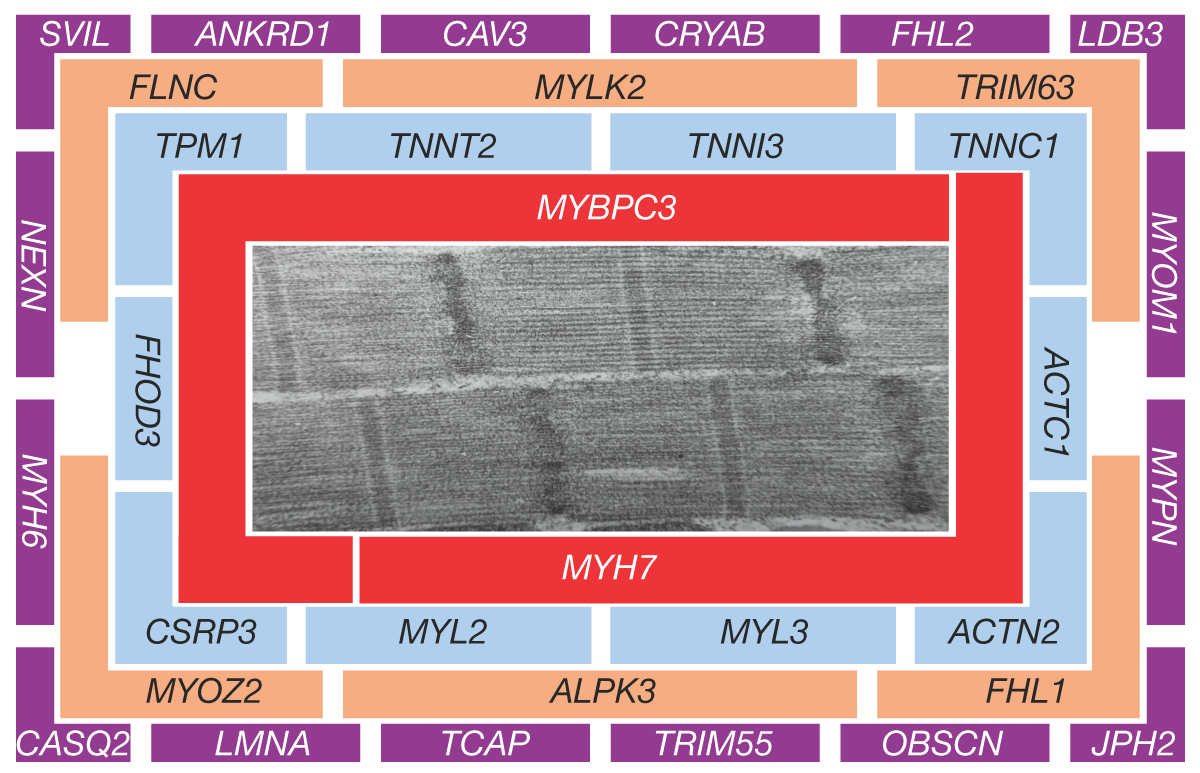
Figure 1
Hierarchical categories of genes implicated in hypertrophic cardiomyopathy (HCM). An electron micrograph showing sarcomere structure is at the center of the graph along with the partial list of genes implicated in HCM. The genes are categorized based on their frequency in HCM and their proximity to actomyosin interactions. Those at the center (category I) are well-established causal genes, and those in the periphery (category IV) have an unclear causal role.
Table 2
Criteria to assess pathogenicity of a variant in hypertrophic cardiomyopathy. HCM: hypertrophic cardiomyopathy; PV: pathogenic variants
| CRITERION | STRENGTHS | WEAKNESSES |
|---|---|---|
| Cosegregation with the disease (linkage analysis) |
|
|
| Expression in cardiac myocytes |
|
|
| Population frequency |
|
|
| Biological plausibility |
|
|
| Tolerance to genetic variation (gene constraint) |
|
|
| In silico prediction |
|
|
Table 3
Categorization of genes involved in HCM according to the strength of the genetic evidence and their frequency in the HCM population. HCM: hypertrophic cardiomyopathy; pLoF: predicted loss of function; NFAT: nuclear factor of activated T-cells; SERCA2 A: sarcoplasmic reticulum Ca2+ ATPase 2a; ATP2 A2: ATPase sarcoplasmic/endoplasmic reticulum Ca2+ transporting 2
| CATEGORY | GENE | PROTEIN | GENETIC AND BIOLOGICAL PLAUSIBILITY |
|---|---|---|---|
| I | MYBPC3 | Myosin binding protein C3 |
|
| MYH7 | Myosin heavy chain 7 |
| |
| II | ACTN2 | α-actinin 2 |
|
| CSRP3 | Cysteine and glycine-rich protein 3 (muscle LIM protein) |
| |
| FHOD3 | Formin homology 2 domain-containing 3 |
| |
| MYL2 | Myosin light chain 2 |
| |
| MYL3 | Myosin light chain 3 |
| |
| TNNC1 | Cardiac troponin C |
| |
| TNNI3 | Cardiac troponin I |
| |
| TNNT2 | Cardiac troponin T |
| |
| TPM1 | a-Tropomyosin 1 |
| |
| III | ALPK3 | a-protein kinase 3 |
|
| FHL1 | Four-and-a-half LIM domains 1 |
| |
| FLNC | Filamin C |
| |
| MYLK2 | Myosin light chain kinase 2 |
| |
| MYOZ2 | Myozenin 2 (calsarcin 1) |
| |
| TRIM63 | Tripartite motif containing 33 (muscle ring finger protein 1) |
| |
| IV | ANKRD1 | Ankyrin repeat domain 1 (CARP) |
|
| CASQ2 | Calsequestrin 2 |
| |
| CAV3 | Caveolin 3 |
| |
| CRYAB | Crystalin a/b |
| |
| FHL2 | Four-and-a-half LIM domains protein 2 |
| |
| JPH2 | Junctophilin2 |
| |
| LDB3 | Lim domain binding 3 (ZASP, CYPHER) |
| |
| LMNA | Lamin A (lamin A/C) |
| |
| MYH6 | a-myosin heavy chain, MYH6 |
| |
| MYOM1 | Myomesin 1 |
| |
| MYPN | Myopalladin |
| |
| NEXN | Nexilin |
| |
| OBSCN | Obscurin |
| |
| PLN | Phospholamban |
| |
| SVIL | Supervillin |
| |
| TCAP | Tcap (Telethonin) |
| |
| TRIM55 | Tripartite motif containing 55 (muscle ring finger protein 2, MuRF2) |
| |
| TTN | Titin |
| |
| VCL | Vinculin |
|
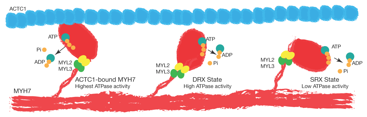
Figure 2
States of MYH7 molecules. The cartoon depicts the three states of the MYH7 molecule in the sarcomeres. A small fraction of MYH7 is in a bound state to ACTC1, has the highest ATPase activity, and generates the energy for contraction by converting ATP to ADP and inorganic phosphate. Approximately 40% to 50% of the MYH7 molecule is poised to bind to ACTC1 during actomyosin interaction. This state, which has a high ATPase activity, is referred to as the DRX state. The remaining MYH7 molecules are in resting states with their globular head folded away from ACTC1. This subset does not interact with ACTC1 during a cardiac cycle unless they are activated in response to a stimulus, such as an inotropic agent. This state is referred to as the super-relaxed (SRX) state. Mutations in HCM are thought to switch a fraction of the MYH7 molecules from an SRX to a DRX state.