
Definition
Hypertrophic cardiomyopathy (HCM), a primary disease of cardiac myocytes, is a clinical entity characterized by left ventricular hypertrophy in the absence of a known cause, such as pressure overload (unexplained cardiac hypertrophy). Classically, the triad of “unexplained” cardiac hypertrophy, a small left ventricular cavity size, and a preserved or enhanced left ventricular ejection fraction defines HCM.1,2
Diagnosis
The diagnosis of HCM is based on the detection of unexplained cardiac hypertrophy, which is often detected first on an electrocardiogram and verified, subsequently, by an imaging modality, most commonly echocardiography.2,3 A maximum left ventricular wall thickness of 13 mm or greater in adults is considered evidence of cardiac hypertrophy. In children, a Z-score of 2 or greater, as compared to the normal range of wall thickness in age- and sex-matched children, is used to indicate cardiac hypertrophy. Thus, in familial cases, a wall thickness of ≥ 13 mm suggests HCM. Additional findings, such as electrocardiographic abnormalities and the detection of a pathogenic variant (PV), would confirm the diagnosis in such cases.3 In sporadic cases, a stricter criterion of ≥ 15 mm for the maximum LV wall thickness is recommended for the diagnosis of HCM, which is designed to avoid overdiagnosis of HCM.3 The current diagnosis of HCM requires exclusion of other causes of cardiac hypertrophy, such as systemic arterial hypertension or aortic stenosis.
Cardiac hypertrophy is commonly restricted to the left ventricle but could be regional, such as in apical HCM, whereby the apex is the predominant or exclusive site of involvement. In about 35% to 50% of patients with HCM, the interventricular septum is the predominant site of hypertrophy, which is referred to as asymmetric septal hypertrophy.4,5
Prevalence
The estimated prevalence of HCM varies according to the detection methods and the population characteristics. Considering HCM is a genetic disease that affects sarcomere proteins, one can estimate the prevalence of HCM from the population frequency of pathogenic (PVs), defined as variants that are highly likely to cause a disease (Table 1), in genes encoding sarcomere proteins (see Table 1 for terminology). In the UK Biobank population, the estimated prevalence of PVs in genes encoding sarcomere proteins was 0.25% or 1:400.6 At the clinical level, the estimated prevalence of HCM varies from approximately 1:100 to 1:1000, depending on the diagnostic criteria and the population characteristics.4,7,8,9 The estimate is much higher when a maximum left ventricular wall thickness of ≥ 13 mm is used to diagnose HCM as opposed to ≥ 15 mm. The prevalence of HCM is also higher in males than in females.7
Table 1
Terminologies used in the article and their definitions. *The distinction between pathogenic and likely pathogenic variant is often unclear. In addition, pathogenicity is context-dependent. Therefore, in this article, the term “pathogenic variants” is often used rather than pathogenic and likely pathogenic variants.
| Mutation | Mutation is a change in the DNA nucleotide sequences that typically occurs during DNA replication. In clinical terms and in this article, a variant whose causal role in HCM is well-established is considered a mutation. |
| Pathogenic variant* | A variant that is expected to cause a disease. Pathogenic variant is used in the population context and whenever the causal role of the variant in the disease cannot be ascertained unambiguously. This class of variants typically includes variants that affect the stop codons (gain or loss), induce a frame shift (insertion/deletion, or splicing variant) and a subset of missense variants. |
| Likely pathogenic variant* | A variant that is likely to cause a disease, as in the pathogenic variants, but the degree of certainty of its causal role is less than that of the pathogenic variant. |
| Predicted loss-of-function variant | These variants are expected to change protein structure and function, such as non-sense, frameshift and splicing variants, which are expected to lead to premature truncation of the encoded protein. |
| Tolerance and intolerance to genetic variation | Ratio of observed to expected loss-of-function variants in a gene. The ratio is high for the tolerant and low for intolerant genes. |
Current genetic-based diagnosis, ie, the population frequency of candidate PVs, could lead to underestimating the prevalence of HCM, as PVs in the genes known to cause HCM may account for only a fraction of the cases, and PVs in yet-to-be-identified genes also cause HCM. On the other hand, the clinical diagnosis, ie, detection of a maximum left ventricular wall thickness of ≥ 13 mm, could lead to overdiagnosis of HCM because of the non-specificity of cardiac hypertrophy. However, it will also lead to underdiagnosis of the mutation carriers, particularly for PVs with low penetrance. To expand, penetrance of the causal variants, defined as the presence of any feature of the phenotype, is age-dependent. Consequently, an individual may carry the causal variant but not exhibit cardiac hypertrophy at a given age or exhibit only a mild degree of cardiac hypertrophy and be considered unaffected. In cases where cardiac hypertrophy is mild, an integrated approach that considers family history, the presence or absence of a PV, electrocardiographic tissue Doppler, and strain abnormalities, among others, should be considered in making the diagnosis of HCM.
The disparity in the prevalence of HCM between men and women is likely because of differences in the factors that affect the phenotypic expression of cardiac hypertrophy between the sexes, as HCM is not a sex chromosome-linked disease.7,10,11 HCM, except for rare phenocopy conditions, is an autosomal disease and affects males and females equally.7,10,11 A similar notion also applies to the differences in the phenotypic expression of HCM among races and ethnicities.
Molecular Genetic Basis of HCM
The familial nature of HCM and its autosomal dominant mode of inheritance were recognized more than half a century ago.12,13,14 However, identification of the first causal gene and mutation for HCM had to await the development of modern genetic linkage and sequencing technologies. Christine and Jon Seidman were the first to map the causal gene in a large Canadian family to chromosome 14 and identify the p.Arg403Gln mutation in the MYH7 gene, encoding myosin heavy chain 7 (MYH7) protein, aka the b-MYH, as the first mutation in HCM in 1990.15 The discovery had a watershed effect as it soon led to the identification of about a dozen causal genes for familial HCM,16 Soon, MYBPC3, encoding myosin binding protein C, and MYH7 were established as the two most common causal genes for familial HCM (Figure 1). Subsequently, mutations in genes encoding additional sarcomere proteins, namely TNNT2, TNNI3, and TPM1, encoding cardiac troponin T, troponin I, and a tropomyosin, respectively, were identified as causes of familial HCM.17,18 Collectively, these initial findings established familial HCM as a disease of cardiac sarcomere protein.
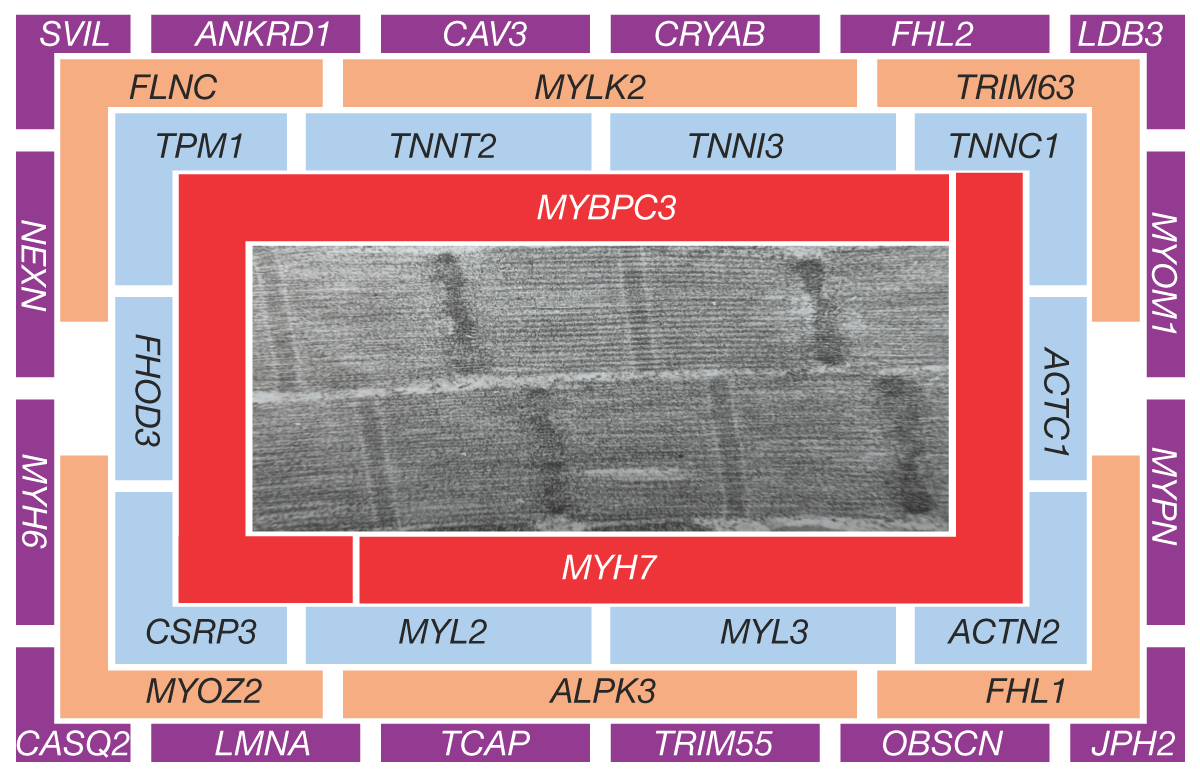
Figure 1
Hierarchical categories of genes implicated in hypertrophic cardiomyopathy (HCM). An electron micrograph showing sarcomere structure is at the center of the graph along with the partial list of genes implicated in HCM. The genes are categorized based on their frequency in HCM and their proximity to actomyosin interactions. Those at the center (category I) are well-established causal genes, and those in the periphery (category IV) have an unclear causal role.
In sporadic cases, however, identifying causal mutations and genes has been more challenging because of the presence of a very large number of genetic variants in each genome, which makes unambiguous ascertainment of causality difficult.19,20 Consequently, the causal genes in a significant subset of patients with HCM are unknown.
Advances in sequencing technologies have also led to the identification of two or more P/LPV in a subset of HCM cases, supporting the oligogenic and even polygenic nature of this subset.21,22,23,24 Establishing causality in this subset of patients with HCM has also been difficult due to the small size of the families and the sporadic nature of the cases.
Hierarchical Categorization of Genes Implicated in HCM
Over three dozen genes have been implicated as causes of HCM. However, as noted above, the strength of the evidence varies from robust to weak. Several criteria are considered when evaluating the causal role of a gene in HCM (Table 2).
Table 2
Criteria to assess pathogenicity of a variant in hypertrophic cardiomyopathy. HCM: hypertrophic cardiomyopathy; PV: pathogenic variants
| CRITERION | STRENGTHS | WEAKNESSES |
|---|---|---|
| Cosegregation with the disease (linkage analysis) |
|
|
| Expression in cardiac myocytes |
|
|
| Population frequency |
|
|
| Biological plausibility |
|
|
| Tolerance to genetic variation (gene constraint) |
|
|
| In silico prediction |
|
|
The most important evidence for the causal role of a gene or a variant is genetic, mainly evidence of cosegregation of the variant with the phenotype in the family. The evidence of cosegregation is most robust in large families but less compelling in small families and not feasible in the sporadic cases.
Along the same lines, a gene or a variant that is implicated in multiple small families or cases with HCM (recurring variant in independent families and cases) is more likely to be a causal gene or variant than one that is identified in a single small family.
A variant in a gene that is constrained to genetic variation throughout evolution is more likely to cause a disease than a variant in a gene that is tolerant to genetic variation.
A variant in a gene that encodes a protein that participates in sarcomere structure and function, particularly the generation of the contractile force, is more likely to cause HCM than a gene whose product is indirectly or not at all relevant to sarcomere function.
An important factor to consider is the expression of the candidate gene in cardiac myocytes, which is a prerequisite for its pathogenic role in HCM.
The latter two criteria, ie, the expression of the involved gene in cardiac myocytes along with the involvement of the encoded protein in cardiac sarcomere structure and function, reflect the biological plausibility of the candidate gene being causal in HCM. Based on these criteria, the implicated genes in HCM are categorized into four categories (Table 3, Figure 1).
Table 3
Categorization of genes involved in HCM according to the strength of the genetic evidence and their frequency in the HCM population. HCM: hypertrophic cardiomyopathy; pLoF: predicted loss of function; NFAT: nuclear factor of activated T-cells; SERCA2 A: sarcoplasmic reticulum Ca2+ ATPase 2a; ATP2 A2: ATPase sarcoplasmic/endoplasmic reticulum Ca2+ transporting 2
| CATEGORY | GENE | PROTEIN | GENETIC AND BIOLOGICAL PLAUSIBILITY |
|---|---|---|---|
| I | MYBPC3 | Myosin binding protein C3 |
|
| MYH7 | Myosin heavy chain 7 |
| |
| II | ACTN2 | α-actinin 2 |
|
| CSRP3 | Cysteine and glycine-rich protein 3 (muscle LIM protein) |
| |
| FHOD3 | Formin homology 2 domain-containing 3 |
| |
| MYL2 | Myosin light chain 2 |
| |
| MYL3 | Myosin light chain 3 |
| |
| TNNC1 | Cardiac troponin C |
| |
| TNNI3 | Cardiac troponin I |
| |
| TNNT2 | Cardiac troponin T |
| |
| TPM1 | a-Tropomyosin 1 |
| |
| III | ALPK3 | a-protein kinase 3 |
|
| FHL1 | Four-and-a-half LIM domains 1 |
| |
| FLNC | Filamin C |
| |
| MYLK2 | Myosin light chain kinase 2 |
| |
| MYOZ2 | Myozenin 2 (calsarcin 1) |
| |
| TRIM63 | Tripartite motif containing 33 (muscle ring finger protein 1) |
| |
| IV | ANKRD1 | Ankyrin repeat domain 1 (CARP) |
|
| CASQ2 | Calsequestrin 2 |
| |
| CAV3 | Caveolin 3 |
| |
| CRYAB | Crystalin a/b |
| |
| FHL2 | Four-and-a-half LIM domains protein 2 |
| |
| JPH2 | Junctophilin2 |
| |
| LDB3 | Lim domain binding 3 (ZASP, CYPHER) |
| |
| LMNA | Lamin A (lamin A/C) |
| |
| MYH6 | a-myosin heavy chain, MYH6 |
| |
| MYOM1 | Myomesin 1 |
| |
| MYPN | Myopalladin |
| |
| NEXN | Nexilin |
| |
| OBSCN | Obscurin |
| |
| PLN | Phospholamban |
| |
| SVIL | Supervillin |
| |
| TCAP | Tcap (Telethonin) |
| |
| TRIM55 | Tripartite motif containing 55 (muscle ring finger protein 2, MuRF2) |
| |
| TTN | Titin |
| |
| VCL | Vinculin |
|
Category I genes benefit from the strongest evidence in all criteria, including genetic linkage and biological plausibility. They are the most common causes of HCM. The genes in category I include MYBPC3 and MYH7, which together are responsible for about 35% to 40% of HCM cases, particularly in large families.10,25,26
The genes in category II also benefit from strong genetic evidence for their causal role in HCM as well as biological plausibility. However, some are tolerant to genetic variations, and all are uncommon causes of HCM. The list includes TNNI3, encoding troponin I, TNNT2, encoding cardiac troponin T, and MYL2, encoding myosin light chain 2, among others. Genes in this category account for about 10% of HCM cases.25,26
Genes in category III either do not benefit from strong genetic evidence or lack a strong biological plausibility. The genes in this category are typically tolerant to genetic variation and are rarely implicated in HCM. These genes code for proteins that are associated with sarcomere structure and function and therefore benefit from biological plausibility.
Finally, the genes in category IV include those that are typically associated with HCM but do not have an established and defined causal role. These genes are typically tolerant of genetic variation or do not benefit from a strong biological plausibility.
The Common Causal Genes for HCM
The familial nature of HCM and its transmission in an autosomal dominant Mendelian pattern, indicating a monogenic etiology, was recognized by Pare more than half a century ago.14 Linkage analysis in the family described by Pare led to identification of the first causal gene, namely MYH7, for HCM and consolidated HCM as a single gene disorder.15 In about two-thirds of patients, HCM is a familial disease and, by and large, monogenic, ie, a single gene disorder.27 In a single-gene disorder as opposed to oligogenic conditions, a single mutation is sufficient to cause the disease, even though the expression of the phenotype depends on the penetrance of the mutation and is often determined by numerous genetic and environmental modifiers.28 As a single-gene disorder, HCM is genetically heterogeneous, meaning that a single mutation in several distinct genes is sufficient to cause HCM. This contrasts with oligogenic HCM, whereby two or more genetic variants, typically in different genes, cause HCM. In monogenic HCM, the established causal genes encode sarcomere proteins, indicating that it is a disease of sarcomere proteins.
MYBPC3: Mutations in the MYBPC3 gene are the most common cause of HCM because they are responsible for about 20% of cases.25,26 The gene encodes MYBPC3, which is a large sarcomere protein that interacts with MYH7, cardiac α actin (ACTC1), and giant sarcomere protein titin (TTN).29 These interactions, which are regulated by post-transcriptional modifications of MYBPC3, stabilize sarcomere structure and regulate its function.30,31
Several hundred mutations in the MYBPC3 gene have been identified in patients with HCM. The population frequency of each MYBPC3 mutation in HCM is less than 1%, except for the founder mutations, which typically occur in isolated populations.10,25,26,32,33,34 The majority of MYBPC3 mutations are heterozygous nonsense, frameshift indels, and splice site variants.10,25,26 Such mutations introduce a premature stop codon and lead to the expression of unstable truncated transcripts, which are degraded by the RNA quality control mechanisms, such as nonsense-mediated decay.35,36 Transcripts that escape the nonsense-mediated decay are degraded by the ubiquitin-proteasome system.35 Therefore, the full-length MYBPC3 protein is expressed from one copy of the MYBPC3 gene but not from the mutant copy of the gene, and hence, haplo-insufficiency.37,38,39
Functionally, the MYBPC3 protein inhibits actomyosin interactions and the generation of the contractile force.40,41,42 Accordingly, MYBPC3 binds to the neck and tail regions of the MYH7 protein, sequesters the MYH7 globular head in a folded position, and prevents the binding of MYH7 to ACTC1.43 Reduced level of the MYBPC3, such as in the haploinsufficiency state, increases the number of the MYH7 globular heads that are available to bind to ACTC1 and generate the contraction force during a cardiac cycle.44,45 Consequently, the deficiency of MYBPC3 removes the inhibitory effect of MYBPC3 on actomyosin interactions and leads to increased cardiac myocyte contractility.44,45 The mechanisms by which an increased force of contraction induces a hypertrophic response of the myocardium is not well-defined but likely involves the activation of several signaling pathways upon the expression of stress-responsive trophic and mitotic factors.
MYH7: The MYH7 gene codes for the MYH7 protein, which is the most abundant sarcomere protein in cardiac myocytes. Mutations in the MYH7 gene, the first gene identified for HCM, are found in about 15% of HCM cases, making it the second most common causal gene in HCM.10,15,25,26
Most mutations in the MYH7 gene, in contrast to those in the MYBPC3 gene, are missense mutations that are mainly located in the globular head and neck domains of the protein. Mutations in the rod domain are rare causes of HCM and are typically associated with skeletal myopathy.46,47,48,49 As in the other causal genes, the population frequency of each mutation in HCM is low, except for those identified in isolated populations.25,26,50
MYH7 is the motor unit of the sarcomeres. It utilizes adenosine triphosphate (ATP) to move the thin filament and generate the force of contraction. Mutations in the MYH7 protein could affect various components that are involved in actomyosin interactions, such as ATPase activity, displacement of the globular head of myosin over the thin filaments, and the release of MYH7 from the thin filament.
In a working cardiac muscle, the MYH7 hexameric molecule (2 MYH7, 2 MYL2, and MYL3 proteins) exists in three different states in terms of ATPase activity (Figure 2). A small fraction of the MYH7, estimated to be < 10%, is bound to ACTC1 at any given time during a cardiac cycle. This subset of the MYH7 molecule has the highest ATPase activity.51,52,53 Approximately 40% to 50% of the MYH7 molecules are poised to bind to ACTC1, have a high level of ATPase activity, and are ready to contribute to the generation of the contractile force during a cardiac cycle.51,52 This state of the MYH7 molecule is referred to as the disordered-relax state (DRX).53 The remaining MYH7 molecules have their globular head domains folded away from the thin filaments and hence do not interact with ACTC1 in a resting state. This subset of MYH7 is in the super-relaxed state (SRX) and has a very low ATPase activity.51,52,53 The SRX state of MYH7 is presumed to serve as a reserved capacity that interacts with ACTC1 in response to stimuli requiring a higher force of contraction.
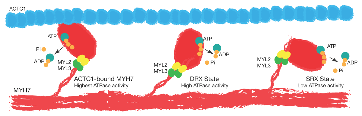
Figure 2
States of MYH7 molecules. The cartoon depicts the three states of the MYH7 molecule in the sarcomeres. A small fraction of MYH7 is in a bound state to ACTC1, has the highest ATPase activity, and generates the energy for contraction by converting ATP to ADP and inorganic phosphate. Approximately 40% to 50% of the MYH7 molecule is poised to bind to ACTC1 during actomyosin interaction. This state, which has a high ATPase activity, is referred to as the DRX state. The remaining MYH7 molecules are in resting states with their globular head folded away from ACTC1. This subset does not interact with ACTC1 during a cardiac cycle unless they are activated in response to a stimulus, such as an inotropic agent. This state is referred to as the super-relaxed (SRX) state. Mutations in HCM are thought to switch a fraction of the MYH7 molecules from an SRX to a DRX state.
The prevailing hypothesis in the pathogenesis of HCM caused by MYH7 mutations posits that the mutations increase the number of MYH7 molecules that are in the DRX state and shift their distribution across the sarcomere.41,44,45,54,55,56,57 Consequently, the shift increases the number of MYH7 heads that are ready to interact with ACTC1 and generate the contraction force. The shift also leads to a higher ATPase activity and the depletion of myocardial ATP levels, which impairs the release of MYH7 from the thin filaments and hence impairs myocardial relaxation.58,59,60
The partial elucidation of the molecular pathogenesis of HCM has led to the development of specific therapies that target MYH7 conformational changes, which are called myosin inhibitors or modulators.55,61,62 Two myosin inhibitors, namely mavacamten and aficamten, have been approved and shown to be effective in reducing the left ventricular outflow tract (LVOT) gradient.63,64 However, their clinical efficacy in improving patients’ symptoms and exercise capacity is modest and restricted to a subset of patients with HCM with LVOT obstruction.63,64 These drugs are ineffective in HCM without LVOT obstruction.65
MYL2 and MYL3 genes: These genes encode the regulatory and essential light chain proteins, also known as MYL2 and MYL3, respectively, which are the components of the thick filaments of sarcomeres. Functionally, MYL2 and MYL3 bind to the lever arm of MYH7 and contribute to contractile force generation.66 Mutations in the MYL2 and MYL3 genes are rare causes of HCM.67 Mechanistically, mutations in these two genes seem to affect MYH7 conformation and shift the SRX toward DRX state.56
Genes encoding the components of the thin filaments: The thin filaments are comprised of ACTC1 and the troponin/tropomyosin complex. The latter complex is comprised of cardiac troponin T, troponin I, troponin C, and a tropomyosin. Mutations in the genes coding for the thin filament proteins collectively account for < 10% of the HCM cases.17,18,68
The TNNT2 gene was among the first set of genes identified for HCM.17 The TNNT2 protein is the largest subunit of the troponin-tropomyosin complex and positions the complex on ACTC1.69 The TNNI3 protein is the inhibitory protein of the complex. The C-terminal domain of TNNI3 binds to ACTC1 and occupies the binding site of the MYH7 protein, blocking the binding of MYH7 to ACTC1. The TNNC1 protein is the calcium-sensing unit of the troponin complex. It is activated upon binding of calcium and induces conformational changes in the inhibitory arm of TNNI3, shifting it away from the binding site of ACTC1 to MYH7. The shift enables binding of MYH7 to ACTC1 and the flexion of the MYH7 globular head over thin filaments.69 Finally, the TPM1 protein forms a dimeric coiled-coil α helical structure that overlays the ACTC1 in the thin filament and places the troponin/tropomyosin complex on the ACTC1 filaments.
The ACTC1 protein, encoded by the ACTC1 gene, is the key MYH7 partner in the generation of the contractile force. The process is initiated upon calcium entry into the cytosol, which binds to TNNC1 and induces conformational changes that affect its binding to TNNI3. The binding induces further conformational changes in TNNI3, which exposes the binding domain MYH7 on the ACTC1 protein. The process induces the flexion of the MYH7 globular head over the actin filaments and the displacement of the latter, which generates the contraction force. The ACTC1 gene is strictly intolerant to genetic variation (ie, it has a very low ratio of observed/expected predicted LoF variants) and is rarely implicated in HCM.70,71
Selected gene encoding Z-disc proteins: A near-complete list of genes implicated in HCM is presented in Table 2. Notable among the remaining genes are a few that encode Z-disc proteins, such as ACTN2, CSRP3, FHL1, and MYOZ2.
The ACTN2 gene encodes actinin α2, which cross-links ACTC1 and TTN to Z discs.72 PVs in the ACTN2 gene are associated with HCM as well as skeletal myopathy, which is in agreement with the high level of expression of this gene in cardiac and skeletal muscles.72,73,74
The CSRP3 gene codes for cysteine and glycine-rich protein 3 (CSRP3) protein, which is also known as muscle LIM protein. The CSRP3 gene has been linked to HCM through genetic linkage studies in a large family with HCM.75 However, PVs in the CSRP3 gene are very rare in patients with HCM.
The FHL1 gene, located on the X chromosome, encodes the four-and-a-half LIM domain 1 protein, which is also a Z disk protein involved in sarcomere organization and function.76 Expression of the FHL1 gene is upregulated in the heart in HCM.77 PVs in the FHL1 gene have been associated with skeletal myopathies and rarely with HCM.78,79,80
The data implicating the MYOZ2 gene in HCM is based on a suggestive linkage score and case reports.81 The encoded Z-disc protein myozenin 2 (MYOZ2), also known as calsarcin 1, interacts with other Z-disc proteins such as α-actinin and regulates the calcineurin signaling pathway.82 However, the calcineurin pathway does not seem to be involved in the pathogenesis of HCM associated with the MYOZ2 variants.83,84
Genes encoding sarcomere-associated proteins: The FHOD3 gene encodes formin homology 2 domain-containing 3, which binds to ACTC1 and MYBPC3 and regulates ACTC1 organization and sarcomere function.85 Heterozygous and homozygous PVs in the FHOD3 gene have been associated with cardiomyopathies, including HCM, through population studies including genome-wide association studies (GWAS).86,87,88,89,90
PVs in the FLNC1 gene have been associated with cardiomyopathies, including HCM.91,92 The encoded filamin C protein is expressed in cardiac myocytes and skeletal muscle and is implicated in maintaining the structural integrity of the sarcomeres.93
Post-translational modifications of sarcomere proteins, including MYL2, in addition to MYBPC3, as discussed earlier, regulate sarcomere function. Myosin light chain kinase 2 (MYLK2), encoded by the MYLK2 gene, is a calcium/calmodulin-dependent serine/threonine protein kinase and a post-translational regulator of sarcomeres, as it phosphorylates MYL2, which affects the SRX states of MYH7.56,94 Rare PVs in the MYLK2 gene are associated with HCM.95
Oligogenic HCM: Advances in DNA sequencing technologies shifted the genetic studies from linkage analysis in large families to sequencing the candidate genes, initially, and whole-exome sequencing subsequently, particularly in small families and sporadic cases. The approach has led to the identification of two or more PVs in patients with HCM, rendering a subset of HCM as an oligogenic disease21,23,24,96,97 Overall, the data suggest that approximately 5% of HCM cases carry at least two PVs in genes implicated in HCM.21,23,24,96,97 The oligogenic HCM encompasses only genes that are already known to cause HCM and is challenging to extend it to the new genes because establishing the causality of the variants unambiguously in cases and small families is almost impossible. The ambiguity arises from the presence of over 4 million genetic variants in each genome, including PVs in genes implicated in HCM, which makes the ascertainment of the causal and non-causal variants very challenging.98
Genes identified through GWAS: The established causal genes for HCM, discussed above, account for approximately half of the HCM cases.25,26,99,100,101 In the remainder, the causal gene and variant have not been identified. The so-called “missing causal genes,” along with the recognition that a subset of HCM is oligogenic and likely polygenic, led to genome-wide association studies to identify the susceptibility loci for HCM.
A partial list of genes implicated in HCM through GWAS is provided in Table 2 and Figure 2, mostly under Category IV genes. Overall, the results of the GWAS studies have been unremarkable as the mapped loci either contain a known causal gene, such as MYBPC3, FLNC, and FHOD3, or do not identify a biologically or genetically plausible candidate gene. In addition, because of the presence of over 4 million genetic variants, including PVs in each genome, establishing the causal role of the loci and variants identified through GWAS studies is very challenging.19,20,102,103 Moreover, the mapped loci typically contain several genes and are compounded by the presence of large linkage disequilibrium in the human genomes, which makes identification of the susceptibility allele extremely challenging.104
Among the GWAS candidate genes in HCM, besides the known ones, are TRDN, encoding Triadin, and TMEM182, encoding transmembrane protein 182, which are expressed in cardiac and skeletal muscles and regulate calcium release and muscle differentiation, respectively.90 Another notable example is the SVIL gene, which encodes supervillin, an actin-binding protein.90 The protein, which is expressed mainly in skeletal and smooth muscles and to some extent in cardiac myocytes, anchors ACTC1 to the plasma membrane and is implicated in cytokinesis.105,106 Truncating variants in the SVIL gene are estimated to increase the risk of developing HCM by tenfold.90
Genetic Testing
Genetic testing is now part of the routine clinical evaluation of patients and their genetically related family members (cascade screening). The approach is targeted gene sequencing, comprised of about 100 genes that are implicated in cardiomyopathies. Pathogenicity of the variants identified in genetic screening is analyzed using various algorithms and literature/database searches to determine whether the variant has been reported in HCM. In addition, biological and biochemical characteristics of the variants, and tolerance of the gene and domain to genetic variation, among others, are considered.
Genetic testing provides several benefits. The extension of the genetic screening to the family members not only enables establishing the causal role of the variants through Cosegregation analysis but also provides the opportunity to identify family members who carry the PVs and, therefore, are at an increased risk of developing HCM. The identification enables close monitoring of the evolving phenotype and intervening as needed. The approach has the potential to reduce the risk of unexpected outcomes, including sudden cardiac arrest.
Genetic testing also enables discerning HCM caused by mutations in genes encoding sarcomere proteins from the phenocopy conditions. The distinction is not trivial, as it has both prognostic and therapeutic implications. For example, it is increasingly recognized that characteristics of patients with HCM caused by mutations in genes encoding sarcomere proteins differ from those in whom no PV is identified.10,99,100,101 Likewise, the pathogenesis and treatment of HCM differ from the HCM phenocopy conditions, such as storage diseases.
The diagnostic yield of genetic testing remains relatively low, with pathogenic variants identified in only about one-third of cases.107,108,109 This limited yield reflects, in part, the current focus of genetic screening on known HCM associated genes and variants that are expected to significantly alter protein structure or function. For example, the regulatory and the non-coding regions, which contain more than 98% of the identified genetic variants in the human genome, are not analyzed.103,110 There are a few other technical issues that limit the yield of genetic testing.111
The clinical interpretation of genetic testing results and discerning the causal role of the identified variants are often challenging. Several criteria are considered to assess the pathogenicity of the PVs that are identified through genetic testing in HCM, as discussed earlier (Table 2). The strongest evidence for the causal role of a variant in HCM is cosegregation of the variant with the disease (linkage analysis) in several related family members. Unfortunately, the family size is often too small, and cosegregation in small families could occur by chance alone. Likewise, penetrance is typically age-dependent, and a low penetrance could complicate cosegregation analysis. Considering the above, genetic screening should be extended to all informative family members to strengthen the evidence for the cosegregation or refute it.
The candidate gene carrying the PV must be expressed in cardiac myocytes, as HCM is mainly a disease of cardiac myocytes even though the ensuing phenotype is a multicellular biological process. The population frequency of the variant is also an important factor, particularly for monogenic HCM. Considering the genetic heterogeneity of HCM, the population frequency of the candidate variant is expected to be much less common than the population frequency of HCM. In general, rare variants typically exert large effect sizes, whereas the common variants typically have no discernible or small effect sizes.98
HCM is considered mainly a disease of genes encoding sarcomeres or sarcomere-associated proteins. Consequently, a PV in a gene that encodes a sarcomere protein or a protein that is known or expected to affect the sarcomere structure and function is more likely to cause HCM than those that are not related to the sarcomere. Likewise, a variant in a gene that is intolerant to genetic variation is more likely to be pathogenic than a variant in a gene that is tolerant to variation. Finally, the existing knowledge on amino acid and protein biochemistry has led to the development of in silico algorithms to predict the pathogenicity of the genetic variants. The programs include changes in the amino acid structure and function and the predicted effects of the amino acid change on the structure and function of the involved protein. The in silico algorithms, while powerful, are not conclusive. They should not be used alone to determine the causal role of a variant in HCM.
Perspective
In roughly half of HCM cases, the causal gene has not been identified, a pattern most commonly seen in small families and sporadic presentations. The likely explanation for these “missing causal genes” is that establishing a definitive causal variant in isolated cases or limited pedigrees is extremely difficult with current methods. As a result, even when a pathogenic variant is detected, its causal role cannot be confirmed unless it is a well-established variant in a gene known to cause HCM. Given these constraints, it may be more appropriate to shift from a deterministic framework—labeling a variant as definitively disease causing—to a probabilistic one, in which a pathogenic variant in a gene encoding a protein involved in sarcomere structure or function is considered a likely causal contributor.
Current genetic screening programs focus on the protein-coding regions of genes and do not typically include the regulatory regions and structural variants, including copy number variants. Consequently, the approach could lead to missing a fraction of the causal genes and variants that are not located in the protein-coding regions. This deficiency may contribute to a relatively low yield of genetic testing, which is currently about 30%.107,108,109
Given the disparity in the prevalence of HCM, diagnosed based on genetic and clinical criteria, it is also possible that the clinical diagnosis of HCM, based on the detection of unexplained cardiac hypertrophy, lacks specificity and leads to its overdiagnosis. This point might be particularly relevant for the mild cases of cardiac hypertrophy. The notion is supported by recent data that suggest HCM caused by mutations in genes encoding sarcomere proteins has a distinct set of characteristics from those lacking mutations in such genes.10,99,100,101 Given these findings, considering that the pathogenesis of HCM and the clinical outcomes might vary according to the causal gene, it might be prudent to shift from a clinically defined to a genetically defined HCM. Likewise, given our increasing understanding of the differences in the underpinning mechanisms involved in the pathogenesis of HCM, it might be preferable to define HCM according to the causal genes, such as MYH7-HCM, MYBPC3-HCM, or TNNI3-HCM.112 The latter is also important as gene therapy and base-editing technologies are rapidly evolving and are increasingly being applied to the cure of human genetic disorders, including HCM.113,114
Speculations
To overcome the current limitations in the diagnosis and treatment of HCM, new approaches are expected to emerge. At the genetic level, the screening approach is expected to shift from the candidate gene and exome sequencing approaches, which have significant limitations, to whole-genome sequencing approaches using low-error-rate techniques.111 To identify the likely causal variants, the whole-genome sequencing output will be subjected to meticulous algorithms that are built upon biological and genetic plausibility and utilize a probabilistic approach assisted by artificial intelligence. The genetic findings will be complemented with multiomics data, including cardiac and circulating metabolomics, to gain insights into the involved pathogenic pathways. The findings will be used not only to support the causal role of the genetic variants but also to identify new therapeutic targets in HCM. These advances are expected to shift the diagnosis of HCM from a clinical-based to a genetic-based approach.
Elucidation of new and detailed mechanisms involved in the pathogenesis of HCM would be expected to lead to the development of new mechanistic-based drugs besides the existing MYH7 inhibitors that mainly target the contraction force.55,62 Moreover, given that current medications have modest, if any, effects on diastolic dysfunction, new medications are expected to be developed to improve cardiac diastolic function in HCM. Mechanistic studies have already provided a major potential target, namely, the dissociation sensitivity of the actomyosin complex to intracellular ATP concentration.115 It is plausible that impaired sensitivity of actomyosin dissociation in response to ATP concentration, which is already depleted in HCM, is the underpinning mechanism of diastolic dysfunction.58,116 This mechanism is likely a more potent determinant of clinical symptoms in HCM than LVOT obstruction or increased contractility. Thus, by and large, the current myosin inhibitors, which target sarcomere force generation, do not address the main mechanism in HCM and hence have limited clinical utility.63,64 The next generation of medications would be expected to increase the dissociation rate of MYH7 from ACTC1 in response to a given ATP concentration, thereby improving diastolic function in HCM. Likewise, interventions to replenish the depleted cellular ATP in HCM would be expected to enhance actomyosin dissociation and hence improve cardiac diastolic function. Nevertheless, a heterogeneous set of mechanisms, partly dependent on the underlying causal variant(s) and the involved gene(s), are likely to be involved in the pathogenesis of HCM, mandating specific genes and mutation-specific therapies. The latter would likely advance the base-editing approaches to the forefront of individualized treatment of HCM patients. Thus, the road ahead is long and winding, and the journey is tedious. To quote Sir Winston Churchill, “Every step may be fruitful. Yet there will stretch out before you an ever-lengthening, ever-ascending, ever-improving path. You know you will never get to the end of the journey. But this, so far from discouraging, only adds to the joy and glory of the climb.”
Key Points
Hypertrophic cardiomyopathy (HCM) is a classic single-gene disorder.
Mutations in over three dozen genes are implicated as causes of HCM.
HCM is an oligogenic disease in a subset of patients (caused by two or more mutations).
The MYBPC3 and MYH7 genes, encoding myosin binding protein C3 and myosin heavy chain 7, are the two most common causal genes for HCM.
The established genes code for sarcomere and sarcomere-associated proteins.
Genetic testing, including cascade screening, is now part of the routine care of patients with HCM and helps identify the causal mutation in about one-third of patients with HCM.
Elucidation of the molecular genetic basis of HCM has paved the way for specific therapies, ushering in the era of gene therapy and base-editing.
Acknowledgements
The following public databases were used in this article to extract population frequency of the PVs and gene expression: